www.issisglobal.org 99
Journal of International Society for Simulation Surgery 2014;1(2):99-102
서 론
두개골 결손은 선천성인 원인 뿐 아니라, 두부 외상, 신경외과 수술 이후에 드물지 않게 발생하며, 이러한 두개골 결손 재건은 오랫동안 성형외과, 신경외과영역에서 다양한 방법으로 이루어 져왔다 (1-3).
두개골 재건을 위해서 전통적으로 자가조직을 이용하는 것이 가장 일반적으로 알려져 왔다. 두개골의 정상측 공여부에서 두 개골을 채취하여 얇은 두 조각으로 나누어 한 조각은 수혜부에 이식하고, 나머지 한 조각을 공여부에 다시 이식함으로써 두개골 결손을 치료하거나, 장골등에서 내측골편이나 늑골을 이용하는 방법이 표준적인 방법으로 오랜기간 사용되어 왔다. 하지만, 90년
대부터 각종 골 지지체의 발전으로 Hydroxyapatite, Tricalcium phosphate, PMMA (polymetylmetacrylate) 등이 자가골의 대용으로 사용되기 시작하여 많은 부분에서 사용되고 있다. 그럼 에도 자가골에 비해 이물질 반응이 많고, 염증이나 노출시 상처 치유가 되지 않는 등의 다양한 문제가 있어 이상적인 대체재로 서는 인정되기 어려운 상황이다.
이에 반해 약 30여년간 의료에 사용되어온 티타니움은 그 생 체적합성이 상대적으로 가장 우수하여 타 물질에 비해 큰 부작 용 없이 사용되어 오고 있다. 하지만, 물성 자체를 기존 뼈의 두께 만큼 만들기 어렵고, 모양을 미리 생성하는데 어려움이 있어온 것이 사실이다 (4, 5).
1990년부터 3D Printing의 발전으로 의료에 술전 계획 및 진
Case Report
3D Printed Titanium Implant for the Skull Reconstruction:
A Preliminary Case Study
Jong-Woo Choi, M.D., Ph.D., M.M.M., Jae-Sung Ahn, M.D., Ph.D.
1Department of Plastic and Reconstructive Surgery, 2Neurosurgery College of Medicine, Ulsan University, Asan Medical Center, Seoul, Korea
The skull defect can be made after the trauma, oncologic problems or neurosurgery. The skull reconstruction has been the chal- lenging issue in craniofacial fields for a long time. So far the skull reconstruction with autogenous bone would be the standard.
Although the autogenous bone would be the ideal one for skull reconstruction, donor site morbidity would be the inevitable problem in many cases. Meanwhile various types of allogenic and alloplastic materials have been also used. However, skull re- construction with many alloplastic material have produced no less complications including infection, exposure, and delayed wound healing. Because the 3D printing technique evolved so fast that 3D printed titanium implant were possible recently.
The aim of this trial is to try to restore the original skull anatomy as possible using the 3D printed titanium implant, based on the mirrored three dimensional CT images based on the computer simulation. Preoperative computed tomography (CT) data were processed for the patient and a rapid prototyping (RP) model was produced. At the same time, the uninjured side was mir- rored and superimposed onto the traumatized side, to create a mirror-image of the RP model. And we fabricated Titanium im- plant to reconstruct three-dimensional orbital structure in advance, using the 3D printer. This prefabricated Titanium-implant was then inserted onto the defected skull and fixed.
Three dimensional printing technique of titanium material based on the computer simulation turned out to be very successful in this patient. Individualized approach for each patient could be an ideal way to manage the traumatic patients in near future.
Key WordsZZSkull defect ㆍThree-dimensional surgery ㆍCalvarial reconstruction ㆍSkull reconstruction ㆍPre-surgical simulation ㆍThree-dimensional simulation ㆍD printing ㆍ3D printed titanium implant ㆍTitanium.
Received: November 24, 2014 / Revised: November 25, 2014 / Accepted: December 1, 2014 Address for correspondence: Jong-Woo Choi, M.D., Ph.D., M.M.M.
Department of Plastic and Reconstructive Surgery, Seoul Asan Medical Center, 88 Olympic-ro, 43-gil, Songpa-gu, Seoul 138-736, Korea Tel: 82-2-3010-3604, Fax: 82-2-476-7471, E-mail: [email protected]
pISSN 2383-5389 / eISSN 2383-8116
100
Journal of International Society for Simulation Surgery █ 2014;1(2):99-102
단에 큰 도움이 되었고, 최근에는 이러한 3D printer로 직접 티 타니움 소재를 우리가 원하는 모양으로 출력할 수 있게 되어, 이 를 두개골 결손환자에서 사용한 증례를 소개하고자 한다 (4-6).
증 례 보 고
57세의 여자 환자는 2년전 후교통 동맥에서 기원한 지주막하 출혈로 신경외과에서 응급 개두술 및 좌측 후교통 동맥류 결찰 술을 시행받았고, 이후 두부 상처에서 감염소견 보여 두개골 절 제술 및 창상 감염 세척술을 시행하였다. 우측 두개골 결손 부위 는 장기화된 감염소견을 바로 재건이 불가능하였으며, 2년뒤 신 경외과와 성형외과 협진으로 두개골 재건을 하기로 계획하였다.
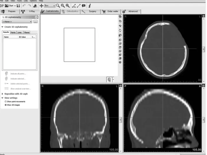
이에 CT 촬영을 포함한 통상의 술전 검사를 시행하였으며 3 차원 물체로 재구성한 환자의 두개안면골에 대해 악안면기형 진단 및 분석을 시행하였다. 삼차원 CT scan을 촬영하였고, CT data를 Simplant (simplant pro, MaterialsⓇ, Belgium)를 이용 하여 분석한 후 컴퓨터 시뮬레이션 수술을 계획하였다 (Fig. 1).
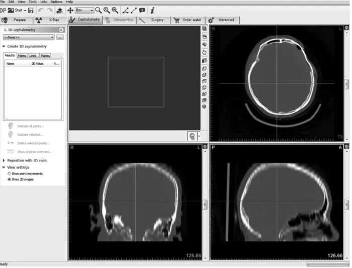
CT DICOM 파일을 기반으로 환자의 두개골을 3차원 물체로 재구성 하였으며, 좌측의 정상두개형태의 CT data를 mirroring 기법 (Mimics Z software: Materialize, Leuven, Belgium) 을 이용하여 우측에도 적용하여 환자가 다치기 전의 양측 두개 골의CT 이미지를 얻었다. 이를 3D 프린팅을 통해 제작함으로써 실제 수술 중 두개골 결손을 복원하기 위한 3D printer를 이용 한 titanium implant을 위치 시키기 위한 기준으로 사용하였다 (Fig. 2).
Titanium 소재로 제작된 3차원 티타니움 임플란트를 이용 하여 통상의 두개골 복원수술을 진행하였으며 기존의 두피절개 선을 이용하여, 두개골 결손부위를 노출시키고, coagulator와 blade로 경막과 두피를 박리하였다. 3차원 컴퓨터 시뮬레이션을 이용해 사전에 제작하여둔 Titanium implant를 삽입하여 두개 골을 복원하였다. 환자는 수술 후 감염, 혈종 등의 합병증이 발생 하지 않았으며, 특이한 문제 없이 술 후 5일째 퇴원하였다. 술 후 3일째 3개월째 술 후 CT scan을 통해 수상전과 유사하게 복원 되었음을 확인할 수 있었다 (Fig. 3).
Fig. 1. Preoperative CT scan.
3D Printed Titanium Implant for the Skull Reconstruction: A Preliminary Case Study █ Choi JW, et al
www.issisglobal.org 101
고 찰
두개골의 형태는 각 개인, 인종, 성별에 따라 다른 것으로 알 려져 있으며, 두개골 역시 각 개인마다 조금씩 그 크기가 뼈의
위치 및 각도가 다르다. 하지만, 지금까지 일반적으로 수술을 함에 있어 이러한 환자 개인의 차이가 수술에 반영되기는 어려 웠다 (1). 특히나, 두개골 복원 수술의 경우에는 절대 손상이 되 어서는 안 되는 뇌의 경막과 인접되어 수술이 진행되어야 하고, 특히 두개골의 측두부는 다양한 굴곡을 가진 복잡한 형태의 골 조직이어서, 자가골을 이용한 재건시에도 매우 큰 어려움이 따르 는 부분이다. 따라서 이러한 두개골 결손의 복원을 위해 사용되 는 임플란트의 경우에는 미리 그 모양을 제작하여 삽입하는 형태 로 수술이 진행될 수 있다면 가장 이상적이다. 하지만, 이러한 사 전 제작을 위해서는 환자의 원래 두개골의 형태를 알 수 있어야 하는데, 동측은 이미 결손부위가 되어 정확한 이미지를 얻을 수 없으므로, 반대편 건측 두개골이 가장 좋은 대안이 될 것이 다. 이를 이용한 기법이 Mirroring technique이다. 본 증례에 서 우측 두개골 복원을 위해 이 미러링 기법을 이용하였는데, 만 족할 만한 결과를 얻을 수 있었다. 이는 통상 대부분의 경우 양 측 얼굴이 2mm 이내의 대칭성을 가지고 있는 것으로 알려져 있 고, 반대측 건측이 가장 좋은 landmark가 될 수 있기 때문이다.
자가골이 현재까지 가장 이상적인 골 결손 재료임은 이론의 Fig. 2. Postoperative CT scan.
Fig. 3. Postoperative picture.
102
Journal of International Society for Simulation Surgery █ 2014;1(2):99-102
여지가 없으나, 두개골 결손의 경우 공여부에 문제가 생길 여지 가 있으며, 측두부와 같이 골조직의 형태가 복잡한 경우에는 이 를 정확하게 재건하는데 한계가 있는 것이 사실이다 (1).
또한, 기존의 다양한 골 대체재 등을 사용하는 경우에도 감 염 및 노출시의 창처치유 지연 등의 문제가 크다. 게다가 충분한 골전도가 이루어 지지 못하는 경우가 많고, 파우더 형태의 지지 체의 경우에는 큰 골 결손은 재건하는데 한계가 있다.
이에 반해 티타니움 제재는 30여년간 의료의 다양한 분야에 서 실제 인체에 적용되어서 큰 문제 없이 사용되어 온 물질로, 골대체재 alloplastic material 중에는 비교적 생체적합성이 우수한 것으로 알려져 있다 (4, 5, 7). 하지만, 주로 티타니움 메 쉬형태나, 플레이트, 스크류 형태 등으로 주로 사용되어 왔고, 커 다란 골 결손시 사용될 경우에도 메쉬형태의 막을 재건하는 정도 가 최선이었다.
90년대부터 발전되기 시작한 3D Printing 기법이 최근 의료에 더욱 활발하게 적용되면서, 이제는 티타니움 소재 자체를 3D printer로 바로 출력할 수 있는 기술까지 도입되었다. 이를 임상 에 적용한 초기의 경험으로, 본 증례를 바탕으로 살펴보면, Ti- tanium-implant를 이용하여 두개골을 재건함으로써 술후 CT scan에서 반대편 건측과 유사하게 우측의 두개골이 잘 재건되었 음을 확인할 수 있었고, 본 거울영상을 이용한 3차원 컴퓨터 시뮬 레이션 기법을 이용해 환자 본인의 두개골을 최대한 원래에 가 깝게 복원할 수 있었으며, 이러한 접근은 미래의 individualized medicine의 현실적인 접근방법의 하나로 간주 될 수 있을 것으 로 판단된다.
결 론
두개골 결손은 전통적으로 술자의 경험에 의존하여 주변 두
개골을 노출 시킨 후, 결손 부위를 자가골이나 다양한 임플란트 로 대체하는 형태의 수술이 주로 이루어져왔으나, 자가골, 골 대 체재 등의 사용에 있어 문제점과 한계가 있음을 감안하였을 때, 이러한 3차원 컴퓨터 시뮬레이션을 기초로 3D Printed Titanium implant를 이용한 두개골 재건은 individualized medicine의 첫걸음이자, 매우 유용한 이상적인 수술 방식임을 확인 할 수 있 었다.
참 고 문 헌
1. Chang TJ, Choi JW, Ra YS, Hong SH, Cho YH, Koh KS. Changes in graft thickness after skull defect reconstruction with autogenous split calvarial bone graft. The Journal of Craniofacial Surgery 2014;
25(4):1241-1244.
2. Habal MB. Repair of a skull defect by a split cranial bone graft. The Journal of Craniofacial Surgery 1992;3(4):230-234.
3. Liao CC, Kao MC. Cranioplasty for patients with severe depressed skull bone defect after cerebrospinal fluid shunting. Journal of clini- cal neuroscience: Official Journal of the Neurosurgical Society of Australasia 2002;9(5):553-555.
4. Vignes JR, Jeelani N, Dautheribes M, San-Galli F, Liguoro D. Cra- nioplasty for repair of a large bone defect in a growing skull fracture in children. Journal of cranio-maxillo-facial surgery: official publi- cation of the European Association for Cranio-Maxillo-Facial Sur- gery 2007;35(3):185-188.
5. Yamamoto Y, Minakawa H, Yoshida T, Igawa H, Sugihara T, Ohura T, et al. Role of bone graft in reconstruction of skull base defect: is a bone graft necessary. Skull Base Surgery 1993;3(4):223-229.
6. Ventola CL. Medical Applications for 3D Printing: Current and Pro- jected Uses. P & T: A Peer-Reviewed Journal for Formulary Man- agement 2014;39(10):704-711.
7. Stoodley MA, Abbott JR, Simpson DA. Titanium cranioplasty us- ing 3-D computer modelling of skull defects. Journal of clinical neu- roscience : official journal of the Neurosurgical Society of Australasia 1996;3(2):149-155.