• 검색 결과가 없습니다.

The Effects of Exercise of Diverse Intensities on the Recovery of Articular Cartilage in Osteoarthritic Rats

N/A
N/A
Protected

Academic year: 2021

Share "The Effects of Exercise of Diverse Intensities on the Recovery of Articular Cartilage in Osteoarthritic Rats"

Copied!
10
0
0

로드 중.... (전체 텍스트 보기)

전체 글

(1)

Research Article Open Access

다양한 운동 강도가 골관절염 흰쥐의 관절 연골 회복에 미치는 영향

박수진⋅최영철

1†

⋅김진상

214)

선린대학교 물리치료과,

1

대구대학교 대학원 재활과학과,

2

대구대학교 재활과학대학 물리치료학과

The Effects of Exercise of Diverse Intensities on the Recovery of Articular Cartilage in Osteoarthritic Rats

Soo-Jin Park, PT, PhD, Young-Chul Choi, PT, MS

1†

, Jin-Sang Kim, D.V.M, PhD

2

Department of Physical Therapy, Sunlin College

1

Department of Rehabilitation Science, Graduate School of Daegu University

2

Department of Physical Therapy, College of Rehabilitation Science, Daegu University

Received: February 6, 2013 / Revised: February 19, 2013 / Accepted: February 20, 2013

ⓒ 2013 Journal of the Korean Society of Physical Medicine

| Abstract |

PURPOSE: This study examined the effects of exercise of diverse intensities on the recovery of articular cartilage in osteoarthritic rats.

METHODS: Over a period of four weeks, the authors applied treadmill exercise programs of diverse intensities to Sprague-Dawley rats, to which intra-articular injection of monosodium iodoacetate(MIA, 3㎎/50㎕, diluted in saline) was applied to the right knee joint to induce osteoarthritis. The four-week exercise program was not carried out with the control group(CG, n=10). Exercise programs of applicable intensities were applied to the low-intensity exercise group(LEG, n=10), moderate-intensity exercise group(MEG, n=10), and high-intensity exercise group(HEG, n=10) over the four weeks. Observations were made of morphological changes in the rats’ articular cartilage, using hematoxylin and eosin stains.

†Corresponding Author : [email protected]

This is an Open Access article distributed under the terms of the Creative Commons Attribution Non-Commercial License (http://creativecommons.org/licenses/by-nc/3.0) which permits unrestricted non-commercial use, distribution, and reproduction in any medium, provided the original work is properly cited.

RESULTS: there were significant differences(p<.05) in the comparison of articular damage scores between the four groups involved. Articular cartilage damage scores were found to be significantly lower in the LEG, MEG, and HEG than in the CG, indicating that exercise helped with the recovery of cartilage. Of these latter three groups, the MEG showed the highest level of recovery, while the HEG showed the lowest.

CONCLUSION: These study results suggest that exercise is effective in treating OA. They also indicate that in prescribing exercise to treat osteoarthritic patients, exercise of moderate intensity is most suitable to patients’ physical conditions, rather than low or high intensity, maximizes, and so should be used to maximize the effects of therapy.

Key Words: Osteoarthritis, Articular cartilage, Exercise intensity

Ⅰ. 서 론

골관절염은 만성 기능 장애를 초래하는 퇴행성 관절

질환으로(Brandt와 Mazzuca, 2005; Pelletier 등, 2001),

(2)

체중을 지지하는 무릎관절(knee joint)이나 엉덩관절 (hip joint)에서 많이 발생하고(Combe 등, 2004), 관절 연골의 파괴와 연골 밑뼈(subchondral bone)의 경화증 (sclerosis), 뼈곁돌기(osteophyte) 형성과 같은 뼈의 변화 가 특징이다(Janusz 등, 2002).

골관절염을 치료하기 위한 방법으로 현재 비스테로 이드성 또는 스테로이드성 약물 및 면역 억제제와 같은 약물 치료가 많이 이루어지고 있는데, 이들 약물은 주 로 염증 감소나 진통 효과가 우수하다 . 그러나 이 약물 들을 장기간 사용할 경우, 비스테로이드성 약물은 위장 계통과 신장 계통에 자극을 주고 , 스테로이드성 약물은 부신과 뇌하수체의 기능을 저하시킬 뿐만 아니라 , 무기 력 , 부종, 창상 치유 지연, 면역 억제, 과도한 털의 성장, 부정맥 등의 부작용을 유발한다 (Park과 Kim, 2009). 따 라서 이러한 부작용이 발생되지 않는 새로운 치료 방법 이 개발되어야 하지만, 관절 치환술을 하기 전까지는 이를 효과적으로 해결할 수 있는 탁월한 치료 방법이 현재까지는 많지 않고, 효과도 그리 명확하지가 않다 (Fernihough 등, 2004; Schuelert와 McDougall, 2009).

골관절염 치료에서는 뼈를 보호하여 관절의 기능을 유지하는 것이 중요한데(곽한복 등, 2007),이러한 효과 를 기대할 수 있는 방법 중에 하나가 운동 요법이다.

운동은 슬관절 주변의 근육과 인대를 강화하고, 관절 연골의 충격 흡수 능력과 재생력을 향상시키며 , 더불어 체중을 감소시키는 효과가 있다(Lapveteläinen 등, 2001). Park 등(2008)은 운동이 골관절염 환자의 근육과 관절 주변 조직의 신축성을 증가시키고, 관절 가동성을 향상시킨다고 하였고, Oh (2003)과 Choi (2010)은 적절 한 강도의 운동은 면역 기능을 향상시켜 관절염에 도움 을 준다고 하였다. Ikenoue 등(2003)은 적절한 생리학적 범위의 기계적 부하는 관절의 항상성을 유지해주는 효 과가 있다고 하였는데, Roos와 Dahlberg (2005)는 적절 한 강도의 운동이 관절의 기능을 향상시킬 뿐만 아니 라 , 관절 연골 내 glycosaminoglycan (GAG) 함량을 증가 시켜 연골 재생에 효과적이라는 연구를 통해 이론적으 로 운동의 효과를 증명하였다.

골관절염은 관절 연골(articular cartilage) 조직의 항상 성 파괴로 인해(Seidel 등, 2010) 연골세포 괴사, 연골의

섬유화, 프로테오글리칸 소실, 연골 기질 파괴 등 조직 학적 변화가 나타나는데(Goldring과 Goldring, 2007;

Guzman 등, 2003; Janusz 등, 2002), 발생 기전이 복잡하 고 다양한 연골의 이러한 변화를 연구하기 위해서는 조직학적 실험 기법이 매우 중요하다. 따라서 많은 연 구자들이 중재 적용과 관리가 용이한 실험동물을 대상 으로 조직학적 실험을 통해 골관절염의 유발 원인과 치료 방법에 따른 연골 회복 효과를 연구하고 있으며 (Park, 2006; Jang, 2007; Appleton 등, 2007; Janusz 등, 2001; Janusz 등, 2002), 그 결과를 임상 치료에 대한 기초 자료로 활용하고 있다.

운동을 환자들에게 처방할 때는 운동 강도가 구체적 으로 명시되어야 하는데 (Pakr 등, 2008), 대부분 치료사 들은 환자에게 적합한 알맞은 강도를 정확히 제시해 주지 못하고 있다. 또한 골관절염 치료를 위한 운동 치료의 효과에 대한 연구들에서도 운동과 그 외 다른 치료법의 효과 차이를 비교한 연구는 많지만, 동일한 운동 방법을 이용한 치료에서 운동 강도의 차이에 따른 치료 효과를 비교한 연구는 아직까지 부족하다고 볼 수 있다.

따라서 본 연구에서는 동일한 운동 방법에서 운동 강도가 골관절염 관절의 연골 회복에 어떠한 영향을 미치는지 알아보기 위해 monosodium iodoacetate (MIA) 를 무릎 관절에 주입하여 골관절염을 유발한 흰 쥐를 대상으로 다양한 강도에서 트레드밀 운동을 적용한 후 , 헤마톡시린-에오진 염색(Hematoxylin & Eosin stain)법 을 통해 연골의 형태학적 변화를 비교하고자 하였다.

Ⅱ. 연구 방법

1. 연구 대상

본 연구에서는 생후 8∼10주, 체중 250∼300g의 건강

하고 성숙한 Sprague-Dawley계 수컷 흰쥐 40마리를 대

상으로 실험하였다 . 실험 기간 중 실험동물들은 먹이와

물을 자유스럽게 섭취하였고, 사육실의 환경을 온도

25±2℃, 습도 60±5%로 최적의 상태를 유지하였으며,

사육장의 광⋅암주기를 각각 12시간(광주기 08:00∼

(3)

20:00, 암주기 20:00∼08:00)으로 조절하였다. 실험동물 들은 2주간의 환경 적응 기간을 거친 후 무작위 표본 추출에 의해 골관절염을 유발한 후 운동을 실시하지 않은 대조군(contral group, CG, n=10), 골관절염을 유발 한 후 저강도 운동을 실시한 저강도 운동군 (low intensity exercise group, LEG, n=10), 유발 후 중강도 운동을 실시 한 중강도 운동군(moderate intensity exercise group, MEG, n=10), 유발 후 고강도 운동을 실시한 고강도 운동군(high intensity exercise group, HEG, n=10)으로 구 분하였다.

2. 실험방법

1) Monosodium iodoacetate (MIA) 주입 골관절염 유 발 모델

MIA 주입 골관절염 동물 모델은 사람의 관절염과 조직학적 및 형태학적으로 비슷한 양상을 보여줄 뿐만 아니라(Bove 등, 2003), MIA 농도에 따라 골관절염의 심한 정도를 다양하게 유발할 수 있고, 동물 모델을 관리하기가 용이하여(Schuelert와 McDougall, 2009) 관 절염에 대한 병리생리학 연구나 약물의 치료 효과를 검증하기 위한 연구에 많이 이용되고 있다(Bove 등, 2003). MIA는 glyceraldehye-3-phosphate dehydrogenase 의 활동을 억제하여 해당 작용의 파괴를 초래해 연골 세포의 대사 과정을 파괴하고, 연골의 퇴행성 변화를 유발하며, 이런 퇴행성 변화가 진행되면서 연골밑뼈가 노출되고, 윤활막이 손상을 입게 되는데(Combe 등 2004; Fernihough 등, 2004), 이러한 과정을 통한 연골 세포의 소실 과정과 관절 연골의 형태학적 변화가 사람 에서 볼 수 있는 과정과 매우 유사하다 (Janusz 등, 2001).

본 연구에서는 트레드밀 운동을 실시하기 3주 전에 Park과 Kim (2009), Fernihough 등(2004), McGaraughty 등(2010), Schuelert와 McDougall (2009)의 연구를 토대 로 무릎 관절의 관절강 내에 MIA를 주입하여 골관절염 을 유발시켰다. 관절염을 유발하기 위해 우선 실험동물 들에게 졸레틸(Zoletil, Virbac Laboratories)과 럼푼 (Rompun, 바이엘코리아)을 1:1의 비율로 혼합한 전신 마취제를 복강주사(2㎖/㎏)하여 마취한 후, 오른쪽 무

릎 관절강 내에 MIA(Monosodium iodoacetate, 3㎎/50㎕, diluted in saline, Sigma, St Louis, MO, 미국)를 26 gauge 주사기를 이용하여 주입하였다. MIA가 관절강 내로 잘 퍼지도록 하기 위해 약물을 주입한 후 약 5분 정도 신전과 굴곡을 반복하였다. MIA를 주입하고 3주가 지 난 후 무릎에 열이 나고 부종이 있고 , 압통 반응을 보이 면 관절염이 유발된 것으로 판정하였다.

2) 트레드밀 운동 적용

본 연구에서는 Rodrigues 등(2007)과 Byun (2010), Lee (2009), Kim (2010), Han (2006)등의 연구에서 적용한 운동 강도를 응용하여 운동 프로그램을 설정하였고 (Table 1), 소형 동물용 트레드밀(JD-A-09 type, JEUNGDO Bio & Plant Co., Ltd., 한국)을 이용하여 실험동물들에게 운동을 적용시켰다.

골관절염이 유발된 실험동물들은 2일 동안 경사도 0%, 속도 8m/min로 20분간 트레드밀 운동에 대한 적응 훈련 을 실시하였고, 1일 휴식 후 본 운동을 실시하였다.

운동 시간은 실험동물의 생체 리듬을 유지하기 위해 활동이 왕성한 야간 시간대인 오후 8시에 운동을 실시 하였으며 , 실험동물이 달리지 않고 운동을 중단할 때에 는 트레드밀의 벨트 하단에 설치된 장치로 10 volts의 전기 자극이 가해지도록 하여 계속해서 운동을 할 수 있도록 유도하였다.

Group N Duration (weeks)

Frequency (days/week)

Intensity Exercise time (min) Speed

(m/min) gradient

(%)

CG 10 4 5 0 0 0

LEG 10 4 5 8 0 30

MEG 10 4 5 16 0 30

HEG 10 4 5 25 0 30

CG; control group

LEG; low intensity exercise group

MEG; moderate intensity exercise group

HEG; high intensity exercise group

Table 1. Protocol of treadmill exercise

(4)

3. 결과 측정 방법

1)조직 절편 제작

조직 채취를 위해 졸레틸(Zoletil, Virbac Laboratories) 과 럼푼(Rompun, 바이엘코리아)을 1:1의 비율로 혼합 한 전신 마취제를 실험동물에게 복강주사(2㎖/㎏)하여 마취시킨 후, 0.9% NaCl 용액으로 심장 관류를 하여 희생시키고, 4% paraformaldehyde(pH 7.4)로 조직을 전 고정한 후, 오른쪽 하지의 정강뼈를 채취하였다. 채취 한 정강뼈는 12시간 동안 4% paraformaldehyde (pH 7.4) 에 담궈 후고정(4℃,overnight)을 한 후, 증류수 90㎖와 40% 포르말린 10㎖에 5.5g의 EDTA-2Na가 함유된 탈회 액에 침수시켜 3주간 탈회(decalcification)시켰으며, 탈 회액은 매일 새 용액으로 갈아 주었다. 탈회가 완료된 조직을 동결 보호(Cryoprotection,20% sucrose,4℃,overnight) 를 거친 다음 일반적 제작 과정에 따라 탈수(dehydration), 청명(cleaning)과정을 거친 뒤 파라핀 포매(paraffin embedding)를 실시하였고, 미세 절단기(microtome, BRIGHT5040, 미국)를 사용하여 전두면상(frontal section)에서 10㎛ 두께 로 잘라 조직 절편을 제작하였으며, 40°C의 건조기 (C-SLS,Chang Shin ScienceCo., 미국)에서 하루 동안 건 조시킨 후 헤마톡시린-에오진 염색을 시행하였다.

2) 헤마톡시린-에오진 염색(Hematoxylin & Eosin stain) 연골 조직의 형태학적 변화를 관찰하기 위해 헤마톡 시린-에오진 염색을 실시하였다. 탈파라핀(deparaffin) 과 함수(hydration)과정을 거친 조직 절편을 흐르는 물 에 5분간 수세(washing)한 후 헤마톡시린에 5분 동안 침수시킨 다음 다시 흐르는 물에 1분간 수세하였다.

그 후 1% 염산 알코올(HCl-alcohol)용액에 담갔다 빼기 를 4회 반복한 후 흐르는 물에 5분간 수세하고, 암모니 아 용액에 담갔다 빼기를 4회 반복 후 흐르는 물에 5분 간 수세하고 나서 에오진에 2분간 침수 후 탈수와 청명 과정을 거쳐 봉입(mounting)을 하였다.

3)손상 정도 분석 방법

관절 연골의 손상 정도를 평가하기 위하여 Jang (2007)과 Janusz 등(2001; 2002)의 연구에서 사용된 방법 을 응용하여, 정강뼈의 관절면(tibial plateau)에서 관절

연골의 손상 정도를 관찰하였다 (Table 2). 손상의 깊이 에 따라 연골의 표층에 국한된 경우에는 1점, 심층에 있는 경우를 5점으로 간주하였으며, 손상 너비를 점수 화하기 위해 정강뼈의 무릎 관절 측 관절면을 중앙부에 서 내측 가장자리가지를 3등분하여 1/3 손상이 있는 경우 1배, 2/3 손상이 있는 경우를 2배, 3/3 손상이 있는 경우를 3배하여 총점을 평가하였다.

Score State of cartilage damage 0 No lesion

1 Minimal superficial zone only

2 Mild extends into the upper middle zone 3 Moderate well into the middle zone

4 Marked into the deep zone but not to tidemark 5 Severe full thickness degeneration to tidemark Table 2. The articular cartilage damage score

4. 자료 분석

본 연구에서는 운동 강도에 따른 관절 연골의 변화를 비교하기 위해 SPSS Win. 12.0 for window를 이용하여 일원배치분산분석(One-way ANOVA)을 통해 집단 간 의 차이를 확인하였으며, 사후 검정으로 LSD 사후분석 을 실시하였다. 통계학적 유의수준 α는 .05로 하였다.

Ⅲ. 결 과

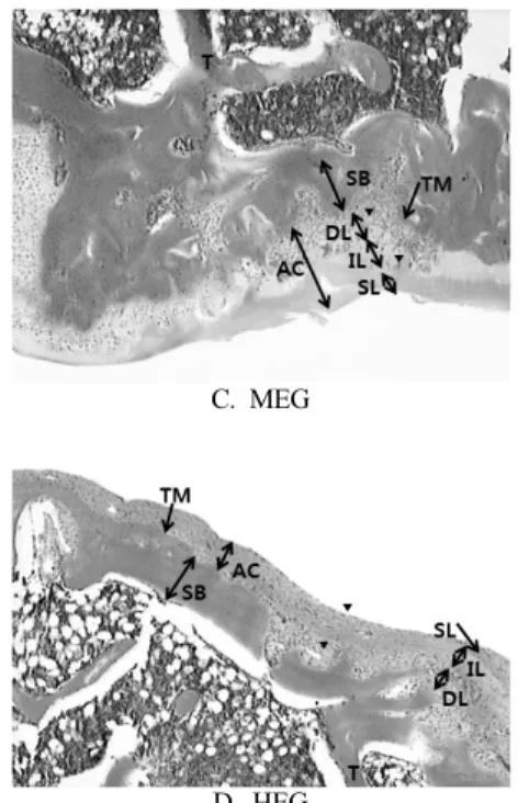
1. 헤마톡시린-에오진 염색을 통한 관절 연골의 형태 학적 변화 비교

본 연구에서는 운동 강도가 골관절염 유발 흰쥐의

연골 회복에 어떠한 영향을 미치는지 알아보기 위해

헤마톡시린-에오진 염색을 통해 형태학적 변화를 관찰

하였다 . 그 결과 CG 군은 연골 일부 부분에서 연골밑뼈

까지 손상된 부위를 확인할 수 있었다(Fig. 1A). LEG

군에서는 연골의 바탕질의 두께가 CG군에 비해 약간

두터워 진 것을 볼 수 있었고(Fig. 1B), MEG군에서는

연골의 바탕질이 상당히 회복된 것을 볼 수 있었으며

(Fig. 1C), HEG 군은 심층과 중간층에서 연골세포가

(5)

조금씩 관찰되었지만, 다른 두 운동군들에 비해 연골 두께가 비교적 얇고, 연골 세포의 회복 수준도 미미한 것을 확인할 수 있었다(Fig. 1D).

집단 간의 정도 차이를 확인하기 위해 일원배치분산 분석을 실시한 결과 통계적으로 유의한 차이가 나타났 고(p<.05)(Table 3), LSD 사후검정을 실시한 결과, 각 집단 사이에서도 서로 유의한 차이가 나타난 것을 확인 할 수 있었다(p<.05)(Table 3).

Cartilage damage score (Mean±SE)

Group (n=40)

CG (n=10)

LEG (n=10)

MEG (n=10)

HEG

(n=10) F p 11.58±.

27

7.73±.34

†§∥

5.20±.24

†‡∥

9.53±.30

†‡§

86.86 .00

*

*

p< .05

= significant difference from CG. p<.05

= significant difference from LEG. p<.05

§

= significant difference from MEG. p<.05

= significant difference from HEG. p<.05 Mean±SE; mean±standard error

Abbreviations: See Table 2.

Table 3. The comparison of articular cartilage damage score between four groups

unit; score

A. CG

B. LEG

C. MEG

D. HEG

AC: articular cartilage SL: superficial layer IL:

intermediate layer

DL: deep layer TM: tide mark T: trabecular SB:

subchondral bone Arrow head: chondrocyte Abbreviations: See Table 2.

Fig. 1. The histological changes of tibial articular cartilage in rats with osteoarthritis (×40)

Ⅳ. 고 찰

노인들에게서 많이 발병하고 있는 골관절염은 관절 연골의 퇴행성 변화가 가장 큰 특징으로, 그동안 이 질환을 치료하기 위한 치료법이 많이 연구되어 왔다.

하지만 골관절염의 발병 원인에 대해서는 아직 명확히 밝혀진 바가 없어 근본적인 치료를 할 수 없는 상황이다 (Galois 등, 2004). 더군다나 장기간의 약물 치료는 부작 용을 발생시켜 합병증을 유발할 수도 있는 문제점이 있다(Kim과 Lee, 2009). 그런데 운동은 적절한 강도로 실시할 경우 , 부작용 없이 관절의 기능과 항상성을 증진시 키는 효과가 있다 (Ikenoue 등, 2003; Roos와 Dahlberg, 2005).

환자에게 운동을 처방하기 위해서는 환자의 현재

(6)

상태를 더 이상 악화시키지 않고, 부작용 없이 긍정적 인 효과를 유도하기 위한 적절한 강도가 처방되어져야 한다. 따라서 본 연구에서는 운동 강도가 관절 연골의 회복에 미치는 영향을 분석하고, 어느 정도의 강도가 골관절염 연골 회복에 가장 효과적인지를 알아보기 위 해 골관절염이 유발된 흰쥐를 저강도 운동군(LEG), 중 강도 운동군(MEG), 고강도 운동군(HEG)으로 나누어 4주간 트레드밀 운동을 실시한 후, 헤마톡시린-에오진 염색을 통해 4주간 운동을 실시하지 않은 NEG군과 연골의 형태학적 변화를 비교하였다.

그 결과 운동을 실시하지 않은 CG군은 대부분의 영역에서 연골 밑뼈가 그대로 노출되어 있을 정도로 연골 손상이 심한 상태였지만, 운동을 한 LEG군과 MEG군, HEG군은 연골의 바탕질이 회복되었고, 연골 밑뼈가 연골에 의해 덮혀 있을 정도로 두께가 두꺼워지 고 있었으며, 연골 세포의 수가 상당히 증가되어 있는 것을 관찰할 수 있었다. 특히 MEG군이 가장 좋은 회복 상태를 보여주었는데 , 연골의 표층 부위까지 연골 세포 가 회복되고 있는 것을 확인할 수 있었다. LEG도 비록 MEG에 비해 낮은 수준이지만, 연골이 중간층까지 회 복된 영역이 CG보가 많이 분포하고 있었으며, 연골 두께도 상당히 두터워진 것을 보아 연골이 회복된 것을 짐작할 수 있었다. 하지만 HEG군은 CG군에 비해서는 유의한 회복 효과를 보여 주었지만, MEG군과 LEG군 과 비교하여 가장 낮은 회복 수준을 나타냈다.

본 연구에서 운동 강도에 따라 이러한 차이가 나타난 것은 선행 연구들에서 근거를 찾을 수 있다. Park 등 (2008)은 최대심박동수가 50%에서 75% 사이, 피로인지 척도가 10∼13 정도로, 운동 중 편하게 말을 할 수 있는 저강도나 중강도의 신체적 운동은 통증을 감소시키고 , 병리적 진행 과정을 억제하여 건강의 약화나 기능적 능력의 저하를 극복하는데 중요한 역할을 한다고 하였다 . 그리고 Baek (2008)과 Oh (2003)은 최대심박수의 40∼

50%의 저강도와 60∼70%의 중강도 운동이 70∼80%의 고강도 운동에 비해 면역 조절기능을 더욱 증가시킨다 고 하였는데 , Helmark 등(2010)등도 적절한 강도의 운동 이 염증성 사이토카인의 생산을 억제하여 염증 반응을 감소시킨다고 제시하였다 . 그리고 관절에 과도한 부담

이 가해지지 않는 강도의 운동은 연골의 충격 흡수 능력 을 향상시키고 (Lapveteläinen 등, 2001), 연골 내 콜라겐 섬유의 리모델링(remodeling)을 촉진시킬 뿐만 아니라 (Julkunen 등, 2010), glycosaminoglycan (GAG)의 함유 비율을 증가시켜(Roos와 Dahlberg, 2005) 프로테오글리 칸의 합성률이 향상되어 연골의 퇴행 변화의 진행을 억제하고, 연골 세포에 의한 골형성과 골흡수가 서로 균형을 회복해 정상적인 본래의 모습으로 유지되도록 도와주며, B 세포에서 히알루론(hyaluronan)의 합성을 일정하게 조절하여 관절 내 활액의 양이 정상적으로 유지된다는 것이(Otterness 등, 1998) 선행 연구들에 의 해 밝혀졌다.

Cifuentes 등(2010) 본 MIA를 주입하여 골관절염을 유발한 흰쥐에게 중강도의 운동인 VO2max의 60-70%

에 해당하는 강도인 16m/min의 속도로 트레드밀 운동 을 30분간 적용한 결과 운동을 하지 않은 군에 비해 항산화 효소인 superoxide dismutase (SOD)가 증가하였 고 , 이 SOD로 인해 산화 스트레스에 저항 기전이 활성 화되어 활성 산소에 의한 세포 손상이 감소되었으며, 이로 인해 관절의 표층과 중간층의 회복이 증가하였다 고 하였다.

이처럼 많은 선행 연구들에서 저강도나 중강도의

운동이 고강도의 운동에 비해 골관절염 치료에 효과적

이라는 것을 뒷받침해주고 있다 . 그런데 본 연구에서는

저강도에 비해 중강도의 운동이 연골 회복에 더 효과적

이라는 결과가 나왔는데, 이는 선행 연구에서 근거를

찾아볼 수 있다. Galois 등(2004)은 골관절염 유발 흰쥐

를 대상으로 운동을 하지 않은 군 , 30㎝/s의 속도를 15분

간 적용한 저강도 운동군 , 30㎝/s의 속도를 30분간 적용

한 중강도 운동군, 30㎝/s의 속도를 60분간 적용한 고강

도 운동군으로 나누어 트레드밀 운동을 5일간 4주간

실시한 결과, 저강도 운동군과 중강도 운동군에서 운동

을 하지 않은 군에 비해 연골의 회복이 유의하게 향상되

었고, 중강도 운동이 가장 좋은 회복 효과를 보여주었

으며, 고강도 운동은 연골 손상이 더 악화되었다고 하

였다. 이는 중강도의 운동이 caspase 3의 활성 억제와

열충격단백질 70(HSP 70)의 발현 증가에 가장 많은 영

향을 주었으며, 이로 인해 연골 세포의 세포사를 억제

(7)

해 연골 세포가 회복되었다고 하였다.

본 연구에서 HEG가 다른 운동군들에 비해 관절 연 골의 회복 수준이 가장 낮았는데, 이는 Choi (2010)의 연구에서처럼 고강도 운동이 에너지 대사를 촉진시켜 체내 산소 소비량을 증가시키고, 활성 산소 증가로 인 한 항산화 방어 체계의 불균형을 초래하여 IL-1β, IL-6, TNF-α와 같은 염증성 사이토카인을 증가시켜 체내 염 증 반응을 유발하였기 때문일 것이다. 염증 반응으로 인해 증가된 사이토카인은 퇴행을 유발하는 효소의 분 비를 증가시켜 활막 섬유모세포(synovial fibroblast)와 연골세포 (chondrocyte)에서 콜라겐 분해 효소(collagenase) 및 중성 프로테아제(protease)의 생산을 유도하고, 이 효소들이 콜라겐과 프로테오글라이칸(proteoglycan)을 파괴하여 관절연골의 퇴행을 더욱 가속화시키게 되며 (Cho와 Jeong, 2004; Bondeson 등, 2006). 프로스타글란 딘(prostaglandin)을 증가시켜 통증을 증가시키게 된다 (Pelletier 등, 2001).

본 연구에서 운동을 실시한 군들이 운동을 실시하지 않은 CG군에 비해 관절 연골에서의 연골 두께 증가와 연골 세포 수 증가를 관찰할 수 있었고, 연골 손상 척도 점수가 감소한 것으로 보아 운동으로 인해 관절 연골이 회복되었다고 할 수 있다. 그 중에서 MEG군이 가장 좋은 회복 상태를 보여주었는데, 이를 통해 중강도의 운동이 연골 회복에 가장 효과적인 운동 강도라고 할 수 있을 것이다.

Ⅴ. 결 론

본 연구에서는 운동 강도가 골관절염이 유발된 흰쥐 의 연골에 어떠한 영향을 미치는지 알아보기 위해 저강 도와 중강도, 고강도의 트레드밀 운동을 적용한 후, 헤 마톡시린 -에오진 염색을 통해 연골의 형태학적 변화를 관찰하였다. 본 연구의 결과를 전체적으로 볼 때 MEG 군에서 가장 높은 회복 수준을 보여 주어 골관절염 치료 가 가장 효과적인 것으로 나타났고, LEG 또한 MEG에 비해 회복 수준이 낮지만, 긍정적인 치료 효과를 유도 한다는 것을 확인할 수 있었다 . 그러나 HEG 군은 운동

을 하지 않은 CG 군에 비해 양호한 회복 정도를 보여 주었지만, MEG군과 LEG군에 비해서는 치료 효과가 떨어지는 것으로 나타났다.

비록 고강도 운동이 치료 효과는 있었지만, 이 운동 을 장시간 할 경우 연골에 가해지는 부하로 인해 환자의 통증이 오히려 증가될 수 있을 것이라 생각되며, 따라 서 환자들에게 운동 치료를 처방할 경우에는 여러 선행 연구를 통해서 그 효과가 입증이 되고, 치료 효율성도 높은 저강도나 중강도의 운동을 적용하는 것이 환자에 게 부담을 덜어 주면서 치료 효과도 크게 기대할 수 있을 것이다.

또한 앞으로의 연구에서는 운동 속도에 따른 강도 차이가 아니라 운동 시간에 따른 강도 차이, 그리고 운동 속도와 운동 시간의 혼합에 따른 치료 효과를 연구 한다면 더욱 골관절염 환자들을 위한 더욱 정확한 운동 처방 강도를 설정할 수 있을 것이다.

참고문헌

Appleton CT, McErlain DD, Henny JL et al. Molecular and histological analysis of a new rat model of experimental knee osteoarthritis. Ann NY Acad Sci. 2007;1117(1):

165-74.

Baek KY. The effect of different exercise intensity on the blood pressure and inflammatory markers change in pre-hypertension leveled male. Graduate School of Chonnam National University. Doctor’s thesis. 2008.

Bondeson J, Wainwright SD, Lauder S et al. The role of synovial macrophages and macrophage-produced cytokines in driving aggrecanases, matrix metalloproteinases, and other destructive and inflammatory responses in osteoarthritis. Arthritis Res Ther. 2006;8(6):187-98.

Bove SE, Calcaterra SL, Brooker RM et al. Weight bearing as a measure of disease progression and efficacy of anti-inflammatory compounds in a model of monosodium iodoacetate-induced osteoarthritis.

Osteoarthritis Cartilage. 2003;11(11):821-30.

(8)

Brandt KD, Mazzuca SA. Lessons learnde from nine clinical trials of disease-modifying osteoarthritis drugs.

Arthritis Rheum. 2005;52(11):3349-59.

Byun KS. Effect of exercise intensity on GLUT-4 expression of skeletal muscle and insulin resistance in type 1 diabetic rats. Graduate School of Hannam University.

Doctor’s thesis. 2010.

Cho SY, Jeong CS. Rheumatoid arthritis and cytokine. Duksung Bull Pharm Sci. 2004;15(1):83-90.

Choi JK. The Effects of high intensity exercise and DA-9601 administration on antioxidant enzyme activities and TNF-α, IL-1β mRNA expression in rats. Graduate School of Kyungpook National University. Master’s thesis. 2010.

Cifuentes DJ, Rocha LG, Silva LA et al. Decrease in oxidative stress and histological changes induced by physical exercise calibrated in rats with osteoarthritis induced by monosodium iodoacetate. Osteoarthritis Cartilage.

2010;18(8):1088-95.

Combe R, Bramwell S, Field MJ. The monosodium iodoacetate model of osteoarthritis: a model of chronic nociceptive pain in rats?. Neurosci Lett. 2004;370(2-3):236-40.

Fernihough J, Gentry G, Malcangio M et al. Pain related behaviour in two models of osteoarthritis in the rat knee. Pain. 2004;112(1-2):83-93.

Galois L, Etienne S, Grossin L et al. Dose-response relationship for exercise on severity of experimental osteoarthritis in rats: a pilot study. Osteoarthritis Cartilage.

2004;12(10):779-86.

Goldring MB, Goldring SR. Osteoarthritis. J Cell Physiol.

2007;213(3):626-34.

Guzman RE, Evans MG, Bove S et al. Mono-iodoacetate- induced histologic changes in subchondral bone and articular cartilage of rat femorotibial joints: an animal model of osteoarthritis. Toxicol Pathol. 2003;31(6):

619-24.

Han DY. mRNA expression on Neuropeptide(NPY) to exercise intensity and recovery time. Graduate School of

Dankook University. Doctor’s thesis. 2006.

Helmak IC, Mikkelsen UR, Børglum J et al. Exercise increase interleukin-10 levels both intraarticularly and peri-synovially in patients with knee osteoarthritis:

a randomized controlled trial. Arthritis Res Ther.

2010;12(4):126-36.

Ikenoue T, Trindade MC, Lee MS et al. Mechanoregulation of human articular chondrocyte aggrecan and type II collagen expression by intermittent hydrostatic pressure in vitro. J Orthop Res. 2003;21(1):110-6.

Jang SK. The effect on intra-articular administration of hyaluronic acid and growth hormone on arthritis in osteoarthritis rat model. Graduate School of Dongshin University. Master’s thesis. 2007.

Janusz MJ, Hookfin EB, Heitmeyer SA et al. Moderation of iodoacetate-induced experimental osteoarthritis in rats by matrix metalloproteinase inhibitors. Osteoarthritis Cartilage. 2001;9(8):751-60.

Janusz MJ, Bendele AM, Brown KK et al. Induction of osteoarthritis in the rat by surgical tear of the meniscus:

Inhibition of joint damage by a matrix metalloproteinase inhibitor. Osteoarthritis Cartilage. 2002;10(10):785-91.

Julkunen P, Halmesmäki EP, Iivarinen J et al. Effects of growth and exercise on composition, structural maturation and appearance of osteoarthritis in articular cartilage of hamsters. J Anat. 2010;217(3):262-74.

Kim SS, Lee EN. Effects of the progressive exercise program on flexibility, grip strength, pain, and fatigue for osteoarthritis patients. J Muscle Joint Health.

2009;16(2):184-92.

Kim WH. The effects of treadmill exercise on blood glucose and expression of UCP-2 of brown adipose tissue and TNF-α of soleus muscle in obese zucker rats.

Graduate School of Chonnam National University.

Doctor’s thesis. 2010.

Kwak HB, Kwon DS, Jang SJ et al. Roles of sphingosine-1

phosaphate during pathogenesis of bone destruction

and inflammation in rheumatoid arthritis mice model.

(9)

The Korean J Anat. 2007;40(4):277-86.

Lapveteläinen T, Hyttinen M, Lindblom J et al. More knee joint osteoarthritis(OA) in mice after inactivation of one allele of type II procollagen gene but less OA after lifelong voluntary wheel running exercise.

Osteoarthritis Cartilage. 2001;9(2):152-60.

Lee JY. Expression of apoptosis-related proteins in muscle fiber types Ⅰ&Ⅱ of aged rats depending on the exercise intensity. Graduate School of Dankook University. Doctor’s thesis. 2009.

McGaraughty S, Chu KL, Perner RJ et al. TRPA1 modulation of spontaneous and mechanically evoked firing of spinal neurons in uninjured, osteoarthritic, and inflamed rats. Mol Pain. 2010;6:14-24.

Oh SD. The effect of intensity exercise on the changes T-cell and cytokines following submaximal exercise.

Exercise Science. 2003;12(4):693-701.

Otterness IG, Eskra JD, Bliven ML et al. Exercise protects against articular cartilage degeneration in the hamster.

Arthritis Rheum. 1998;41(11):2068-76.

Park ES. The effect of pulsed electromagnetic fields on degeneration and pain in rat knee articular cartilage.

Graduate School of Daegu University. Master’s thesis.

2006.

Park HS, Kim JS. Clinical article : The effects of low intensity of ultrasound on monosodium iodoacetate(MIA)

induced athritis in rats. The Korean Journal of Sports Medicine. 2009;27(1):27-32.

Park IH, Lee EN, Park SY. Development of a progressive exercise program for older adults with osteoarthritis.

J Muscle Joint Health. 2008;15(2):155-65.

Pelletier JP, Martel-Pelletier J, Abramson SB. Osteoarthritis, an inflammatory disease: potential implication for the selection of new therapeutic targets. Arthritis Rheum. 2001;44(6):1237-47.

Rodrigues B, Figueroa DM, Mostarda CT et al. Maximal exercise test is a useful method for physical capacity and oxygen consumption determinatio in streptozotocin- diabetic rats. Cardiovasc Diabeto. 2007;6(1):38-44, 1-7.

Roos EM, Dahlberg L. Positive effects of moderate exercise on glycosaminoglycan content in knee cartilage: a four-month, randomized, controlled trial in patients at risk of osteoarthritis. Arthritis Rheum. 2005;52(11):

3507-14.

Schuelert N, McDougall JJ. Grading of monosodium iodoacetate- induced osteoarthritis reveals a concentration-dependent sensitization of nociceptors in the knee joint of the rat. Neurosci Lett. 2009;465(2):184-8.

Seidel MF, Herguijuela M, Forkert R et al. Nerve growth factor in rhumatic diseases. Semin Arthritis Rheum.

2010;40(2):109-26.

(10)

수치

Table 3. The comparison of articular cartilage damage score between four groups

참조

관련 문서

The effect of Combined Exercise program on Body composition, Physical fitness and thickness of subcutaneous fat in. Obese

Taken together, by applying physical strength exercise program to the elite table tennis players we can conclude that physical strength exercise program has positive effects

Wrestler anaerobic exercise on body composition and function by analyzing the impact of physical man wrestler necessary for the efficient exercise

The purpose of this research is to confirm whether voluntary early morning physical exercise effects on the emotional and behavioral development of

Changes in the level of physical fitness of obese women who performed complex exercise were significantly different in muscle endurance,

The purpose of this study is to investigate how participation in 8-week swimming and resistance exercise programs affects changes in health fitness and

The purpose of this study was to investigate how the 8-week aerobic exercise program affects the physical strength and stress of obese women in the

Contents of total cholesterol in the epididymal adipose tissue of STZ-induced diabetic rats containing barley leaf powder for 4 weeks ··· 147 Fig.. The change of growth rate