DOI : 10.3341/jkos.2008.49.12.1917
수정체유화술에서 프리챠퍼 사용이 초음파 에너지량의 감소에 미치는 영향
조경진․이현수․주천기 가톨릭대학교 의과대학 안과 및 시과학교실
목적: 수정체유화술에 앞서 Prechopper를 이용하여 미리 수정체를 2등분 또는 4등분으로 나누는 것의 효율과 안전 성을 기존의 수정체유화술과 비교해 알아 보고자 한다.
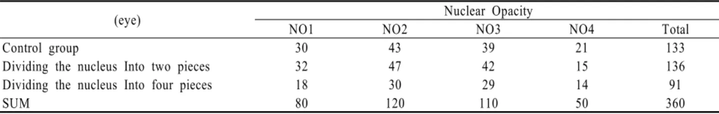
대상과 방법: 백내장 수술을 시행 받은 360안을 핵경화도에 따라 4군으로 나누고 각 군을 다시 수정체 적출 방법에 따라 3군으로 나누었다(대조군, Prechopper를 사용하여 유화술 전에 수정체 핵을 2등분한 군, 4등분한 군)(NO1 80 안(대조군 30안, 수정체 핵을 2등분 한 32안, 수정체핵을 4등분한 18안), NO2 120안(43안, 47안, 30안), NO3 110안(39안, 42안, 29안), NO4 50안(21안, 15안, 14안). 같은 핵경화도 내에서 각 군간에 수술 2개월 후의 각 막내피세포밀도의 감소율, 육각형 세포비율, 사용된 초음파 시간과 에너지량, 그리고 합병증 발생률을 비교하였다.
결과: 모든 군에서 수술 중 또는 수술 후 합병증은 발생하지 않았다. 각막내피세포의 감소율은 Prechopper를 사용하 였을 때 핵경화도 2, 3군에서 대조군에 비해 유의하게 낮았다. 육각형 세포비율은 Prechopper를 사용한 핵경화도 3 군에서 대조군에 비해 유의하게 많았다. 초음파 시간은 핵경화도 2, 3군에서 Prechopper를 사용하였을 때 대조군에 비해 유의하게 적었다. 총 초음파 에너지량은 Prechopper를 사용하였을 때 대조군에 비해 핵경화도 2, 3, 4군에서 적었다.
결론: Prechopper를 이용한 수정체유화술은 안전하며 Prechopper를 사용하지 않는 기존의 방법에 비해 각막내피 세포의 손상 적었고, 초음파 시간 및 초음파 에너지량을 감소시킬 수 있었다.
<대한안과학회지 2008;49(12):1917-1922>
<접수일 : 2008년 5월 20일, 심사통과일 : 2008년 9월 2일>
통신저자 : 주 천 기
서울시 서초구 반포동 505 가톨릭대학교 강남성모병원 안과 Tel: 02-590-2615, Fax: 02-590-7405 E-mail: [email protected]
* 본 논문의 요지는 2007년 대한안과학회 제98회 추계학술대회 에서 구연으로 발표되었음.
백내장 수술방법의 하나인 수정체유화술은 술 후 빠 른 회복과 적은 난시발생 등의 이유로 최근에 많이 시 행되고 있으나 초음파에너지가 각막내피세포에 전달되 어 고식적인 백내장 낭외적출술보다 각막내피세포에 손 상을 많이 일으킬 가능성이 있다. 또한 각막내피의 손 상은 심할 경우 각막 대상부전을 초래할 수 있다. 이런 단점을 보완하기 위하여 현재 수정체유화술을 낭내에서 시행하고 있고, 물리적인 수정체핵분리술과 수력분층술 을 같이 행하고 있다. 또한 유화기 첨단부의 단면이 각막 내피세포층과 반대 방향을 향하는 드릴 초음파(phaco
drill)의 사용을 통해 초음파 유화술에 따른 내피세포 의 손상을 줄였다는 보고도 있다.1,2
Phaco prechop 방법은 Akahoshi가 개발한 것으 로 수정체유화술을 시행하기 전에 특수 제작된 감자인 phaco prechopper를 이용해서 수정체핵을 분리시키 는 방법으로 초음파유화기로 수정체핵을 분리하는 것보 다 수술시간과 초음파의 사용시간이 단축되고, 초음파 유화기의 첨단부(tip)에서 발생하는 열과 기계적 조작 에 의한 안내조직 손상, 각막내피세포의 손상, 후낭파 열 등의 합병증 을 감소시킬 수 있는 수술법이라고 보 고된 바 있다.3
본 연구는 수정체유화술에 있어서 Prechopper를 이용하여 미리 수정체 핵을 2등분 또는 4등분으로 나누 는 것이 수정체유화술의 시간과 세기의 단축 및 임상결 과에 미치는 영향을 알아보고자 하였다.
대상과 방법
본원 안센터에서 한 명의 술자에 의하여 2002년 3월
(eye) Nuclear Opacity
NO1 NO2 NO3 NO4 Total
Control group 30 43 39 21 133
Dividing the nucleus Into two pieces 32 47 42 15 136
Dividing the nucleus Into four pieces 18 30 29 14 91
SUM 80 120 110 50 360
Table 1. A classified table of subjects
부터 2007년 5월까지 수정체유화술을 이용한 백내장 수술을 시행받은 360안을 백내장의 정도평가인 LOCS III 분류법을 이용하여 핵경화도(Nuclear opacity, NO)를 기준으로 NO1 (1군), NO2 (2군), NO3 (3 군), NO 4 (4군)로 나누고 각 군을 다시 3개의 하위 군 즉, 수정체유화술 전에 수정체 핵을 나누지 않은 (prechopper를 사용하지 않은) 대조군, Akahoshi prechopper를 사용하여 유화술 전에 수정체 핵을 2등 분한 군, 4등분한 군으로 나누었다(NO1 (1군) 80안 (수정체핵을 나누지 않은 30안, 수정체 핵을 2등분 한 32안, 수정체핵을 4등분한 18안), NO2 (2군) 120안 (43안, 47안, 30안), NO3 (3군) 110안(39안, 42안, 29안), NO4 (4군) 50안(21안, 15안, 14안))(Table 1). 같은 핵경화도 내에서 각 군별로 초음파 시간(sec), 사용된 총 초음파 에너지량(%·sec), 수술 2개월 후의 각막내피세포밀도(Number/mm2)의 감소율, 육각형 세포비율(%)의 변화 그리고 후낭파열 등의 합병증 발 생율을 비교하였고 술 중 사용된 총 초음파 에너지량은 절대 초음파 시간(Absolute phaco time=average phaco power×phaco time)으로 하였다.
각막질환, 녹내장, 망막질환 등 시력 및 각막내피 세 포에 영향을 줄 수 있는 환자는 제외하였으며 술 후 망 막박리, 수포각막병증, 낭포황반부종, 녹내장 등의 합 병은 발생하지 않았다. 수정체유화술은 각막 윤부에 접 선되게 이측부 투명각막 절개를 하고 점탄물질 주입후 에 전낭원형절개술을 시행하였으며 크기는 인공수정체 의 광학부 크기보다 약간 작게 하였으며 평형염액 (balanced salt solution : BSS)으로 수력분리술을 시행한 후 phaco prechop을 하였다. Phaco prechop 을 하기 전 전방 내에 점탄물질을 충분히 채워 전방내 prechopper가 들어 갈 공간을 확보하였고, 혼탁된 수 정체 피질을 수정체 핵 표면에서 밀어내어 수정체핵 안으로 prechopper가 정확히 들어가게 하였고, pre- chopper가 수정체 핵내에 들어갈 때 깊이와 진행 방 향을 더 잘 보이게 하였다. Phaco prechopper의 끝 부위를 닫은 채로 수정체 섬유의 주행 방향으로 핵의 중심부를 향하게 삽입시켰고, 깊이는 prechopper의 끝을 벌렸을 경우에 수정체핵을 2등분 할 수 있을 정도 로 수정체 핵의 가장 단단한 중심부위까지 하였다. Pre-
chopper를 삽입한 후 끝 부위를 천천히 벌려서 수정체 핵을 2등분하였고 90° 돌려서 또 4등분 하였다. 그 후 초음파수술기구(Millennium, Stortz, Louis, USA) 를 이용하여 초음파유화술을 시행하였다. 인공수정체는 연성재질의 광학부를 갖고 있는 렌즈를 낭내에 삽입하 였다. 수술이후 각막은 봉합하지 않고 저절로 치유되게 하였다. 각막내피세포수와 육각 세포의 밀도는 비접촉성 경면현미경(Konan Noncon specularmicroscope, Japan)으로 측정하였다.
통계학적인 분석은 SPSS 13.0 통계 프로그램을 사 용하였다. 같은 핵경화도 내에서 각 처치 방법에 따라 각막내피세포 감소율, 수술 후 육각형 세포비율, 초음 파 이용시간, 수술 중 사용된 총 초음파 에너지량의 평 균에 유의한 차이가 있는지는 일원배치분산분석법(one way ANOVA)을 사용하여 분석하였고, 차이가 있다 면 어느 처치 방법간에 유의한 차이가 있는지의 분석은 다중비교(Multiple comparison)의 방법인 Tukey HSD (Honestly significant difference)와 Duncan 검정법을 사용하였다.
결 과
대상자의 평균연령은 Prechopper를 사용하지 않은 대조군, Prechopper를 사용하여 수정체 핵을 2등분 한 군, 그리고 Prechopper를 사용하여 수정체 핵을 4 등분한 군에서 각각 54.4±15.1세, 54.2±12.2세, 54.5
±12.3세(핵경화도 1군), 67.4±10.5세, 67.1±8.1세, 67.2±11.1세(핵경화도 2군), 67.9±10.8세, 68.1±11.7 세, 68.2±11.2세(핵경화도 3군), 71.1±13.3세, 70.4
±10.4세, 71.2±8.4세(핵경화도 4군)로 각 군간에 연 령의 유의한 차이를 보이지는 않았다.
수술 2개월 후 각막내피세포 감소율(%)은 대조군, Prechopper를 사용하여 수정체 핵을 2등분한 군, 그 리고 4등분한 군에서 각각 7.12±1.28%, 7.11±1.41%, 6.18±1.31% (핵경화도 1군), 13.97±2.14%, 10.91
±2.08%, 10.85±1.09% (핵경화도 2군), 17.21±1.98%, 13.07±2.08%, 13.19±3.14% (핵경화도 3군), 20.41±
3.51%, 18.74±3.59%, 18.82±3.10% (핵경화도 4 군)으로 핵경화도 2, 3군에서 핵을 2등분 및 4등분 한
Figure 1. Comparison of corneal endothelial cell loss rate (%) among various phacoemulsification techniques prior to and two months after cataract operation. *1 P=0.047:
Multiple comparisons (Tukey HSD and Duncan test) for NO2, *2 P=0.048: Multiple comparisons (Tukey HSD and Duncan test) for NO2 , **1 P=0.04 : Multiple comparisons (Tukey HSD and Duncan test) for NO3, **2 P=0.04:
Multiple comparisons (Tukey HSD and Duncan test) for NO3 groups (Control group: Groups not using phaco prechopper).
Figure 2. Comparison of corneal endothelial cell hexagonality (%) two months after cataract operation among various phacoemulsification techniques. *1 P=0.03: Multiple comparisons (Tukey HSD and Duncan test) for NO3, *2 P=0.04: Multiple comparisons (Tukey HSD and Duncan test) for NO3 (Control group: Groups not using phaco pre- chopper).
Figure 3. Comparison of phaco time (sec) between subgroups using different phacoemulsification techniques. *1 P=0.04: Multiple comparisons (Tukey HSD and Duncan test) for NO2, *2 P=0.04: Multiple comparisons (Tukey HSD and Duncan test) for NO2, **1 P=0.04: Multiple comparisons (Tukey HSD and Duncan test) for NO3, **2 P=0.04:
Multiple comparisons (Tukey HSD and Duncan test) for NO3 groups (Control group: Groups not using phaco prechopper).
Figure 4. Comparison of absolute phaco time (Absolute phaco time (%·sec)) delivered between subgroups using different phacoemulsification techniques. The absolute phaco time is calculated by multiplying the phaco power and time.
*1 P=0.04: Multiple comparisons (Tukey HSD and Duncan test) for NO2, *2 P=0.04: Multiple comparisons (Tukey HSD and Duncan test) for NO2 , **1 P=0.04: Multiple comparisons (Tukey HSD and Duncan test) for NO3, **2 P=0.03: Multiple comparisons (Tukey HSD and Duncan test) for NO3 groups, ***1 P=0.04: Multiple comparisons (Tukey HSD and Duncan test) for NO3, ***2 P=0.04:
Multiple comparisons (Tukey HSD and Duncan test) for NO4 groups (Control group: Groups not using phaco prechopper).
군이 대조군에 비해 수술 2개월 후 각막 내피세포가 유 의하게 감소하였다(Fig. 1).
수술 전 육각형 세포비율(%)는 대조군, Precho- pper를 사용하여 수정체 핵을 2등분한 군, 그리고 4등 분한 군에서 각각 61.1±12.1%, 57.3±13.4%, 61.2±
13.1% (핵경화도 1군), 57.1±11.7%, 54.4±11.2%,
53.9±14.1% (핵경화도 2군), 56.4±10.5%, 54.21±
14.1%, 53.21±17.7% (핵경화도 3군), 57.4±7.5%, 54.24±13.4%, 54.17±11.4% (핵경화도 4군)으로 같 은 핵경화도 내에서 각 군간에 통계적으로 유의한 차이 를 보이지 않았다.
반면 수술 후 두 달째 육각형 세포비율(%)는 대조 군, Prechopper를 사용하여 수정체 핵을 2등분한 군, 그리고 4등분한 군에서 각각 59.7±13.2%, 60.1±
13.1%, 60.2±11.2% (핵경화도 1군), 54.8±12.1%, 58.1±9.2%, 58.5±12.1% (핵경화도 2군), 51.7±
14.5%, 57.74±14.1%, 57.47±19.1% (핵경화도 3 군) 50.17±13.5%, 53.31±11.0%, 52.44±12.1%
(핵경화도 4군)으로 핵경화도 3군에서 핵을 2등분 및 4등분한 군이 대조군에 비해 수술 2개월 후에 육각세 포의 밀도가 유의하게 높았다(Fig. 2).
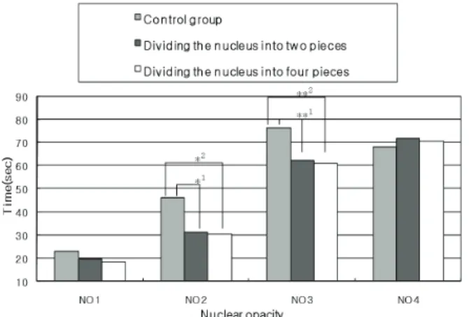
초음파 이용시간(seconds)은 대조군, Prechopper 를 사용하여 수정체 핵을 2등분한 군, 그리고 4등분한 군에서 각각 23.12±13.12 sec, 19.88±19.88 sec, 18.55±10.15 sec (핵경화도 1군), 46.1±20.43 sec, 31.28±17.73 sec, 30.48±15.43 sec (핵경화도 2군), 76.35±50.57 sec, 62.28±17.73 sec, 61.18±15.43 sec (핵경화도 3군) 68.25±23.67 sec, 72.08±28.38 sec, 70.6±29.76 sec (핵경화도 4군)으로 핵경화도 2, 3군 에서 핵을 2등분 또는 4등분 했을 때 대조군에 비해 초 음파 이용 시간이 유의하게 적었다(Fig. 3).
총 초음파 에너지량(Asolute phaco time (%⋅
sec))은 대조군, Prechopper를 사용하여 수정체 핵 을 2등분한 군, 그리고 4등분한 군에서 각각 132.29±
11.30%·sec, 108.86±54.10%·sec, 110.05±34.10%
·sec(핵경화도 1군), 353.29±79.42%·sec, 264.86±
54.10%·sec, 252.58±123.57%·sec (핵경화도 2군), 796.88±102.42%·sec, 518.66±64.47%·sec, 453.28
±58.47%·sec (핵경화도 3군) 803.32±105.44%·sec, 584.62±58.35%·sec, 522.6±212.53%·sec (핵경화 도 4군)으로 핵경화도 NO1, 2, 3군에서 핵을 2등분 또는 4등분 했을 때 대조군에 비해 사용된 총 초음파 에너지량이 유의하게 적었다(Fig. 4).
고 찰
백내장 수술의 한 방법인 수정체유화술은 Kelman 이 소개한 이래로 적은 난시의 발생과 빠른 시력의 회 복 등의 장점으로 인해 여러 가지 방법으로 발전되어 왔다. 그러나 수정체유화술의 경우 각막내피세포의 손 상에 대한 위험성이 있다. 이 때 손상에 영향을 미칠 수 있는 요소들에는 술자의 숙련도, 환자의 연령, 수술방
법, 백내장의 정도, 수술절개창의 크기와 위치 등이 있 고 이중 백내장의 수술방법이 각막내피세포의 손상을 결정하는데 매우 중요한 요소이다.4 이는 수정체유화술 시 유화기에서 나오는 열, 초음파 진동에 의한 물리적인 힘, 유리기(free radical), 관류액에 의해 형성된 와 류, 수정체조각이나 수술기구에 의한 직접적인 손상 등 이 내피세포손상에 영향을 미치는 것으로 생각된다.5-7 따라서 이러한 손상을 방지하기 위한 다양한 방법이 고 안되어왔는데 전방에서의 초음파의 충격을 줄이고 내피 세포를 보호하기 위한 방법의 일환으로 점탄 제재를 사 용하게 되었으며,8-11 초음파 에너지 양을 줄이기 위한 시도로서 드릴초음파 등의 고안으로 수술적인 방법에 변화를 주거나12-14 단속적인 에너지,15 또는 레이저16,17 등을 사용하기도 하였다.
본 연구에서는 수정체유화술에 있어서 Prechopper 를 이용하여 미리 수정체 핵을 2등분 또는 4등분으로 나누었을 때 수정체유화술의 효율과 안전성을 기존의 수정체유화술과 비교해 알아 보았다.
경면현미경으로 각막내피세포의 양적인 변화(quan- titative analysis)인 세포의 밀도(cells/mm2)와 각 막내피세포의 안정과 기능에 가장 중요한 지표가 되는 세포의 모양변화(morphometric parameter) 즉 각 세포 6각형의 모양변화(pleomorphism)를 측정하였
다.18-20 수술 후 각막내피세포가 손상을 받고 재편성
(remodeling) 되는 기간동안은 내피세포의 고유 형태 인 6각형 모양이 다각형의 다양한 모양(pleomorphic polygonal pattern)이 되나 각막내피세포가 안정 상 태로 되면, 세포의 밀도가 낮아지고 세포면적이 다소 넓어지며, 6각형 모양이 점차적으로 회복된다. 즉, 세 포모양의 변화가 정상화되었다는 것은 내피세포의 안정 화를 의미한다.21,22 각막의 정상 두께와 투명도는 각막 내피피세포의 방어기능과 능동적인 펌프작용에 의해서 유지되는데 이 기능들은 세포모양들이 정상화되기 전에 회복되는 것으로 알려져 있어 각막두께 변화로 각막내 피 기능을 평가하는 것은 부적절한 것으로 알려져 있기
때문에23,24 본 연구에서도 수술 중 사용된 초음파 에너
지 및 기졔적 자극에 의한 각막내피세포의 손상정도를 알아보기 위해 수술 후 각막내피세포 감소율(%)과 수 술 전⋅후 육각형 세포비율(%)을 측정하였다. 연구 결 과 수술 2개월 후 각막내피세포 감소율(%)은 핵경화도 2, 3군에서 핵을 2등분 및 4등분 한 군이 대조군에 비 해 유의하게 적었다.
수술 전 육각세포의 밀도(%)는 같은 핵경화도 내에 서 각 군간에 통계적으로 유의한 차이를 보이지 않았으 나 수술 후 두 달째 육각세포의 밀도(%)는 핵경화도 3 군에서 핵을 2등분 및 4등분한 군이 대조군에 비해 유
의하게 높았는데 이는 각막내피세포가 손상을 적게 받 았거나 빠르게 안정상태로 회복되었음을 의미한다.
Walkow et al은 수정체유화술 이용 시간이 각막내피 세포의 감소에 중요한 인자로 작용한다고 했는데,25 본 연 구에서도 같은 결과를 보여주고 있다. 하지만 핵경화도 4군에서는 유의한 차이를 보이지 않았는데, Hayashi et al26은 각막내피세포 손상에 가장 중요한 인자는 수 정체 핵의 경화정도라고 했으며 이는 수정체 핵 조각의 기계적 접촉에 의해 각막내피세포가 손상을 받기 때문 이라고 했다. 따라서 본 연구에서도 핵경화도 4군에서 단단한 수정체 핵 조각의 기계적 접촉에 의한 각막내피 세포의 손상이 컸을 것으로 생각된다.
초음파 이용시간(seconds)은 핵경화도 2, 3군에서 핵을 2등분 또는 4등분 했을 때 대조군에 비해 유의하게 적었다. 수술 중 사용된 총 초음파 에너지량(Asolute phaco time (%·sec))은 핵경화도 2, 3, 4군에서 핵 을 2등분 또는 4등분 했을 때 대조군에 비해 유의하게 적었다. 따라서 수정체유화술 전 Phaco prechopper 를 사용하여 수정체를 미리 2조각 또는 4조각을 내면 수정체유화술 중 발생하는 각막내피세포의 손상을 감 소시킬 수 있을 것으로 생각된다. 초음파 이용시간 (seconds)이 핵경화도 4군에서는 Phaco prechopper 사용에 따른 통계적으로 의미있는 감소가 없었는데 이 는 핵경화가 심할 경우 요구되는 초음파 양의 변동량이 크기 때문일 것으로 생각되며, 또한 본 연구에서 사용 된 개체의 수가 적었기 때문일 수도 있다.
결론적으로 Phaco prechopper를 이용하여 수정체 핵을 분리한 후 수정체유화술을 시행하는 방법은 핵경 화도 NO2, NO3인 백내장에서 기존의 단순 수정체유 화술에 비해 후낭파열 등 합병증의 증가 없이 초음파의 사용시간 및 총 초음파 에너지량을 단축시키고, 수정체 유화기의 첨단부에서 발생하는 열과 기계적 조작에 의 한 안내 조직 및 각막내피세포의 손상을 감소시킬 수 있는 효과적이고 안전한 수술 법이라 사료된다.
참고문헌
1) Kim YH, Joo CK. Phacoemulsification by Phaco-drill Method with Microseal Phaco Tip. J Korean Ophthalmol Soc 1997;
38:1566-71.
2) Kim YH, Joo CK. Short-term Change of Corneal Endothelium after Phacoemulsification using Phaco-drill Technique. J Korean Ophthalmol Soc 1999;40:81-7.
3) Akahoshi T. Phaco prechop: manual nucleofracture prior to phacoemulsification. Operative Techniques in Cataract and Refractive Surgery 1998;11:69-91.
4) Park YJ, An GJ, Woo CH. Corneal Endothelial Cell Change after Small Incision Cataract Surgery J Korean Ophthalmol
Soc 1998;39:487-94.
5) Polack M, Sugar A. The phacoemulsification procedure Ⅲ.
Corneal complications. Invest Ophthalmol Vis Sci 1977;16:
39-46.
6) Gwin RM, Warren JK, Samuelson DA, Gum GG. Effects of phacoemulsification and extracapsular lens removal on corneal thickness and endothelialcell density in the dog. Invest Ophthalmol Vis Sci 1983;24:227-36.
7) Strobel J, Jacobi KW. Phacoemulsification and planed ECCE:
Intraoperative differences in intraocular heating. Eur J implant Ref Surg 1991;3:135-8.
8) Behndig A, Lundberg B. Transient corneal edema after phacoemulsification : comparison of 3 viscoelstic regimens. J Cataract Refract Surg 2002;28:1551-6.
9) Davis EA, Lindstorm RL. Corneal thickness and visual acuity after phacoemulsification with 3 viscoelastic materials. J Cataract Refract Surg 2000;26:1505-9.
10) Arshinoff SA. Dispersive-cohesive viscoeastic soft shell technique. J Cataract Refract Surg 1999;25:167-73.
11) Arshinoff SA. Using BSS with viscoadaptives in the ultimate soft-shell technique. J Cataract Refract Surg 2002;28:1509-14.
12) Pirazzoli G, D'Eliseo D, Ziosi M, Acciarri R. Effects of phacoemulsification time on the corneal endothelium using phacofracture and phaco chop techniques. J Cataract Refract Surg 1996;22:967-9.
13) Joo CK, Kim YH. Phacoemulsification with a bevel-down phaco tip: phaco-drill. J Cataract Refract Surg 1997;23:1149-52.
14) Fine IH, Packer M, Hoffman RS. Use of power modulations in phacoemulsification; choo-choo chop and flip phacoemulsi- fication. J Cataract Refract Surg 2001;27:188-97.
15) Badoza D, Fernández Mendy J, Ganly M. Phacoemulsification using the burst mode. J Cataract Refract Surg 2003;29:1101-5.
16) Fine IH, Packer M, Hoffman RS. New phacoemulsification technologies. J Cataract Refract Surg 2002;28:1054-60.
17) Durán S, Zati M. Erbium: YAG laser emulsification of the cataractous lens. J Cataract Refract Surg 2001;27:1025-32.
18) Doughty MJ. Toward a quantitative analysis of corneal endothelial cell morphology: a review of technique and their application. Optom Vis Sci 1989;66:626-42.
19) Matsuda M, Suda T, Manabe R. Serial alterations in endothelial cell shape and pattern after intraocular surgery. Am J Ophthalmol 1984;98:313-9.
20) Kok JH, Dunnebier EA, Nieurwendaal CP, Kijlstra A.
Polymegethism of the corneal endothelium in an eye with long-standing ptosis. Arch Ophthalmol 1992;110:1529-30.
21) Díaz-Valle D, Benítez Del Castillo Sanchez JM, Toledano N, et al. Endothelial morphological and functional evaluation after cataract surgery. Eur J Ophthalmol 1996;6:242-5.
22) Kraff MC, Sanders DR, Lieberman HL. Specular microscopy in cataract and intraocular lens patients. Arch Ophthalmol 1980;98:1782-4.
23) Mishima S. Clinical investigations on the corneal endothelium -XXXVIII Edward Jackson Memorial Lecture. Am J Ophthalmol
1982;93:1-29.
24) Yee RW, Geroski D, Matsuda M, et al. Correlation of corneal endothelial pump site density, barrier function and morphology in wound repair. Invest Ophthalmol Vis Sci 1985;26:1191-201.
25) Walkow T, Anders N, Klebe S. Endothelial cell loss after
phacoemulsification: relationto preoperative and intraoperative parameters. J Cataract Refract Surg 2000;26:727-32.
26) Hayashi K, Hayashi H, Nakao F, Hayashi F. Risk factors for corneal endothelial injury during phacoemulsification. J Cataract Refract Surg 1996;22:1079-84.
=ABSTRACT=
The Effectiveness and Safety of the Phaco Prechopper Technique Before Lens Phacoemulsification in Cataract Surgery
Kyong Jin Cho, M.D., Hyun Soo Lee, M.D., Choun-Ki Joo, M.D., Ph.D.
Department of Ophthalmology and Visual Science, Kangnam St. Mary’s Hospital, College of Medicine, The Catholic University of Korea, Seoul, Korea
Purpose: To compare the effectiveness and safety of the phaco prechopper technique dividing lenses into 2 or 4 pieces before phacoemulsification with the effectiveness and safety of conventional phacoemulsification during cataract surgery.
Methods: This study included 360 eyes, which were divided into 4 groups according to nuclear opacity each group was subdivided into 3 groups according to the lens extraction technique (control, dividing the lens into two pieces using a phaco prechopper, and dividing it into four pieces) (Table 1). The following parameters were compared between the groups that had the same degree of nuclear opacity: changes in the endothelial cell count and hexagonality 2 months postoperatively, phaco time as well as the total phaco energy used for the operation, and the complication rates.
Results: There were no intra- and post-operative complications in any group. The endothelial cell loss rate was significantly less in nuclear opacity groups 2 and 3 with the prechopper technique than in the control group. Hexagonality was significantly higher in the nuclear opacity group 3 with the prechopper technique than in the control 2 months after the operation. Phaco time was significantly less in the nuclear opacity groups 2 and 3 with the prechopper technique than in the control. Total phaco energy was significantly less in the nuclear opacity groups 2, 3, and 4 with the prechopper technique than in the control.
Conclusions: Phacoemulsification using the prechopper technique is safe, resulting in significantly less endothelial cell damage, and requires less phaco time and less total phaco energy compared to those of the conventional phacoemulsification technique.
J Korean Ophthalmol Soc 2008;49(12):1917-1922 Key Words: Absolute phaco time, Phaco prechopper
Address reprint requests to Choun Ki Joo, M.D., Ph.D.
Department of Ophthalmology and Visual Science, Kangnam St. Mary’s Hospital College of Medicine
#505 Banpo-dong, Seocho-gu, Seoul 137-040, Korea
Tel: 82-2-590-2615, Fax: 82-2-590-7405, E-mail: [email protected]