ISSN 0378-6471 (Print)⋅ISSN 2092-9374 (Online)
https://doi.org/10.3341/jkos.2019.60.12.1307
Case Report
소세포신경내분비암종의 안와 전이로 진단된 전체외안근마비
Metastatic Small Cell Neuroendocrine Carcinoma to the Orbit Manifesting as Total Ophthalmoplegia
이주황1⋅이은철1⋅이현욱2⋅오신엽1
Joo Hwang Lee, MD1, Eun Chul Lee, MD1, Hyoun Wook Lee, MD, PhD2, Shin Yeop Oh, MD1
성균관대학교 의과대학 삼성창원병원 안과학교실1, 성균관대학교 의과대학 삼성창원병원 병리학교실2
Department of Ophthalmology, Samsung Changwon Hospital, Sungkyunkwan University School of Medicine1, Changwon, Korea Department of Pathology, Samsung Changwon Hospital, Sungkyunkwan University School of Medicine2, Changwon, Korea
Purpose: To report a case of primary ureteral small cell neuroendocrine carcinoma metastasis in the left orbital wall, with a re- view of the literature.
Case summary: A 79-year-old male visited our clinic with a 10 day history of ocular pain, ptosis, and ophthalmoplegia in the left eye. He had been diagnosed with diabetes mellitus and was being treated for prostate adenocarcinoma. The corrected visual acuity was 0.8 in the right eye and 0.5 in the left eye. An extraocular movement test showed total ophthalmoplegia, mild exoph- thalmos, and ptosis in the left eye. Orbital computed tomography (CT) and magnetic resonance imaging showed an irregular mass-like enhancement in the superolateral orbital wall of the left eye, suggesting infectious and inflammatory orbital disease.
After antibiotic treatment, high dose systemic steroids were administered. However, there was no improvement and the orbital CT was again performed, with no changes. A surgical biopsy for differentiating orbital tumors was performed and diagnosed as a metastatic orbital small cell neuroendocrine carcinoma. Using positron emission tomography, he was later diagnosed with a metastatic orbital small cell neuroendocrine carcinoma with multiple metastases.
Conclusions: When the patient has a mass-like lesion on imaging with ocular pain, ophthalmoplegia, or ptosis, a surgical biopsy should be performed to make a diagnosis and determine the optimal management.
J Korean Ophthalmol Soc 2019;60(12):1307-1311
Keywords: Exophthalmos, Ophthalmoplegia, Orbital metastasis, Small cell carcinoma
■Received: 2019. 9. 23. ■ Revised: 2019. 10. 28.
■Accepted: 2019. 12. 6.
■Address reprint requests to Shin Yeop Oh, MD
Department of Ophthalmology, Samsung Changwon Hospital, Sungkyunkwan University School of Medicine, #158 Paryong-ro, Masanhoewon-gu, Changwon 51353, Korea Tel: 82-55-233-5400, Fax: 82-55-233-5419
E-mail: [email protected]
*Conflicts of Interest: The authors have no conflicts to disclose.
ⓒ2019 The Korean Ophthalmological Society
This is an Open Access article distributed under the terms of the Creative Commons Attribution Non-Commercial License (http://creativecommons.org/licenses/by-nc/3.0/) which permits unrestricted non-commercial use, distribution, and reproduction in any medium, provided the original work is properly cited.
전이안와종양은 전체 암환자의 약 2-3%에서 발생하는 드문 질환이다.1 전이에 의한 안와 종양은 안와에서 발생하 는 종양 중 1-13%로 알려져 있고,2 60세 전후에 많이 발생
하며 여성에서는 유방암이 제일 흔하고 남성의 경우 폐암 과 전립선암이 제일 흔하다. 다른 원발병소로는 피부, 소화 기, 신장 순으로 보고되고 있다.3 안와 내로 전이된 종양은 대부분 주변 조직으로 침윤하는 양상으로 안구돌출이나 안 구운동제한, 통증 등을 일으키며 다른 염증성 질환들과 유 사한 양상으로 나타날 수 있어 진단 및 치료에 주의가 필요 하다.4 한편 이전 보고들을 통해 신경내분비암종의 안와 전 이는 드물다고 알려져 있다.5
신경내분비암종은 광범위한 스펙트럼의 신경내분비 상피 세포 악성 신생물로 구성되며, 전형(고분화도) 유암종, 비 전형(중등도의 분화도) 유암종 그리고 소세포 및 대세포 신
Figure 1. Clinical photograph showing severe ptosis, mild proptosis and conjunctival vessels engorgement in his left eye. -3.5 limitation
of ocular movement in medial, superior and inferior gaze and -1.5 limitation of lateral gaze is noted in his left eye at initial visit.A B
Figure 2. Coronal and axial orbit computed tomography scans (axial [A] and coronal [B] views) showing irregular mass like soft tis-
sue density of anterio temporal pole and superolateral orbital wall with well contrast enhancement and underlying bone enhancement at the left.경내분비암종(저분화 신경내분비 암종)으로 나누어진다.6 소세포신경내분비암종의 제일 흔한 원발병소는 폐며 다른 원발병소로는 소화기, 췌장, 비뇨생식기, 임파선, 흉선과 복 막에서 매우 드물게 발생한다.7 이에 저자들은 안구돌출 및 전체 외안근마비를 유발한 요관 기원으로 고려되는 전이성 소세포신경내분비암종 1예를 보고하고자 한다.
증례보고
79세 남자 환자가 10일 전부터 발생한 양안 복시와 좌측 안와부 통증, 눈주위 부종 및 안검하수를 주소로 내원하였 다. 당뇨 및 3년 전 전립선샘암종으로 호르몬 치료 및 방사 선 치료를 받고 안정 상태인 병력 이외 외상력과 다른 신경
학적 증상은 없었다. 본원 내원 7일 전부터 외부 병원에서 스테로이드 20 mg을 복용 후 통증은 완화되었다고 하였다.
양안 최대교정시력은 우안 0.8, 좌안 0.5이고, 안압은 우안 20 mmHg, 좌안 18 mmHg이었다. 전안부검사상 좌안 결막 충혈, 결막부종 및 중등도의 백내장 소견이 관찰되었고, 동 공반응검사 및 상대구심동공운동장애검사에서도 특이 소 견은 없었다. Krimsky 검사상 정위에 가까운 소견이나 안 구운동검사상 좌안 -3.5의 상전, 하전, 내전 제한 및 -1.5의 외전 제한 소견이 관찰되었다(Fig. 1). 안구돌출계검사에서 우안 13 mm, 좌안 17 mm로 좌안의 안구돌출을 보였다. 안 와 전산화단층촬영검사에서 좌측 안와 외측으로 불규칙한 종괴 모양의 조영증강을 보이는 연조직음영 및 안와외벽의 조영증강 소견이 관찰되었으며(Fig. 2), 안와 자기공명영상
A B
Figure 4. Microscopic examination showing infiltrating tumor cells of hyperchromatic round or oval nuclei and extremely scanty cy-
toplasm resembling lymphocyte in hematoxylin and eosin staining (A, ×400). The tumor cells were positive for synaptophysin (B,×400).
A B
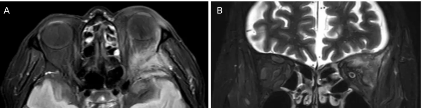
Figure 3. Brain magnetic resonance image (T2 Fluid Attenuated Inversion Recovery) showing lining pattern increased signal in-
tensity with enhancement of dura at the left hemisphere. Axial (A) and coronal (B) views.검사에서는 좌측 안와부 상외측 안와골의 불균일한 저강도 조영증강 소견이 관찰되었다(Fig. 3). 통증이 동반되는 진행 성의 전체 외안근마비와 영상검사 결과 및 고령의 조절되 지 않는 당뇨력을 고려하여 감염성 안와질환을 의심하여 4일 간 Ceftriaxone sodium (Triaxone; Hanmi Pharm Co., Ltd, Seoul, Korea) 2 g 및 Vancomycin (Vanco Kit inj, UK ChemiPharm, Incheon, Korea) 1 g을 하루 2회 정맥주사하 였다. 항생제 투약 4일 이후 호전 소견이 없어 감염성 질환을 배제하고, 염증성 안와질환 진단하에 Methylprednisolone (Solumedrol inj 125 mg, Pfizer Pharmaceuticals Korea, Seoul, Korea) 125 mg을 하루 1회 5일간 정맥투여 이후 62.5 mg을 하루 1회 2일간, 총 7일간 750 mg을 정맥투여하 였다. 그러나 임상 소견의 호전이 없어 치료 시작 2주 뒤 안와 단층촬영검사를 재시행하였고, 초진 시와 변화 없는 소견임을 알 수 있었다.
이에 전립선암 병력과 고령인 점을 감안하여 안와종양의 감별 진단을 위해 전신마취하, 좌측 안와부 상외측 절개 후
안와 조직생검을 시행하였다. 수술 소견 상 좌측 안와후부 에 명확한 종괴 형태가 관찰되지 않아 5곳에서 조직생검을 실시하였으며, 그중 1검체의 면역조직화학염색에서 신경내 분비 표지자인 시넵토파이신(synaptophysin) 및 상피세포 표 지자인 사이토케라틴(cytokeratin)에 양성을 보였다(Fig. 4).
소세포신경내분비암종의 전이성 안와종양 소견으로 추가 적인 평가를 위해 혈액종양내과로 전과하여 시행한 양전자 방출단층촬영검사에서 우측 원위 요관의 종괴, 다발성 골 전이, 다발성 폐전이, 흉막전이 및 흉막삼출, 림프절전이 소 견이 관찰되었다. 다발성 전이의 종양 말기 소견으로 환자 가 추가 검사 진행을 원하지 않아 원발병소 확진을 위한 추 가 조직검사는 시행하지 못하였다. 그러나 양전자방출단층 촬영검사상 다발성 폐전이는 미세결절 소견이며, 우측 요 관은 대사과다의 종괴 소견이 관찰되어 요관 원발성으로 고려되는 소세포신경내분비암종의 안와 전이로 진단되었 다.
고 찰
안와는 종양의 드문 전이병소이지만1 최근 진단 기술의 발전과 암환자의 생존율 증가에 따라 전이안와종양의 빈도 가 증가하고 있다.2 안와 전이를 일으키는 전이성암은 대부 분 샘암종이며, 유방이 제일 흔한 원발병소이고 그 외에도 전립선, 결장, 폐에서 전이가 가능하다.3 Ferry and Font8가 보고한 227명의 눈과 안와의 전이성 암종에 대한 분석을 보면 전이성 안와암종으로 진단받은 28예 중 유방암이 8예, 폐암이 4예, 신장암이 2예, 고환과 전립선암이 각각 1예였 고, 소화기암 중에서는 췌장암이 1예였다. 신경내분비암종 의 안와전이는 매우 드물며,5 임상양상이 봉와직염이나 그 레이브스 안병증처럼 나타나는 경우가 많다고 보고되었 다.9,10 이 중에서 나쁜 분화도를 가지는 소세포신경내분비 암종은 주로 폐에서 발생하며 원발 폐암종의 20%를 차지 하는 데 반해,7 폐외 소세포신경내분비암종의 총 발생률은 모든 종양 중에서 0.1-0.4%를 차지한다.11 안와전이가 동반 된 암 환자의 약 30%에서 원발암이 진단되기 전에 안와 전 이암이 발견된다고 알려져 있으며, 전이성 안와 종양의 25%에서는 안구 증상이 다른 전신 증상에 선행하여 초기 에 나타난다고 알려져 있다.12 일반적으로 복시와 안구돌출이 안와전이의 가장 흔한 증상 및 징후이며, Yeh and Foroozan13 의 연구에 따르면 전이성 안와병변을 가지는 70명의 환자 중 38명(54%)에서 안구운동제한을 보인다고 하였다.
본 증례의 경우에도 통증을 동반한 안검하수와 안구운동 제한을 주 증상으로 안과에 내원한 경우이다. 이처럼 통증 이나 안구돌출이 동반된 안구운동제한 소견이 관찰된다면 안와의 이상 유무를 확인하기 위해 전산화단층촬영 및 자 기공명영상 등의 영상검사가 필요하다.3 그리고 본 증례에 서처럼 안와 주변의 불규칙한 조영증강 소견을 보이는 경 우에 있어서는 감염, 염증 그리고 종양에 대한 감별이 필요 하다. 그러나 이들의 감별은 쉽지 않고 감염, 염증 및 림프 종 등과 같은 일부 종양에서는 초기 스테로이드 치료에 반 응을 보이기 때문에 치료 반응을 통한 감별 진단 또한 어렵 다.14 그러나 증상이 급성으로 발생하여 빠르게 진행하는 양상을 보인다면 염증성 질환에 초점을 두고 스테로이드 치료를 시도해 볼 수 있으며, 이후 호전이 없다면 영상검사 를 통해서 혈관성 질환을 배제한 후 조직생검을 고려해 볼 수 있겠다.15 본 환자의 경우에도 본원 내원 전 steroid 복용 후 증상이 완화된 소견과 급성으로 발생한 통증을 동반한 안구운동제한 소견으로 미루어 감염이나 염증성 질환을 우 선 고려하였다. 또한 초기 영상검사에서 안와 주변의 전형 적인 종괴음영이 관찰되지 않고 불규칙한 조영증강 소견이 관찰되어 종양보다는 감염성이나 염증성 질환 의심하에 항
생제 및 스테로이드 치료를 시행하였고, 이러한 과정으로 인해 최종 진단이 늦어지게 되었다.
본 증례는 소세포신경내분비암종의 안와 전이로 인해 안 구돌출, 안구운동제한 및 안검하수 등 안과적 증상이 첫 증 상으로 나타나 영상검사 및 안와 조직생검을 통해 진단된 경우이다. 이전 전립선암의 과거력이 있지만 호르몬 치료 와 방사선 치료를 통해 잘 조절되고 있는 환자였으며, 조직 생검을 통하여 새로운 암종인 소세포신경내분비암종의 안 와전이로 진단할 수 있었다. 그리고 환자가 추가적인 검사 를 원하지 않아 원발병소 확진을 위한 추가 검사는 시행하 지 못하였으나, 양전자방출단층촬영검사에서는 요관이 원 발병소로 가능성이 높은 소견이 관찰되었다. 소세포신경내 분비암종의 제일 흔한 원발병소는 폐로 알려져 있으나,7 본 증례에서는 다발성 폐전이는 미세결절 소견이며 이에 비해 우측 요관의 종괴는 저명한 대사과다 소견이 관찰되어 요 관이 원발병소일 가능성이 높을 것으로 고려되는 바이다. 이에 요관 기원의 소세포신경내분비암종의 안와전이는 국 내에서 보고된 예가 없었기에 문헌 고찰과 함께 이를 보고 하고자 한다. 또한 안구돌출, 안구운동제한 등의 안과적 소 견으로 내원한 환자에서 감별 진단 시, 특히 고령의 환자에 서는 전이성 종양을 염두하고 진료를 시행하여야 하겠다.
REFERENCES
1) Bloch RS, Gartner S. The incidence of ocular metastatic carcinoma.
Arch Ophthalmol 1971;85:673-5.
2) Ahmad SM, Esmaeli B. Metastatic tumors of the orbit and ocular adnexa. Curr Opin Ophthalmol 2007;18:405-13.
3) Char DH, Miller T, Kroll S. Orbital metastasis: diagnosis and course. Br J Ophthalmol 1997;81:386-90.
4) Papathanassiou M, Nikita E, Theodossiadis P, Vergados I. Orbital metastasis secondary to breast cancer mimicking thyroid-associated ophthalmopathy. Clin Exp Optom 2010;93:368-9.
5) Goldberg RA, Rootman J, Cline RA. Tumors metastatic to the or- bit: a changing picture. Surv Ophthalmol 1990;35:1-24.
6) Barker JL Jr, Glisson BS, Garden AS, et al. Management of non- sinonasal neuroendocrine carcinomas of the head and neck. Cancer 2003;98:2322-8.
7) Fisseler-Eckhoff A, Demes M. Neuroendocrine tumors of the lung.
Cancers (Basel) 2012;4:777-98.
8) Ferry AP, Font RL. Carcinoma metastatic to the eye and orbit. I. A clinicopathologic study of 227 cases. Arch Ophthalmol 1974;92:
276-86.
9) Atik A, Krilis M, Shannon K. Small cell neuroendocrine carcino- ma masquerading as cellulitis and causing blindness via bilateral orbital involvement. Orbit 2013;32:197-9.
10) Sira M, Clauss RP, Maclean C, Rose GE. Orbital metastases from neuroendocrine carcinoma, masquerading as graves orbitopathy.
Orbit 2010;29:94-6.
11) Remick SC, Hafez GR, Carbone PP. Extrapulmonary small-cell
= 국문초록 =
소세포신경내분비암종의 안와 전이로 진단된 전체외안근마비
목적: 좌측 안와부에 전이된 요관 원발성으로 고려되는 전이성 소세포신경내분비암종의 1예를 문헌 고찰과 함께 보고하고자 한다.
증례요약: 79세 남자 환자가 내원 10일 전부터 발생한 좌측 안와부 통증과 안검하수 및 안구운동제한 주소로 본원에 내원하였다. 당뇨 및 전립선샘암종으로 치료를 받은 과거력이 있었고, 최대교정시력은 우안 0.8, 좌안 0.5로 좌안의 안구돌출과 안구운동제한 및 안검하 수가 관찰되었다. 안와 단층촬영검사 및 자기공명영상검사에서 좌측 안와부 상외측으로 불규칙한 종괴 모양의 조영증강이 관찰되어 감염성 및 염증성 안와질환이 의심되었다. 항생제 치료 이후 고용량 전신 스테로이드 투여를 시행하였으나 호전이 없어, 재시행한 안와 단층촬영검사상 초진 시와 변화 없는 소견이었다. 이에 안와종양 감별을 위해 시행한 조직검사에서 소세포신경내분비암종으로 진단되었고, 이후 양전자방출단층촬영을 통해 다발성의 전신전이가 관찰되는 전이성 안와 소세포신경내분비암종으로 진단되었다.
결론: 안와부 통증, 안구운동제한, 안검하수와 같은 임상양상을 보이는 환자에서 영상학적 검사를 하여 종괴 소견이 관찰되면 진단 및 치료 방향 결정을 위해 조직검사를 반드시 고려해야 한다.
<대한안과학회지 2019;60(12):1307-1311>
이주황 / Joo Hwang Lee
성균관대학교 의과대학 삼성창원병원 안과학교실 Department of Ophthalmology,
Samsung Changwon Hospital, Sungkyunkwan University School of Medicine carcinoma. A review of the literature with emphasis on therapy and
outcome. Medicine (Baltimore) 1987;66:457-71.
12) Velten IM, Gusek-Schneider GC, Tomandl B. Diplopia as first symptom of a bronchogenic carcinoma. Klin Monbl Augenheikd 2000;217:52-4.
13) Yeh S, Foroozan R. Orbital apex syndrome. Curr Opin Ophthalmol
2004;15:490-8.
14) Goldberg RA, Rootman J. Clinical characteristics of metastatic or- bital tumors. Ophthalmology 1990;97:620-4.
15) Huang YY, Chang A, Chou YY, Hsu WC. Metastatic neuro- endocrine tumor with initial presentation of orbital apex syndrome.
Interdiscip Neurosurg 2017;7:9-11.
![Figure 2. Coronal and axial orbit computed tomography scans (axial [A] and coronal [B] views) showing irregular mass like soft tis- tis-sue density of anterio temporal pole and superolateral orbital wall with well contrast enhancement and underlying bone e](https://thumb-ap.123doks.com/thumbv2/123dokinfo/5338206.178222/2.892.91.809.867.1036/computed-tomography-irregular-temporal-superolateral-contrast-enhancement-underlying.webp)