https://doi.org/10.4174/astr.2017.93.3.125 Annals of Surgical Treatment and Research
Effectiveness of cholangioscopy using narrow band imaging for hepatobiliary malignancies
Ji Woong Jang, Dong Hyo Noh, Kyu-Hyun Paik, Sae Hee Kim, Il-Hyun Paik, Sung Hee Jung
Department of Gastroenterology, Eulji University Hospital, Eulji University College of Medicine, Daejeon, Korea
INTRODUCTION
Cholangioscopy, though invasive, seems to have several advantages when compared with other diagnostic modalities.
It can be useful for the differential diagnosis of focal stricture of the biliary tree [1,2], especially in the intrahepatic ducts [3], as well as for determining the extent of superficial spread of bile duct tumors, especially up to the proximal margin [4,5]. In addi tion, targeted biopsy specimens can provide an accurate histo logic diagnosis, and therapeutic maneuvers can be chosen based on accurate histologic mapping [6-8].
Narrow band imaging (NBI) is a new technology aimed to improve the quality and utility of endoscopic images that helps to differentiate malignant from benign lesions [9]. Cholangio- scopy using NBI has been attempted, which provides more char ac teristic endoscopic imaging of mucosal structures and mucosal vessels in bile duct lesions to differentiate benign from malignant lesions compared with conventional white- light imaging [10]. However, there is few data on percutaneous cholangioscopy using NBI.
We therefore analyzed patients with suspected hepatobiliary malignancies in whom cholangioscopy using NBI played a Purpose: Recently, cholangioscopy using narrow band imaging (NBI) has been used as a diagnostic modality for better visualization in hepatobiliary malignancies; however, there are few reports on it. Our aim is to evaluate the effectiveness of cholangioscopy using NBI in hepatobiliary malignancies.
Methods: Between January 2007 and December 2016, 152 cholangioscopies using percutaneous approach were conducted in total 123 patients. Among these, 36 patients were suspicious of hepatobiliary malignancies. Thirteen patients with an ambiguous margin on endoscopic retrograde cholangiopancreatography (ERCP) or magnetic resonance cholangiopancreatography (MRCP), for whom NBI tipped the balance in diagnosis of lesion and decision of lesion extent by adding NBI, were involved in our study.
Results: Underlying diseases were all malignant in 13 patients (11 bile duct cancers, 1 liver cancer, 1 pancreas cancer with common bile duct invasion). In 7 cases with papillary type tumor, minute superficial spreading tumor was detected by NBI more easily, and NBI provided a better visualization of tumor vessel and margin evaluation in 4 cases with infiltrative tumor. In 2 cases with mucin-hypersecreting tumor, NBI showed better penetration through the mucin and gave us a much clearer image. Nine patients ultimately underwent surgical resection. The margins predicted by NBI cholangioscopy were consistent with the pathological margins on the resected specimens.
Conclusion: In conclusion, cholangioscopy using NBI is very useful for evaluation of suspected hepatobiliary malignancies with an ambiguous margin on ERCP or MRCP. It can give us an accurate pathologic mapping, and this information seems to be essential before deciding on a treatment strategy.
[Ann Surg Treat Res 2017;93(3):125-129]
Key Words: Cholangiography, Narrow band imaging
Received January 10, 2017, Revised March 2, 2017, Accepted March 21, 2017
Corresponding Author: Ji Woong Jang
Department of Gastroenterology, Eulji University Hospital, Eulji University College of Medicine, 95 Dunsanseo-ro, Seo-gu, Daejeon 35233, Korea Tel: +82-42-611-3065, Fax: +82-42-259-1211
E-mail: [email protected]
Copyright ⓒ 2017, the Korean Surgical Society
cc Annals of Surgical Treatment and Research is an Open Access Journal. All articles are distributed under the terms of the Creative Commons Attribution Non- Commercial License (http://creativecommons.org/licenses/by-nc/4.0/) which permits unrestricted non-commercial use, distribution, and reproduction in any medium, provided the original work is properly cited.
crucial role in diagnosis and decision of treatment plan.
METHODS
Patients
Between January 2007 and December 2016, 152 cholangiosco- pies using percutaneous approach were conducted in a total of 123 patients. Among these, 36 patients were sus pi- cious of hepatobiliary malignancies. Thirteen patients with an ambiguous margin on endoscopic retrograde cholan- giopancreatography (ERCP) or magnetic resonance cholan gio- pan creatography (MRCP), for whom NBI tipped the balance in diag nosis of lesion and decision of lesion extent by adding NBI, were involved in our study. All enrolled patients had undergone ERCP or MRCP; however, these were inadequate in determining the extent of the lesions or in differentiating malignant from benign. According to previous literature, our group categorized hepa tobiliary malignancies as nodular, papillary, or infiltrative types based on cholangioscopic findings [11]. Operability was evaluated in all enrolled patients. If the patient underwent sur gical resection, then the pathological margin was compared with the preoperatively evaluated margin.
All patients provided a written informed consent for cholan- gio scopy using percutaneous approach. The study protocol was approved by the Institutional Review Board of our center.
Techniques
Cholangioscopic examination was performed via a percuta- neous transhepatic tract. For cholangioscopy using percutaneous approach, initial percutaneous transhepatic biliary drainage (PTBD) was carried out by using a pigtail catheter (7.5F to 8.0F;
Cook, Bloomington, IN, USA) under fluoroscopic guidance.
Before PTBD, meperidine (50 mg) and midazolam (3 to 5 mg) were given intravenously for control of pain and anxiety. Local anesthesia of the skin and intercostal space was obtained by the injection of 2% lidocaine. Two or 3 days after PTBD, the tract was dilated up to 16F or 18F. Cholangioscopy using percutaneous approach was then performed 7 to 10 days after tract dilatation. This waiting period is required for stabilization and maturation of the wall of the dilated tract.
Cholangioscopy was carried out by using a cholangioscope (CYF 240A, Olympus Optical Ltd., Tokyo, Japan). During cholan- gio scopy, white-light imaging was done at first, after which NBI was used again to evaluate the same biliary lesion. Multiple targeted biopsies were taken with a forceps under direct cholan- gio scopic visualization.
Statistical analysis
Data were reported as mean and standard deviation for conti- nuous variables, and as relative frequencies for categoric vari- ables. The chi-square test, or the Fisher test when necessary,
was used to compare relative frequencies. P-values of less than 0.05 were considered to indicate statistical significance. All an- aly ses were performed using SPSS 18.0 (SPSS Inc., Chicago, IL, USA).
RESULTS
Baseline characteristics of 13 enrolled patients
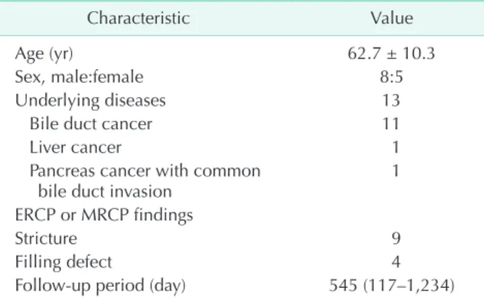
The baseline characteristics of 13 patients who received cholan gioscopy using NBI are shown in Table 1. They included 8 men and 5 women, with a mean age of 62.7 years (standard deviation, ± 10.3). Underlying diseases were all malignant in the 13 patients (11 bile duct cancers, 1 liver cancer, 1 pancreas cancer with common bile duct invasion). Whereas 9 patients showed strictures, 4 showed filling defects on ERCP or MRCP.
The median follow-up period was 545 days (range, 117 to 1,234 days).
The effectiveness of cholangioscopy using NBI according to the categorized tumor types
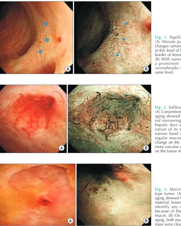
Among the 13 included patients, 7 patients showed papillary tumors on ERCP or MRCP. They showed strictures or filling defects in the tumor-involved site. However, one side of the tumor margins was ambiguous in determining the extent of the lesion. Therefore, cholangioscopy using NBI was performed to evaluate the exact margin of the tumors. Minute superficial spreading of the papillary tumors was observed in the area with strictures or filling defects. After NBI, minute papillary changes were detected more easily not only in the area with strictures or filling defects but also on the area which appeared to be normal on ERCP or MRCP. They changed the treatment plan such as to the extent of surgical resection due to newly detected lesions after cholangioscopy using NBI (Fig. 1).
Table 1. Baseline characteristics of the enrolled 13 patients
Characteristic Value
Age (yr) 62.7 ± 10.3
Sex, male:female 8:5
Underlying diseases 13
Bile duct cancer 11
Liver cancer 1
Pancreas cancer with common
bile duct invasion 1
ERCP or MRCP findings
Stricture 9
Filling defect 4
Follow-up period (day) 545 (117–1,234) Values are presented as mean ± standard deviation, number, or me dian (range).
ERCP, endoscopic retrograde cholangiopancreatography; MRCP, magnetic resonance cholangiopancreatography.
In 4 cases with infiltrative tumor, a definite mucosal mass is not present and the tumors appear as strictures on ERCP or MRCP. During cholangioscopy using NBI, white-light imaging usually showed a tapered luminal narrowing of the bile duct and neovascularization of its surface. NBI cholangioscopy showed a more irregular mucosal surface and color change of the tumor vessel from red to black, and more vascular changes, especially on the tumor margin (Fig. 2).
In 2 cases with mucin-hypersecreting tumor, ERCP or MRCP showed a marked bile duct dilatation. Mucin-like material was
also observed in 2 patients who underwent ERCP. However, the extent of the tumor in the mucosa may not correspond to the segments of the duct that are dilated because the development of mucin plugs also results in dilatation. During cholangioscopy, conventional white-light imaging showed floating mucinous materials, but mucosal changes were difficult to evaluate because of the large amount of mucin. After NBI, both mucin and tumor mass were clearly visualized (Fig. 3).
A B
Fig. 1. Papillary type cancer.
(A) Minute papillary mucosal changes (arrows) were suspected at this level of bile duct. But, the border of lesion was ambiguous.
(B) With narrow band imaging, a prominent papillary lesion (arrow heads) was noted at the same level.
A B
Fig. 2. Infiltrative type cancer.
(A) Conventional white-light im- aging showed a tapered, lumi- nal narrowing of the com mon hepatic duct and neo vas culari- za tion of its surface. (B) After nar row band imaging, more ir- re gular mucosal surface, color change on the tumor vessel, and more vascular changes especially on the tumor margin were noted.
A B
Fig. 3. Mucin-hypersecreting type tumor. (A) White-light im- aging showed floating mucinous material, however, we could not identify any mucosal change be cause of the large amount of mucin. (B) On narrow band im- aging, both mucin and the tumor mass were clearly visible.
Surgical resection and comparison between the preoperatively evaluated margin and the pathological margin
Nine of the 13 enrolled patients ultimately underwent sur- gical resection. Of the 4 patients who did not undergo surgical resec tion, 2 patients were transferred to their hometown hos- pitals for surgery, and 1 patient with a mucin-producing tumor refused surgical resection because of old age and under lying comorbidities. The 1 remaining patient with cholangio car- cinoma was initially scheduled to undergo surgical resection, but NBI cholangioscopy showed a more diffuse involvement of the cancer into both intrahepatic ducts than the extents evaluated by CT and ERCP.
The pathological margins of the 9 patients who underwent sur gical resection were compared with their preoperatively evaluated margins. The margin status predicted by NBI cholan- gio scopy was consistent with the pathological margin in all 9 cases. These results are summarized in Table 2.
DISCUSSION
It is important with bile duct lesions to determine whether filling defects or strictures are benign or malignant because of differences in the approach to treatment. Especially, in resec- table cases, preoperative diagnosis and determination of tumor ex tent are essential before deciding on the extent of resection.
In this study, we investigated whether cholangioscopy using NBI was effective for the identification of hepatobiliary malig-
nan cies. Our results suggested that the depiction ability of the margin of lesions and identification of vessels by NBI is superior to conventional white-light imaging.
While peroral cholangioscopy is a promising method due to recent remarkable developments, technical difficulty and high cost still remain problems to be solved. Also, during bile duct imaging, compared to percutaneous approach, there are limita- tions on proximal margin evaluations of the tumor; also it is still of poor image quality, it is hard to use separate irrigation channels, and there are limits on therapeutic application [12]. There have been some reports [10,13] demonstrating the utility of NBI for diagnosis of bile duct disease using peroral cholan gioscopy, but few studies evaluating NBI utility in cholangioscopy using percutaneous approach.
In papillary tumor, numerous papillary mucosal projections are the characteristic findings [14]. However, the papillary le- sions are sometimes very minute and intermingled with pus and sludge. These minute papillary changes seemed more pro- mi nent after NBI. In mucin-hypersecreting tumor, the extent of the tumor in the mucosa may not correspond to the segments of the duct that are dilated because the development of mucin plugs also results in dilatation. After NBI, mucin is clearly distin guished from the original tumor mass. The most difficult tumor to diagnose was infiltrative tumor. Because this tumor exhibits intramural spread of tumor cells, there may not be any prominent changes in the surface of bile duct. However, with NBI, subtle mucosal elevations and neovascularizations are noted. In cases without applying NBI, tumors can be diagnosed Table 2. Comparison between margin status evaluated radiologically/endoscopically and margin documented pathologically in 9 patients with surgical resection
Patient Diagnosis Margin evaluated by
CT/MRCP ± ERCP Margin reevaluated by
NBI cholangioscopy Surgery Pathological
margin
1 CCC Only CBD CBD, Rt. IHD Rt. hepatectomy + BDR +
hepaticojejunostomy CBD, Rt. IHD
2 CCC Only CBD CBD, Rt. IHD, Lt. IHD Rt. hepatectomy + BDR +
hepaticojejunostomy CBD, Rt. IHD, Lt. IHD
3 CCC Only CBD CBD, Lt. IHD Lt. hepatectomy + BDR +
hepaticojejunostomy CBD, Lt. IHD
4 CCC Only CBD CBD, Rt. IHD Rt. hepatectomy + BDR +
hepaticojejunostomy CBD, Rt. IHD
5 CCC Only CBD CBD, Lt. IHD Lt. hepatectomy + BDR +
hepaticojejunostomy CBD, Lt. IHD
6 CCC CBD, Rt. IHD CBD, Rt. IHD, Lt. IHD Rt. hepatectomy + BDR +
hepaticojejunostomy CBD, Rt. IHD, Lt. IHD
7 CCC Only CBD CBD, Rt. IHD Rt. hepatectomy + BDR +
hepaticojejunostomy CBD, Rt. IHD
8 Pancreas cancer Pancreas head Pancreas head, distal CBD PPPD Pancreas, CBD
9 CCC, mucin-
producing type CBD, Rt. IHD, Lt.
IHD Only CBD BDR + hepaticojejunostomy CBD
MRCP, magnetic resonance cholangiopancreatography; ERCP, endoscopic retrograde cholangiopancreatography; NBI, narrow band imaging; CCC, cholangiocarcinoma; CBD, common bile duct; Rt., right; Lt., left; IHD, intrahepatic duct; BDR, bile duct resection;
PPPD, pylorus-preserving pancreatoduodenectomy.
accurately by white-light imaging and white-light imaging guided biopsy. In these groups of patients, the tumor extent evaluated by white-light imaging and NBI was not significantly dif ferent.
Although not all enrolled patients underwent surgical resec- tion, the pathological margins of the patients who did undergo surgical resection were consistent with the margins evaluated by NBI cholangioscopy. The addition of NBI to cholan gioscopy gave a more precise extent of the lesion for the clinician than the expected extent provided by CT, MRCP, or ERCP.
Our study has some limitations. It includes a small number of only 13 patients. In addition, NBI is not just purely NBI because we evaluated all the lesions with white-light imaging first. If there is unclear imaging, we added NBI imaging. Therefore, the
result of cholangioscopy using NBI was actually produced by combined white-light imaging and NBI.
In conclusion, cholangioscopy using NBI is very useful for evaluation of suspected hepatobiliary malignancies with an ambi guous margin on ERCP or MRCP. It can give us an accurate path ologic mapping by clarifying the fine surface structure of lesions and mucosal vessels, and this information seems to be essential before deciding on a treatment strategy.
CONFLICTS OF INTEREST
No potential conflict of interest relevant to this article was reported.
REFERENCES
1. Nimura Y, K amiya J, Hayakawa N, Shionoya S. Cholangioscopic differen- tia tion of biliary strictures and polyps.
Endoscopy 1989;21 Suppl 1:351-6.
2. Neuhaus H. Cholangioscopy. Endoscopy 1994;26:120-5.
3. Seo DW, Kim MH, Lee SK, Myung SJ, Kang GH, Ha HK, et al. Usefulness of cho lan gio- scopy in patients with fo cal stricture of the intrahepatic duct un related to intra- hepatic stones. Gastrointest Endosc 1999;
49:204-9.
4. Nimura Y, Kamiya J. Cholangioscopy.
Endoscopy 1998;30:182-8.
5. Kim YS, Myung SJ, Kim SY, Kim HJ, Kim JS, Park ET, et al. Biliary papillomatosis:
clini cal, cholangiographic and cholan gio- scopic findings. Endoscopy 1998;30:763-7.
6. Jeng KS. Treatment of intrahepatic biliary
stricture associated with hepatolithiasis.
Hepatogastroenterology 1997;44:342-51.
7. Sheen-Chen SM, Cheng YF, Chou FF, Lee TY. Ductal dilatation and stenting make routine hepatectomy unnecessary for left hepatolithiasis with intrahepatic biliary stricture. Surgery 1995;117:32-6.
8. Jeng KS, Yang FS, Ohta I, Chiang HJ. Dila- ta tion of intrahepatic biliary stric tures in patients with hepatolithiasis. World J Surg 1990;14:587-92.
9. ASGE Technology Committee, Song LM, Adler DG, Conway JD, Diehl DL, Far raye FA, et al. Narrow band imaging and multi band imaging. Gastrointest Endosc 2008;67:581-9.
10. Itoi T, Sofuni A, Itokawa F, Tsuchiya T, Kurihara T, Ishii K, et al. Peroral cholan- gio scopic diagnosis of biliary-tract dis-
eases by using narrow-band imaging (with videos). Gastrointest Endosc 2007;66:730-6.
11. Seo DW, Lee SK, Yoo KS, Kang GH, Kim MH, Suh DJ, et al. Cholangioscopic findings in bile duct tumors. Gastrointest Endosc 2000;52:630-4.
12. Moon JH, Terheggen G, Choi HJ, Neuhaus H. Peroral cholangioscopy: diagnostic and therapeutic applications. Gastroenterology 2013;144:276-82.
13. Kim HK, Moon JH, Choi HJ, Kim HK, Min SK, Park JK, et al. Early bile duct cancer de tected by direct peroral cholangioscopy with narrow-band imaging after bile duct stone removal. Gut Liver 2011;5:377-9.
14. Yoon YS, Kim SW, Jang JY, Park YH. The results of curative reoperation for recur- rent cancer of the extrahepatic biliary tract. J Korean Surg Soc 2003;65:467-73.