ISSN 2234-3806 • eISSN 2234-3814
https://doi.org/10.3343/alm.2017.37.1.71 www.annlabmed.org 71
Ann Lab Med 2017;37:71-73
https://doi.org/10.3343/alm.2017.37.1.71
Letter to the Editor
Diagnostic Hematology
A Family With a Hemoglobin E Variant Including a Thai Immigrant Woman in Korea
Jaehoon Jahng, M.D.1 and Kui Hyun Yoon, M.D.2,3
Departments of Gastroenterology1 and Laboratory Medicine2, Wonkwang University Sanbon Hospital, Gunpo; Institute of Wonkwang Medical Center3, Iksan, Korea
Dear Editor,
A 33-yr-old Thai woman visited Wonkwang University Sanbon Hospital, Korea, with chief complaints of fatigue and dizziness.
Peripheral blood smear examination revealed microcytic hypo- chromic anemia with target cells (Fig. 1). Routine laboratory analysis results were within normal limits (bilirubin, haptoglobin, reticulocyte, lactate dehydrogenase, iron, total iron binding ca- pacity, ferritin, Coombs’ test) except for mild anemia and abnor- mal Hb electrophoresis (Table 1). Hepatosplenomegaly was ab- sent. Hb electrophoresis was performed on an alkaline agarose gel using a Hydrasis 2 (Sebia, Lisses, France), which revealed markedly increased Hb A2 and Hb F fractions with a severely decreased Hb A1 fraction. Hb E, Hb C, and Hb O migrated at the Hb A2 position during alkaline Hb electrophoresis. To rule out hemoglobinopathy, Homo sapiens Hb β (HBB) (NM_
000518.4) gene sequencing was performed. Genomic DNA was extracted from peripheral blood by using a QIAamp DNA mini kit (QIAgen, Hilden, Germany). Two primer sets were uti- lized that included all exons, splice sites, and adjacent intron regions of the HBB gene. DNA was amplified by using a DNA Engine Tetrad 2 Peltier Thermal Cycler (BIO-RAD, Hercules, CA, USA) and sequenced by using an ABI PRISM 3730XL ana- lyzer (Applied Biosystems, Foster City, CA, USA). Sequencing
data were analyzed by using Variant Reporter Software Version 1.1 (Applied Biosystems). The proband’s sequencing data re- vealed a homozygous c.79G>A; p.Glu27Lys (Hb E) variant. A family study revealed Hb E heterozygosity in the proband’s two children (Fig. 1).
Hemoglobinopathies include all genetic Hb diseases, and are divided into the thalassemia and structural Hb variant groups, which contain many subtypes and combined types. The preva- lence of hemoglobinopathies depends on geographic location and ethnicity. Around 7% of the world’s population comprises hemoglobinopathy gene carriers. Hb E is the most prevalent variant in Southeast Asia (Thailand, Myanmar, Cambodia, Laos, Vietnam), where its prevalence is 30-60% [1-4]. This common structural Hb variant results from the substitution of glutamic acid with lysine at codon 26 of the β globin chain. Hb E disor- ders may be heterozygous (AE), homozygous (EE), or compound heterozygous (e.g., Hb E with another Hb variant or thalassemia).
Hb E is usually associated with a normal Hb concentration or mild anemia, but may also be associated with microcytosis, which may be confused with iron deficiency anemia until laboratory studies are completed. Although Hb E alone does not cause any significant clinical problems, its interactions with other hemoglo- binopathies produce a wide range of clinical syndromes. Co-in-
Received: May 10, 2016 Revision received: July 5, 2016 Accepted: September 28, 2016 Corresponding author: Kui Hyun Yoon
Department of Laboratory Medicine, Wonkwang University Sanbon Hospital, 321 Sanbonro, Gunpo 15865, Korea
Tel: +82-31-390-2658, Fax: +82-31-391-2085 E-mail: [email protected]
© The Korean Society for Laboratory Medicine.
This is an Open Access article distributed under the terms of the Creative Commons Attribution Non-Commercial License (http://creativecommons.org/licenses/by-nc/4.0) which permits unrestricted non-commercial use, distribution, and reproduction in any medium, provided the original work is properly cited.
Jahng J, et al.
A family with Hb E variant in Korea
72 www.annlabmed.org https://doi.org/10.3343/alm.2017.37.1.71 Fig. 1. Sanger sequencing of the HBB gene, alkaline Hb electrophoresis (see Table 1) and peripheral blood smear (Wright stain, ×1,000) in the Hb E family. (A) Normal findings. (B) Homozygous Hb E (c.79G>A; p.Glu27Lys) variant, increased Hb F and Hb A2 fractions, and many target cells. (C) Heterozygous Hb E, increased Hb A2 and some target cells. (D) Heterozygous Hb E, increased Hb F and Hb A2, and some target cells.
Husband (Hb AA) A
Proband (Hb EE) B
Daughter (Hb AE) C
Son (Hb AE) D
heritance of Hb E and β-thalassemia, Hb E/β-thalassemia, re- sults in a heterogeneous disease with a phenotype ranging from mild anemia to severe β-thalassemia. To our knowledge, Hb E has not been previously reported in Korea [5, 6]. However, in- ternational marriages with Southeast Asians and immigrants im- portantly contribute to the prevalence of hemoglobinopathy in Korea. This study indicates that clinicians should consider the
possibility of hemoglobinopathy in microcytic anemia.
Authors’ Disclosures of Potential Conflicts of Interest
No potential conflicts of interest relevant to this article were re- ported.
Jahng J, et al.
A family with Hb E variant in Korea
https://doi.org/10.3343/alm.2017.37.1.71 www.annlabmed.org 73
Acknowledgments
This study was supported by the research fund of Wonkwang University (2015).
REFERENCES
1. Kohne E. Hemoglobinopathies: clinical manifestations, diagnosis, and treatment. Dtsch Arztebl Int 2011;108:532-40.
2. Weatherall DJ. Hemoglobinopathies worldwide: present and future. Curr Mol Med 2008;8:592-9.
3. Fucharoen S and Winichagoon P. Thalassemia in Southeast Asia: prob- lems and strategy for prevention and control. Southeast Asian J Trop Med Public Health 1992;23:647-55.
4. Katsanis E, Luke KH, Hsu E, Yates JR. Hemoglobin E: a common he- moglobinopathy among children of Southeast Asian origin. CMAJ 1987;
137:39-42.
5. Park ES, Jung HL, Kim HJ, Park SS, Bae SH, Shin HY, et al. Hereditary hemolytic anemia in Korea from 2007 to 2011: A study by the Korean Hereditary Hemolytic Anemia Working Party of the Korean Society of Hematology. Blood Res 2013;48:211-6.
6. Park SS, Lee YJ, Kim JY, Joo SI, Hattori Y, Ohba Y, et al. Beta-thalasse- mia in the Korean population. Hemoglobin 2002;26:135-45.
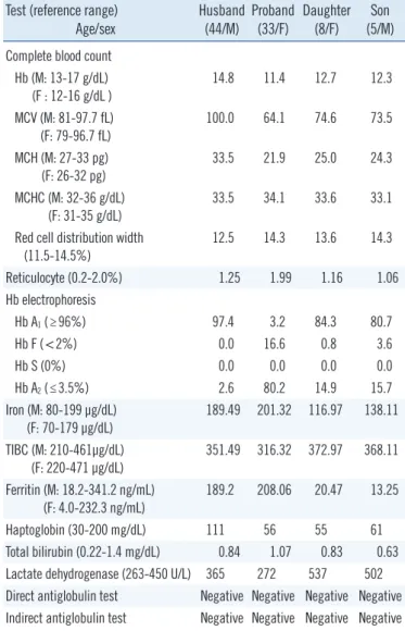
Table 1. Laboratory results of the proband’s family Test (reference range)
Age/sex Husband (44/M) Proband
(33/F) Daughter (8/F) Son
(5/M) Complete blood count
Hb (M: 13-17 g/dL) (F : 12-16 g/dL )
14.8 11.4 12.7 12.3
MCV (M: 81-97.7 fL) (F: 79-96.7 fL)
100.0 64.1 74.6 73.5
MCH (M: 27-33 pg) (F: 26-32 pg)
33.5 21.9 25.0 24.3
MCHC (M: 32-36 g/dL)
(F: 31-35 g/dL) 33.5 34.1 33.6 33.1 Red cell distribution width
(11.5-14.5%) 12.5 14.3 13.6 14.3
Reticulocyte (0.2-2.0%) 1.25 1.99 1.16 1.06
Hb electrophoresis
Hb A1 (≥96%) 97.4 3.2 84.3 80.7
Hb F (<2%) 0.0 16.6 0.8 3.6
Hb S (0%) 0.0 0.0 0.0 0.0
Hb A2 (≤3.5%) 2.6 80.2 14.9 15.7
Iron (M: 80-199 µg/dL) (F: 70-179 µg/dL)
189.49 201.32 116.97 138.11 TIBC (M: 210-461µg/dL)
(F: 220-471 µg/dL)
351.49 316.32 372.97 368.11 Ferritin (M: 18.2-341.2 ng/mL)
(F: 4.0-232.3 ng/mL) 189.2 208.06 20.47 13.25
Haptoglobin (30-200 mg/dL) 111 56 55 61
Total bilirubin (0.22-1.4 mg/dL) 0.84 1.07 0.83 0.63 Lactate dehydrogenase (263-450 U/L) 365 272 537 502 Direct antiglobulin test Negative Negative Negative Negative Indirect antiglobulin test Negative Negative Negative Negative Abbreviations: MCV, mean corpuscular volume; MCH, mean corpuscular he- moglobin; MCHC, mean corpuscular hemoglobin concentration; TIBC, total iron binding capacity.