• 검색 결과가 없습니다.

Analysis of Neurosensory Dysfunction after Dental Implant Surgery JOMP

N/A
N/A
Protected

Academic year: 2021

Share "Analysis of Neurosensory Dysfunction after Dental Implant Surgery JOMP"

Copied!
7
0
0

로드 중.... (전체 텍스트 보기)

전체 글

(1)

pISSN 2288-9272 eISSN 2383-8493 J Oral Med Pain 2014;39(4):133-139 http://dx.doi.org/10.14476/jomp.2014.39.4.133

Analysis of Neurosensory Dysfunction after Dental Implant Surgery

Young-Chan Choi

1

, Eunae S. Cho

1

, Robert L. Merrill

2

, Seong Taek Kim

1

, Hyung Joon Ahn

1

1

Department of Orofacial Pain and Oral Medicine, Dental Hospital, College of Dentistry, Yonsei University, Seoul, Korea

2

UCLA School of Dentistry, Los Angeles, CA, USA

Received August 18, 2014 Revised August 29, 2014 Accepted September 11, 2014

Purpose: There have been reports regarding the various factors associated with the level of discomfort and recovery from neurosensory symptoms in patients with trigeminal nerve injury.

However, the contributing factors remain uncertain and poorly understood. The purpose of this paper was to investigate the possible association between various factors expected to affect neurosensory discomfort and recovery in patients with mandibular nerve injury after dental implant surgery.

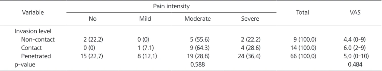
Methods: Eighty-nine post-dental implant surgery patients with mandibular nerve injury were enrolled in this retrospective analysis. A medical records review of the patients was done to de- termine if the patients’ improvement was related to pain intensity, the length of time between the injury and removal of the implant or the depth of penetration of the implant into the man- dibular canal as determined by cone-beam computed tomography.

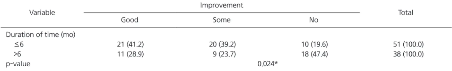
Results: There was no significant linear relationship between pain intensity and symptomatic improvement (p=0.319). There was no significant linear relationship between the level of man- dibular canal penetration and either pain intensity (p=0.588) or symptomatic improvement (p=0.760). There was a statistically significant linear relationship between length of time be- fore the injury was treated, both with pain intensity (p=0.004), and symptomatic improvement (p=0.024).

Conclusions: Our findings indicate that the length of time between nerve injury and initiation of conservative treatment is more closely related to the pain intensity and symptomatic im- provement than other factors, including the level of mandibular canal invasion. Additionally, increased pain intensity and decreased symptomatic improvement can be expected over time, because of this linear trend. Therefore, although direct injury to the nerve is the most impor- tant factor contributing to a neurosensory disturbances, early neurosensory assessment and initiation of conservative treatment should be done to optimize recovery.

Key Words: Dental implants; Mandibular nerve; Trigeminal nerve injuries

Correspondence to:

Hyung Joon Ahn

Department of Orofacial Pain and Oral Medicine, Dental Hospital, College of Dentistry, Yonsei University, Yonsei-ro 50, Seodaemun-gu, Seoul 120-752, Korea

Tel: +82-2-2228-3112 Fax: +82-2-393-5673 E-mail: [email protected]

Although nerve injury during implant surgery is con- sidered rare, unintentional and generally unsuspected se- rious complication do occur, two of the most notable be- ing hemorrhage and infection.

2)

Recent literature suggests that, increased use of mandible implants is associated with greater risk of trauma to the IAN. Results from a prospec- tive multicenter study of partially edentulous patients with mandibular implants suggest that altered sensations may be more prevalent than previously reported,

3)

and there is

INTRODUCTION

Injury to the branches of trigeminal nerve is a well- known risk factor in dental procedures and maxillofacial surgery. Altered sensations or dysesthesias related to nerve injury are recognized complications that may follow dental and surgical procedures of the mandible.

1)

The most com- monly injured branches are the inferior alveolar nerve (IAN), the lingual nerve (LN), and the mental nerve (MN).

2)

JOMP

Journal of Oral Medicine and Pain

Copyright

2014 Korean Academy of Orofacial Pain and Oral Medicine. All rights reserved.

CC

This is an open-access article distributed under the terms of the Creative Commons Attribution Non-Commercial License (http://creativecommons.org/licenses/by-nc/3.0/),

which permits unrestricted non-commercial use, distribution, and reproduction in any medium, provided the original work is properly cited.

(2)

in the Dental Hospital of Yonsei University, Seoul, Korea.

Each patient was examined and received a radiological evaluation with computed tomography. We excluded subject from the study if their injury was due to procedures other than dental implant surgery, if the injury was to the maxil- lary division or if the patient was seen only once. On this basis, five individuals were excluded, and the final sample was comprised of 89 patients who gave a written informed consent, 61 (68.5%) being female and 28 (31.5%) being male. Their mean age was 53.9±9.9 years (range 27-75 years). The study protocol was approved by the institutional review board of Yonsei University Hospital (2-2013-3301).

2. Methods

We reviewed each subject’s medical records to identify confounding factors that could influence neurosensory dys- function or symptomatic improvement after conservative treatment. Those factors were pain intensity, level of inva- sion into mandibular canal in computed tomography (CT), and duration of time from nerve injury to initiation of con- servative treatment.

1) Neurosensory disturbance

At the initial visit, the patients’ neurosensory changes were evaluated using pin-prick, light touch, 2-point dis- crimination, pressure-pain threshold, brush stroke direc- tion test, and current perception threshold (CPT) using Neurometer CPT/C (Neurotron Inc., Baltimore, MD, USA).

The patients with electric or burning pain were asked to grade the severity of their pain on a 10-point visual ana- logue scale (VAS) and the patients were divided into four groups according to VAS: (1) no pain (VAS=0); (2) mild pain (0<VAS<4); (3) moderate pain (4≤VAS<7); (4) severe pain (7≤VAS≤10).

2) Symptomatic improvement after conservative treatment

The conservative treatment group received treatment con- sisting of behavioral, physical, and pharmacologic manage- ment.

16)

This group was divided into three groups: (1) ‘good improvement’—satisfactory improvement and few disabilities on routine daily work; (2) ‘some im prove ment’—unsatisfac- tory improvement with some remaining disabilities during considerable evidence suggesting that IAN injury is the

most common injury experienced by patient undergoing dental implant surgery.

4)

Prospective studies have reported higher incidences of injury to the IAN with implant placement in the posterior mandible. The incidence is variably reported from 3.7% to 39.1% at 1 week postoperative examination.

5-8)

Ellies and Hawker,

9)

in a two-center retrospective study done 2 weeks after implant surgery, reported altered sensation of the IAN in 37% and 36% of the patients respectively. In both cen- ters, these complaints were persistent in 13% of patients.

The incidence of permanent sensory dysfunction (includ- ing dysesthesia, anesthesia or paresthesia) was 1% to 8%

respective to the center, but the incidence of chronic pain is unclear.

10)

In a Korean study, Ryu and Kwon

11)

reported the incidence of dysesthesia after dental implant surgery was 24.6%, and the incidence of permanent dysesthesia was 6.2%.

There have been a number of reports on various factors related to the level of discomfort and recovery of normal sensation in post dental implant patients. Merrill

12)

has sug- gested that nerve damage can lead to transient or persistent alteration in orofacial sensations depending on the severity of the injury. Hillerup

13)

stated that recovery of normal sen- sation was related to the etiology of the injury. Kraut and Chahal

14)

suggested that spontaneous recovery of normal sensation after nerve injury is dependent both on the sever- ity of the injury and the nerve involved. For example, par- tial transection of the LN is less likely to result in spontane- ous resolution of symptoms, compared with similar injury involving the IAN. In another study, Tay and Zuniga

15)

re- ported that there was no significant correlation between the severity of nerve injury and the time elapsed from injury.

In this study of post dental implant surgery patient who sustained mandibular nerve injury, we investigated the pos- sible relationship between factors expected to affect neuro- sensory dysfunction and recovery.

MATERIALS AND METHODS

1. Patients

Ninety-four subjects who had post-dental implant abnor-

mal sensation and/or neuropathic pain in the jaw were re-

ferred to Department of Orofacial Pain and Oral Medicine,

(3)

signed-rank test, Kruskal-Wallis test, Mantel-Haenszel χ

2

test, Mann-Whitney test. Statistical significance was estab- lished at 95%. SPSS Statistics version 17.0 statistical pack- age (SPSS Inc., Chicago, IL, USA) was used for all statistical analyses.

RESULTS

1. Subject Demographics

The sample comprised 89 patients with nerve injury.

Female patients were 61 (68.5%) and male patients were 28 (31.5%). Their mean age was 53.9±9.9 years (range 27-75 years). The mean age by sex distribution was 54.7±9.4 years in female, and 52.1±10.9 years in male.

2. Test Result for Neurosensory Disturbance

As presented in Table 1, both median value of pin-prick and light touch perception were significantly decreased on the injured side (65.0, 0-3,000.0; 60.0, 0-1,000.0) compared with the uninjured side (100.0, p=0.008; 100.0, p=0.004), and median perceived count of brush stroke was signifi- cantly decreased in injured side (29.0, 0-30.0) than in unin- jured side (30.0, 4-30.0; p<0.001). The median distance for 2-point discrimination was significantly increased in in- jured side (13.4, 0-55.0) compared to the uninjured side (10.2, 0-72.4; p<0.001). Median force of pressure-pain thresh- old showed no significant difference between the injured side (160.0, 0-300) and the uninjured side (160.0, 50-270;

p=0.785).

As presented in Table 2, in the frequencies of 2,000 Hz, 250 Hz, and 5 Hz, the median CPT values were statisti- cally significantly increased in injured side compared with uninjured side (2,000 Hz, p<0.001; 250 Hz, p=0.001; 5 Hz, p=0.003).

routine daily work; or (3) ‘no improvement’.

3) Level of invasion into mandibular canal in CT CTs were taken to indirectly confirm implant invasion into the IAN. The confirmed cases were divided into three groups: (1) ‘Non-contact’—the implant was not in contact with the mandibular canal; (2) ‘Contact’—the implant was in close contact with the mandibular canal, but the corti- cal layer of canal was preserved; or (3) ‘Penetrated’—the im- plant penetrated the mandibular canal wall and discontinu- ity of cortical layer was observed.

4) Duration of time from nerve injury

Patients were divided into two groups according to the time from nerve injury to initiation of conservative treat- ment involving behavioral, physical, and pharmacologic management. The criteria for were: (1) within 6 months of the nerve injury; (2) over 6 months from nerve injury.

5) Relation between pain intensity and other factors A validation was performed on the relationship between pain intensity and other factors, i.e., symptomatic improve- ment after conservative treatment, level of invasion into mandibular canal, duration of time from nerve injury.

6) Relation between symptomatic improvement and other factors

An additional validation was performed on the relation- ship between symptomatic improvement after conservative treatment and other factors, i.e., the level of invasion into mandibular canal, duration of time from nerve injury.

3. Statistics

Statistical analyses were performed using the Wilcoxon

Table 1. Result for clinical neurosensory test

Test Injured side (n=89) Uninjured side (n=89) p-value

Pin-Prick (%) Light touch (%)

2-Point discrimination (mm) Pressure-pain threshold (cN) Brush stroke direction (count)

65.0 (0-3,000.0) 60.0 (0-1,000.0) 13.4 (0-55.0)

160 (0-300.0) 29.0 (0-30.0)

100.0 100.0 10.2 (0-72.4)

160 (50-270) 30 (4-30)

0.008*

0.004*

<0.001*

0.785

<0.001*

Values are presented as median (range).

*Statistically significant at significance level of p<0.05.

(4)

improvement, and 6.0 (0-10) in the group reporting no im- provement. There was no significant statistically significant difference between the group (p=0.129), and there was no significant linear-relationship between pain intensity and symptomatic improvement (p=0.319).

4. Relation between the Implant and the Mandibular Canal by CT

As presented in Table 4, there were 9 (10.1%) subject in the non-contact group, 14 (15.7%) subjects in the contact group, and 66 (74.2%) subjects in the penetrated group.

Median VAS score were 4.4 (0-9) in the non-contact group, 6.0 (2-9) in the contact’ group, and 5.0 (0-10) in the penetrated group. There were no significant differences be- tween each groups (p=0.484), and no significant linear- relationship between the level of mandibular canal invasion and pain intensity (p=0.588).

As shown in Table 5, there was no significant linear-re- lationship between the level of mandibular canal invasion and symptomatic improvement (p=0.760).

3. Pain Intensity and Improvement with Conservative Treatment

Table 3 presents the relationship between pain intensity and symptomatic improvement after conservative treat- ment. There were 17 (19.1%) patients who complained of sensory change only, and 72 (80.9%) who complained of both pain and sensory changes.

Thirty two (36.0%) patients reported good improvement after conservative treatment, 29 (32.6%) reported some im- provement, and 28 (31.5%) reported no improvement.

Median VAS scores were 5.0 (0-9) in the group with good improvement, 4.0 (0-9) in the group reporting some

Table 2. Result for current perception threshold test Frequency

(Hz)

Injured side (n=23)

Uninjured side

(n=23) p-value

2,000 250 5

275.0 (138.0-524.0) 120.0 (41.0-292.0)

72.0 (7.0-240.0)

134.0 (89.0-255.0) 51.0 (10.0-188.0) 31.0 (8.0-115.0)

0.000*

0.001*

0.003*

Values are presented as median (range).

*Statistically significant at significance level of p<0.05.

Table 3. Analysis of pain intensity after symptomatic treatment

Variable Improvement

Total p-value

Good Some No

Pain intensity No Mild Moderate Severe Total VAS

7 (41.2) 4 (44.4) 9 (27.3) 12 (40.0) 32 (36.0) 5.0 (0-9)

5 (29.4) 5 (55.6) 14 (42.4) 5 (16.7) 29 (32.6) 4.0 (0-9)

5 (29.4) 0 (0) 10 (30.3) 13 (43.3) 28 (31.5) 6.0 (0-10)

17 (100.0) 9 (100.0) 33 (100.0) 30 (100.0) 89 (100.0)

0.319

0.129 Values are presented as number (%) or median (range).

VAS, visual analogue scale.

Statistically significant at significance level of p<0.05.

Table 4. Analysis of level of invasion into mandibular canal in computed tomography compared with pain intensity

Variable Pain intensity

Total VAS

No Mild Moderate Severe

Invasion level Non-contact Contact Penetrated p-value

2 (22.2) 0 (0) 15 (22.7)

0 (0) 1 (7.1) 8 (12.1)

5 (55.6) 9 (64.3) 19 (28.8) 0.588

2 (22.2) 4 (28.6) 24 (36.4)

9 (100.0) 14 (100.0) 66 (100.0)

4.4 (0-9) 6.0 (2-9) 5.0 (0-10)

0.484 Values are presented as number (%) or median (range).

VAS, visual analogue scale.

Statistically significant at significance level of p<0.05.

(5)

DISCUSSION

Trigeminal neuropathic pain following tissue or nerve in- jury in the nerve distribution is classified as traumatic tri- geminal neuralgia. Traumatic neuralgia is defined as con- tinuous pain following complete or partial damage to a part of peripheral or central nervous system. Local anesthetic blockade may not totally abolish the pain due to central- ization of the pain mechanism and/or a sympathetic com- ponent.

10,17)

Significant neurosensory dysfunction or pain due to the trauma may affect the patient’s quality of life.

Sometimes the patient requires the skill of a clinical psy- chologist or psychiatrist with clinical experience in pain.

18)

In the present study, females outnumbered males at least two to one. This ratio has been consistently noted in stud- ies from various pain centers and is suggestive of a po- tential hormonal link with the tendency to develop neuro- pathic pain after nerve injury. Smith et al.

19)

have reported on the genetic variability associated with increased risk of developing chronic pain after nerve injury. But, Pogrel and Thamby,

6)

Hillerup,

13)

Tay and Zuniga

15)

have suggested that patients referred to tertiary care centers do not represent the true incidence of nerve injury from various causes.

Kraut and Chahal

14)

suggested that when nerve injury is suspected, a thorough neurosensory examination (light 5. Effects of Duration of Time from Nerve Injury to

Initiation of Conservative Treatment

As presented in Table 6, 51 (57.3%) patients were seen within 6 months of the nerve injury and 38 (42.7%) were seen more than 6 months after the nerve injury. Median VAS score were significantly decreased in within 6 months group (4.0, 0-9), than in over 6 months group (6.0, 0-10;

p=0.047).

There was a statistically significant linear-relationship between the duration of time from nerve damage and pain intensity (p=0.004) and, as shown in Table 7, there was sta- tistically significant linear-relationship between duration of time from nerve damage and symptomatic improvement with conservative treatment (p=0.024).

Table 5. Analysis of level of invasion into mandibular canal in computed tomography compared with symptomatic improvement

Variable Improvement

Total

Good Some No

Invasion level Non-contact Close contact Penetrated

4 (44.4) 5 (35.7) 23 (34.8)

1 (11.1) 7 (50.0) 21 (31.8)

4 (44.4) 2 (14.3) 22 (33.3)

9 (100.0) 14 (100.0) 66 (100.0)

p-value 0.760

Values are presented as number (%).

Statistically significant at significance level of p<0.05.

Table 6. Analysis of duration of time from nerve injury to pain intensity

Variable Pain intensity

Total VAS

No Mild Moderate Severe

Duration of time (mo) ≤6

>6

14 (27.5) 3 (7.9)

6 (11.8) 3 (7.9)

19 (37.3) 14 (36.8)

12 (23.5) 18 (47.4)

51 (100.0) 38 (100.0)

4.0 (0-9) 6.0 (0-10)

p-value 0.004* 0.047*

Values are presented as number (%) or median (range).

VAS, visual analogue scale.

*Statistically significant at significance level of p<0.05.

Table 7. Analysis of duration of time from nerve injury to symptomatic improvement

Variable Improvement

Total

Good Some No

Duration of time (mo) ≤6

>6

21 (41.2) 11 (28.9)

20 (39.2) 9 (23.7)

10 (19.6) 18 (47.4)

51 (100.0) 38 (100.0)

p-value 0.024*

Values are presented as number (%).

*Statistically significant at significance level of p<0.05.

(6)

Interestingly, 10.1% (9 cases) of patients didn’t show di- rect contact between affecting implant fixture and man- dibular canal and the distances between fixture and canal wall ranged from 1 mm to 3 mm. In cases such as these, it has been postulated that the damage may be due to im- plant induced indirect trabecular bone compression or con- densation of the nerve,

24)

or thermal injury due to exces- sive heat build-up during the drilling procedure. Hirsch and Brånemark

25)

have suggested the following causes of nerve injury linked to implant surgery: 1) direct mechanical dam- age, 2) pressure on nerve and blood vessel, or 3) formation of hemangioma or osteoma followed by vascular damage.

On the other hand, in some cases of LN and MN injury, the nerve may be traumatized by incision, flap elevation, re- traction, or suturing procedures.

14,26)

Based on these possi- bilities, clinicians shouldn’t ignore the potential of indirect nerve damage when placing a dental implant.

Additionally, current recommendations in are to start the patient on high doses of steroids to decrease the neurogenic inflammation that will further injure the traumatized nerve.

27)

Tay and Zuniga

15)

reported that there was no significant correlation between the severity of nerve injury and the length of time from injury. However, Ryu and Kwon

11)

re- ported that recovery ratio of injured nerve after implant surgery was 71.8%, and 88.2% of recovered group was re- covered within 6 months.

In the present study, there was a significant linear r e lation - ship between duration of time and pain intensity as well as between duration of time and symptomatic improve ment.

Addi tionally, 26.3% of patients who suffered implant place- ment nerve injury over 1 year prior to this study showed sa ti s factory symptomatic improvement after conservative treat ment. Based on this observation, conservative treatment should be initiated for patients who have had an implant- induced nerve injury for over 1 year. In the present study, 36% of the patients showed satisfactory symptomatic im- provement after conservative treatment.

The present analysis was subject to some limitation. In - tensity of pain and improvement of neurosensory symptom were based on self-reported and subjective information. And this analysis can’t include a large number of patients, which resulted in difficulties controlling external factors such as the age and gender distributions. And in future study, touch, pain perception, direction of brush movement, two-

point discrimination, temperature sensitivity) should be performed. Further, any unusual patient reactions during surgery, such as sharp pain or an electrical shock-like sen- sation should be documented. In the present analysis, clini- cal neurosensory testing showed significant differences be- tween the injured side and the uninjured side with the ex- ception of the pressure-pain threshold test. It should be not- ed, however, that these types of tests have some attached difficulties such as the danger of examiner bias or lack of technical equilibration between examiners. Consequently, there is a necessity for more standardized and objective ex- amination procedures to test for neurosensory disturbance.

CPT testing may be performed to assess the damages of peripheral nerve in trigeminal nerve area. The nerve fibers with varied thickness are responsive selectively to the elec- tric stimulation with different frequencies. Therefore sen- sory disturbances such as hyperesthesia or hypoesthesia can be diagnosed when responses outside of these norms are observed.

20,21)

At the onset of this study, we anticipated that pain inten- sity would increase and symptomatic improvement would decrease relative to the level of implant invasion into man- dibular canal. However, there was no significant relation- ship between the level of invasion and pain intensity in this analysis. Additionally, there was no significant relationship between the anatomic invasion depth and symptomatic improvement.

When we studied the CT images, we noted that most of

implant injuries to the mandibular nerve were caused by

direct contact of implant itself with the mandibular ca-

nal. These nerve injuries may occur directly from the drill-

ing procedures, lack of attention to diagnostic information,

and/or direct compression of the nerve during implant in-

sertion.

5,14,22)

Variables such as magnification errors, un-

expected anterior loop of the MN, operator technique, or

anatomical variability can increase the chance of compli-

cations. Additionally, there may be more than one IAN or

canal,

23)

and a plexus of nerve branches may lie superior to

and outside the mandibular canal or infrequently, the prep-

aration drill may be 1 mm longer than implant. Allowances

for these variabilities must be incorporated into the calcula-

tion of available bone height.

5)

(7)

injury related to the difficult impaction. Dent Clin North Am 1979;23:471-488.

13. Hillerup S. Iatrogenic injury to the inferior alveolar nerve: etiol- ogy, signs and symptoms, and observations on recovery. Int J Oral Maxillofac Surg 2008;37:704-709.

14. Kraut RA, Chahal O. Management of patients with trigeminal nerve injuries after mandibular implant placement. J Am Dent Assoc 2002;133:1351-1354.

15. Tay AB, Zuniga JR. Clinical characteristics of trigeminal nerve injury referrals to a university centre. Int J Oral Maxillofac Surg 2007;36:922-927.

16. Park JH, Lee SH, Kim ST. Pharmacologic management of trigemi- nal nerve injury pain after dental implant surgery. Int J Prostho- dont 2010;23:342-346.

17. Graff-Radford SB. Facial pain. Neurologist 2009;15:171-177.

18. Vickers ER, Cousins MJ. Neuropathic orofacial pain part 1--prev- alence and pathophysiology. Aust Endod J 2000;26:19-26.

19. Smith SB, Maixner DW, Greenspan JD, et al. Potential genetic risk factors for chronic TMD: genetic associations from the OP- PERA case control study. J Pain 2011;12(11 Suppl):T92-101.

20. Masson EA, Boulton AJ. The Neurometer: validation and com- parison with conventional tests for diabetic neuropathy. Diabet Med 1991;8 Spec No:S63-S66.

21. Masson EA, Veves A, Fernando D, Boulton AJ. Current perception thresholds: a new, quick, and reproducible method for the assess- ment of peripheral neuropathy in diabetes mellitus. Diabetologia 1989;32:724-728.

22. Lamas Pelayo J, Peñarrocha Diago M, Martí Bowen E, Peñarrocha Diago M. Intraoperative complications during oral implantology.

Med Oral Patol Oral Cir Bucal 2008;13:E239-E243.

23. Carter RB, Keen EN. The intramandibular course of the inferior alveolar nerve. J Anat 1971;108:433-440.

24. Rodríguez-Lozano FJ, Sanchez-Pérez A, Moya-Villaescusa MJ, Rodríguez-Lozano A, Sáez-Yuguero MR. Neuropathic orofacial pain after dental implant placement: review of the literature and case report. Oral Surg Oral Med Oral Pathol Oral Radiol Endod 2010;109:e8-e12.

25. Hirsch JM, Brånemark PI. Fixture stability and nerve function af- ter transposition and lateralization of the inferior alveolar nerve and fixture installation. Br J Oral Maxillofac Surg 1995;33:276- 281.

26. Miloro M, Halkias LE, Slone HW, Chakeres DW. Assessment of the lingual nerve in the third molar region using magnetic resonance imaging. J Oral Maxillofac Surg 1997;55:134-137.

27. Bracken MB, Shepard MJ, Collins WF, et al. A randomized, con- trolled trial of methylprednisolone or naloxone in the treatment of acute spinal-cord injury. Results of the Second National Acute Spinal Cord Injury Study. N Engl J Med 1990;322:1405-1411.

clinical neurosensory test and CPT test should take place regularly for more objective, regular evaluation of neuro sen- sory recovery.

CONFLICT OF INTEREST

No potential conflict of interest relevant to this article was reported.

REFERENCES

1. Bartling R, Freeman K, Kraut RA. The incidence of altered sensa- tion of the mental nerve after mandibular implant placement. J Oral Maxillofac Surg 1999;57:1408-1412.

2. Burstein J, Mastin C, Le B. Avoiding injury to the inferior alveo- lar nerve by routine use of intraoperative radiographs during implant placement. J Oral Implantol 2008;34:34-38.

3. van Steenberghe D, Lekholm U, Bolender C, et al. Applicability of osseointegrated oral implants in the rehabilitation of partial edentulism: a prospective multicenter study on 558 fixtures. Int J Oral Maxillofac Implants 1990;5:272-281.

4. Delcanho RE. Neuropathic implications of prosthodontic treat- ment. J Prosthet Dent 1995;73:146-152.

5. Anderson LC, Kosinski TF, Mentag PJ. A review of the intraos- seous course of the nerves of the mandible. J Oral Implantol 1991;17:394-403.

6. Pogrel MA, Thamby S. The etiology of altered sensation in the inferior alveolar, lingual, and mental nerves as a result of dental treatment. J Calif Dent Assoc 1999;27:531, 534-538.

7. Wismeijer D, van Waas MA, Vermeeren JI, Kalk W. Patients’ per- ception of sensory disturbances of the mental nerve before and after implant surgery: a prospective study of 110 patients. Br J Oral Maxillofac Surg 1997;35:254-259.

8. Zarb GA, Schmitt A. The longitudinal clinical effectiveness of osseointegrated dental implants: the Toronto study. Part III: Problems and complications encountered. J Prosthet Dent 1990;64:185-194.

9. Ellies LG, Hawker PB. The prevalence of altered sensation as- sociated with implant surgery. Int J Oral Maxillofac Implants 1993;8:674-679.

10. Benoliel R, Eliav E. Neuropathic orofacial pain. Alpha Omegan 2012;105:66-74.

11. Ryu JW, Kwon JS. Dysesthesia after tooth extraction and implant surgery reported by dentists. J Oral Med 2007;32:263-272.

12. Merrill RG. Prevention, treatment, and prognosis for nerve

수치

Table 1. Result for clinical neurosensory test
Table 3. Analysis of pain intensity after symptomatic treatment
Table 7. Analysis of duration of time from nerve injury to symptomatic improvement

참조

관련 문서

1 John Owen, Justification by Faith Alone, in The Works of John Owen, ed. John Bolt, trans. Scott Clark, &#34;Do This and Live: Christ's Active Obedience as the

To gain insights about developmental outcomes related to separation experiences, we examined relations between the length of separation from mother and father,

It is believed that people will reconsider our study -the possibility of connection between zinc and for hair loss treatment methods- and especially people would be

Purpose: This study was designed to evaluate the clinical characteristics and the treatment of colonoscopic perforation and the availability of

The purpose of this study was to evaluate surface changes and abrasion of the implant fixture and TiN coated abutment screw after repeated delivery and

With the increase of the dental implant procedure, there are many cases of Maxillary Sinus Floor Elevation Surgery(MSFES). Additionally, its side effect has

Measurement of proximal contact tightness between the left first molar and second molar(dental implant) in the mandible... Clinical dental anatomy, histology,

Evaluating parameters of osseointegrated dental implants using finite element analysis-a two- dimentional comparative study examining the effects of implant