• 검색 결과가 없습니다.

The Association Between Masticatory Function Assessment and Masseter Muscle Thickness in the Elderly JOMP

N/A
N/A
Protected

Academic year: 2021

Share "The Association Between Masticatory Function Assessment and Masseter Muscle Thickness in the Elderly JOMP"

Copied!
7
0
0

로드 중.... (전체 텍스트 보기)

전체 글

(1)

pISSN 2288-9272 eISSN 2383-8493 J Oral Med Pain 2020;45(3):49-55 https://doi.org/10.14476/jomp.2020.45.3.49

The Association Between Masticatory Function Assessment and Masseter Muscle Thickness in the Elderly

Hyo-Jung Jung 1 , Yong-Guang Min 1 , Hyo-Jung Kim 2,3 , Joo-Young Lee 2,3 , Jong-Hoon Choi 1 , Baek-Il Kim 2,3 , Hyung-Joon Ahn 1

1 Department of Orofacial Pain and Oral Medicine, Yonsei University College of Dentistry, Seoul, Korea

2 Department of Preventive Dentistry and Public Oral Health, Yonsei University College of Dentistry, Seoul, Korea

3 BK21 PLUS Project, Yonsei University College of Dentistry, Seoul, Korea

Received June 10, 2020 Revised June 18, 2020 Accepted June 19, 2020

Purpose:

Purpose: This study investigated the association between the objective indicator of mastica- tory function assessment and the masseter muscle thickness (MMT) using ultrasound imaging.

Methods:

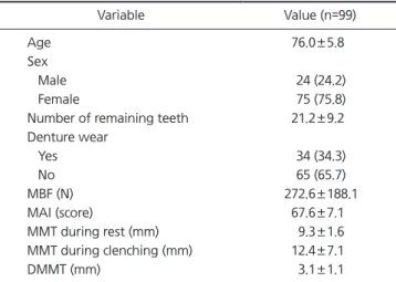
Methods: A total of 99 subjects (males: 24, females: 75, mean age: 76) were analyzed. The maximum bite force (MBF) was measured with a pressure-sensitive sheet and an image scanner. The mixing ability index (MAI) was calculated by image analysis after asking the subjects to chew a wax specimen. The MMT during rest and clenching were obtained with a diagnostic ultrasound system, and the difference in MMT during rest and MMT during clenching was defined as the difference in masseter muscle thickness (DMMT). Multiple re- gression analysis was performed to determine the independent variables affecting MBF and MAI.

Results:

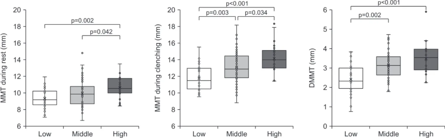
Results: The MBF showed correlation with the number of remaining teeth (β=0.346, p=0.002) and DMMT (β=0.251, p=0.011). The MAI correlated with only the number of re- maining teeth (β=0.476, p<0.001).

Conclusions:

Conclusions: The DMMT reflects the state of masseter muscle contraction, and can be used as a predictor as well as the number of teeth when assessing masticatory function.

Key Words:

Key Words: Bite force; Masseter muscle; Mastication; Ultrasonography

Correspondence to:

Hyung-Joon Ahn

Department of Orofacial Pain and Oral Medicine, Yonsei University College of Dentistry, 50-1 Yonsei-ro, Seodaemun-gu, Seoul 03722, Korea

Tel: +82-2-2228-3112 Fax: +82-2-393-5673 E-mail: [email protected]

https://orcid.org/0000-0001-9669-9781 This work was supported by the Korea Institute of Planning and Evaluation for Technology in Food, Agriculture, Forestry and Fisheries (IPET) through the High Value added Food Technology Development Program, funded by the Ministry of Agriculture, Food and Rural Affairs (MAFRA) (316071031HD020).

JOMP Journal of Oral Medicine and Pain

Copyright Ⓒ 2020 Korean Academy of Orofacial Pain and Oral Medicine. All rights reserved.

CC

This is an open-access article distributed under the terms of the Creative Commons Attribution Non-Commercial License (http://creativecommons.org/licenses/by-nc/4.0/),

INTRODUCTION

Mastication is the main function of the oral cavity, and to process of chewing and swallowing food. The mastication is related to physical and mental functions [1]. It is especially important to accurately evaluate the masticatory function in the elderly because the reduced masticatory ability in the elderly is associated with digestion, malnutrition, aspiration pneumonia, cognitive impairment, and low quality of life [2-5].

The masticatory function can be assessed using a variety of methods. Maximum bite force (MBF) and mixing ability

index (MAI) are useful indicators of objective masticatory function assessment, there are closely related to tooth loss [6,7]. The loss of teeth not only causes difficulty in chew- ing, but also induces the absorption of alveolar bone and reduces masseter muscle mass [8]. In addition, it has been reported that the edentulous group has a thinner masseter muscle thickness (MMT) than the dentulous group [9].

The masseter muscle is a representative masticatory

muscle and plays a key role in the masticatory process. It

has been reported that muscle mass and muscle weakness

due to aging occur not only in skeletal muscles, but also

in masticatory muscles [10]. Therefore, it is necessary to

(2)

consider MMT in the evaluation of masticatory function in the elderly.

Ultrasound imaging has been used to measure MMT as an indicator of muscle size, it was reported as a reliable clinical tool [11,12]. In addition, ultrasound is considered a useful method for clinical evaluation with considerable cost sav- ings and convenience compared to computed tomography or magnetic resonance imaging [12,13].

In previous studies, changes in the MMT according to the tooth loss [14], the relationship between the masseter muscle tension and chewing ability [15], and the role of the masseter muscle size and oral function according to age and sex were reported [16]. However, few studies have ex- amined the relationship between MBF, MAI and MMT, and the effect of MMT on objective masticatory function is un- clear. Therefore, the purpose of this study was to investigate the association between the objective indicator of mastica- tory function assessment and the MMT using ultrasound imaging.

MATERIALS AND METHODS

Informed consent was obtained from subjects for par- ticipation in the study, and the study was approved by the Institutional Review Board of Yonsei University Dental Hospital (IRB no. 2-2016-0034).

1. Subjects

From April 18, 2017 to September 21, 2018, a total of 132 subjects were recruited after visiting the elderly wel- fare facilities in Seodaemun-gu, Seoul, and Seongnam-si, Gyeonggi-do, and performing oral examinations. The crite- ria for selecting the subjects included those who were more than 65 years old, who had no unusual systemic diseases, were able to move on their own, and wanted to participate voluntarily. The following subjects were excluded to reduce the disturbance factors of data collection. i) Subjects with painful caries, ii) Subjects with more than 6mm periodontal pocket, iii) Subjects with pain and symptoms of temporo- mandibular joint, iv) Subjects with masticatory dysfunc- tion, v) Subjects who planned dental treatment (resin filling, prosthetic treatment, extraction, implant placement, etc.) during the study period. A total of 99 data were analyzed,

excluding those who met the exclusion criteria or withdrew consent.

2. Number of Remaining Teeth

The number of existing erupted teeth, excluding the re- sidual roots and third molar was counted, and the denture wear was investigated.

3. Measurement of Maximum Bite Force

The MBF was measured with pressure-sensitive sheets 98mm in thickness (Dental Prescale, 50H type; Fuji Film, Tokyo, Japan) and analyzed by an image scanner (Occluzer, FPD-707; Fuji Film) [17]. Subjects sat comfortably and one’s eyes were toward the front and performed maximal clench- ing in the intercuspal position with a pressure-sensitive film placed between the maxillary and mandibular dental arch- es. Subjects with removable partial dentures kept their den- tures in place during the measurement of the MBF. The bite force was calculated after scanning the sheet with an image scanner (Occluzer), taking into consideration the occlusal contact area and different densities of color. The bite force (N) was determined as the sum of the degree of coloration and the area at each contact point.

4. Measurement of Mixing Ability Index

This study used the MAI reported by Jeong et al. [7] to measure the objective masticatory efficiency. The wax specimens were made to form a 12×12×12 mm cube by ar- ranging red and green utility wax. Three wax specimens were provided to the subjects, that requested to chew the wax specimen ten times using a normal mastication pat- tern. Both sides of the collected wax specimens were pho- tographed with a digital camera and saved as image files.

Using the digital image analyzer (Image-Pro Plus v6.0;

Media Cybernetics Inc., Rockville, MD, USA), the total pro-

jection area, projection area >50 μm in thickness, maxi-

mum length, maximum breadth, red area and green area

of the image data were measured. The MAI value was cal-

culated through the discriminant formula for the measured

information. In order to reduce the variation of data, a sin-

gle examiner conducted the entire process of image analy-

sis. The average score for the three wax specimens chewed

by the subject was determined as the final score (1-100) of

(3)

MAI, and the higher the score, the higher the masticatory efficiency.

5. Measurement of Masseter Muscle Thickness

The MMT was measured using ultrasound system E-cube9 (Alpinion Inc., Seoul, Korea), and linear probe (frequency of 3.0-12.0 MHz) by a dentist. Subjects were instructed to sit with their upper body upright position, scanning was per- formed at the midpoint between the zygomatic arch and mandibular angle, approximately parallel with the Camper’s plane along a line connecting the point under the nasal wing with the tragus of the ear [14]. The MMTs were scan- ning twice on the right and left during rest and during clenching, the thickest part on the image was measured. The difference in MMT during rest and MMT during clenching was defined as the difference in masseter muscle thickness (DMMT) (Fig. 1). To ensure the reliability of the data, intra- class correlation coefficients for MMT during rest and MMT during clenching were 0.794, 0.815 on the right and 0.832, 0.867 on the left. For the MMT value, the mean of the 4 im- aging data (right×2, left×2) was used for analysis.

6. Statistical Analysis

As a result of performing the normality test using the Shapiro-Wilk test, the data were not satisfied with the nor- mal distribution and analyzed in a nonparametric test. MBF and MAI were divided into 3 groups based on quartiles, 1st quartile (25%, Q1) was low group, 2-3 quartiles (50%-75%, Q2-Q3) were middle group, 4th quartile (100%, Q4) was de- fined as High group. To analyze MMT according to MBF and MAI, the Kruskal-Wallis test was performed, post-tested with the Mann-Whitney U-test. The association between MBF, MAI and MMT was investigated by using multiple

regression analysis. For all statistical analysis, the IBM SPSS Statistics for Windows, Version 25.0 (IBM Co., Armonk, NY, USA) program was used, and the statistical significance lev- el was set to 5% (p<0.05).

RESULTS

Table 1 shows the general characteristics of subjects.

There were 99 subjects, 24 males (24.2%) and 75 females (75.8%), and the mean age was 76. The number of remain- ing teeth was 21.2, and the denture wearers was 34 (34.3%).

The mean values of the variables were MBF 272.6 (N), MAI 67.6 (score), MMT during rest 9.3 (mm), MMT during clenching 12.4 (mm) and DMMT 3.1 (mm).

Fig. 2 shows the difference of MMT according to MBF.

The higher the MBF, the thicker the MMT. The MMT dur- ing rest was significantly different in MBF’s low and high

A B

Fig. 1.

Fig. 1. Masseter muscle imaging with an ultrasonic diagnostic equipment. A, masseter muscle thickness during rest;

B, masseter muscle thickness during clenching; B-A, difference in masseter muscle displacement.

Table 1.

Table 1. General characteristics of subjects

Variable Value (n=99)

Age 76.0±5.8

Sex

Male 24 (24.2)

Female 75 (75.8)

Number of remaining teeth 21.2±9.2 Denture wear

Yes 34 (34.3)

No 65 (65.7)

MBF (N) 272.6±188.1

MAI (score) 67.6±7.1

MMT during rest (mm) 9.3±1.6

MMT during clenching (mm) 12.4±7.1

DMMT (mm) 3.1±1.1

MBF, maximum bite force; MAI, mixing ability index; MMT, masseter muscle thickness; DMMT, difference in masseter muscle displacement.

Values are presented as mean±standard deviation or number (%).

(4)

groups (p=0.002), middle and high groups (p=0.042). The MMT during clenching was significantly different in MBF’s low and high group (p<0.001), low and middle group (p=0.003), middle and high group (p=0.034). The DMMT was significantly different in MBF’s low and high groups (p<0.001), low and middle groups (p=0.002).

Fig. 3 shows the difference of MMT according to MAI.

There were no statistically significant differences in MMT during rest, MMT during clenching, and DMMT according to MAI.

Table 2 shows the factors related to the masticatory func- tion assessment. In multiple regression analysis, MMT dur- ing rest and MMT during clenching have a high correlation, so when both variables are input as independent variables, multicollinearity occurs and MMT during clenching is ex- cluded. The regression model using MBF as a dependent

variable was statistically significant (p<0.001). The regres- sion model determination coefficient was R=0.298 and the adjusted coefficient was R 2 =0.261. The MBF increased significantly as the number of remaining teeth (β=0.346, p=0.002) and DMMT (β=0.251, p=0.011) increased. The re- gression model using MAI as a dependent variable was sta- tistically significant (p<0.001). The regression model deter- mination coefficient was R=0.288 and the adjusted coeffi- cient was R 2 =0.250. The MAI increased significantly as the number of remaining teeth (β=0.476, p<0.001) increased.

DISCUSSION

The masseter muscle, temporalis muscle, medial pterygoid muscle, and lateral pterygoid muscle are called mastica- tory muscles. Among them, the masseter muscle is the main

Fig. 2.

Fig. 2. Difference of masseter muscle thickness according to maximum bite force. MMT, masseter muscle thickness; DMMT, difference in masseter muscle displacement. By the Kruskal-Wallis test and Mann-Whitney U-test at α=0.05.

Low 20

18 16 14 12 10 8 6

MMT during rest (mm)

Middle High Low

20 18 16 14 12 10 8 6

MMT during clenching (mm)

Middle High Low

6 5 4 3 2 1 0

DMMT (mm)

Middle High p=0.042

p=0.002

p=0.003 p=0.034 p<0.001

p=0.002 p<0.001

Fig. 3.

Fig. 3. Difference of masseter muscle thickness according to mixing ability index. MMT, masseter muscle thickness; DMMT, difference in masseter muscle displacement. By the Kruskal-Wallis test and Mann-Whitney U-test at α=0.05.

Low 20

18 16 14 12 10 8 6

MMT during rest (mm)

Middle High Low

20 18 16 14 12 10 8 6

MMT during clenching (mm)

Middle High Low

6 5 4 3 2 1 0

DMMT (mm)

Middle High

(5)

elevator muscle of the mandible, it is involved to the mas- ticatory function. Deterioration in muscle strength due to aging may be caused not only in skeletal muscles, but also in facial muscles. In particular, it is thought that changes in masseter muscle will affect mastication. Therefore, the purpose of this study was to investigate the association be- tween the objective indicator of masticatory function as- sessment and the MMT sing ultrasound imaging.

In this study, the mean MMT during rest of the elder- ly was 9.3 mm and MMT during clenching 12.4 mm. In a study by Park et al. [18] the mean MMT during rest of healthy adults aged 20 to 40 years was 14.8 mm and MMT during clenching 17.0 mm. In addition, Radsheer et al.

[19] reported that MMT decreases with age in both men and women, confirming that aging can cause a decrease in MMT (Table 1).

The difference of MMT according to MBF and MAI showed that the higher the MBF, the thicker the MMT (Fig.

2), and there was no statistically significant difference in MMT according to MAI (Fig. 3). In addition, as a result ana- lyzing factors related MBF and MAI using multiple regres- sion analysis, MBF showed the number of remaining and DMMT as predictors, but MAI had an effect only on the number of remaining teeth (Table 2). In a study by Bakke et al. [20] observed that in healthy adults, MMT in contrac- tion was strongly correlated with the number of teeth in contact. In general, the MMT during rest is measured lower

than MMT during clenching. This is because when muscle contracts, muscle fiber filaments slide into each other and become thicker as the fiber diameter increases [21]. DMMT is the difference between MMT during rest and MMT during clenching, and increases with thicker MMT during clench- ing. It suggests that muscle contraction that occurs in the clenching state is related to MBF, and that DMMT is a more important factor in masticatory function than MTT during rest.

However, MAI is a dynamic masticatory state caused by rhythmic movements, and various factors such as move- ment of the mandible, muscle activity, chewing rate, occlu- sion and tooth interference caused by lateral movements are complexly involved [22]. Therefore, the effect of MMT on MAI was weak and limited as a predictor.

One of the main goals of dental treatment is to main- tain a lifelong healthy masticatory function. In a study by Bhoyar et al. [3] MMT of the edentulous patients was in- creased after 3 month of denture wear than the thickness at the denture insertion. It has also been shown that im- plant-supported over-denture were reported to help main- tain MMT, bite force and masticatory efficiency rather than general conventional full denture dentures [23]. Therefore, proper prosthetic restoration is considered to increase the MMT and strengthen the activity, and the improvement of masticatory function can be expected in healthy elderly people.

Table 2.

Table 2. The factors related to the masticatory function assessment

Variable B Standard B t p-value VIF

MBF

Age –0.785 –0.024 –0.274 0.785 1.037

Sex (female) 51.682 0.118 1.133 0.260 1.447

Number of remaining teeth 7.068 0.346 3.122 0.002 1.631

MMT during rest 9.471 0.077 0.752 0.454 1.375

DMMT 56.347 0.251 2.584 0.011 1.254

F=7.914, p<0.001, R=0.298, adjusted R

2

=0.261 MAI

Age –0.018 –0.015 –0.164 0.870 1.037

Sex (female) –0.454 –0.027 –0.261 0.795 1.447

Number of remaining teeth 0.368 0.476 4.260 <0.001 1.631

MMT during rest –0.024 –0.005 0.049 0.961 1.376

DMMT 1.493 0.176 1.792 0.076 1.254

F=7.539, p<0.001, R=0.288, adjusted R

2

=0.250

VIF, variance inflation factor; MBF, maximum bite force; MMT, masseter muscle thickness; DMMT, difference in masseter muscle displacement;

MAI, mixing ability index.

The data was analyzed by multiple regression analysis.

(6)

Since this study was designed as a cross-sectional study, it was difficult to explain the causal relationship between MMT and masticatory function variables. In addition, it may be limited to generalize the results of research on con- venience samples extracted from some regions, and the dis- tribution of subgroups according to gender and tooth loss is uneven. Despite these limitations, this study confirmed that the MMT in the elderly measured by ultrasound imaging can be a predictor of MBF, which is one of the masticato- ry function assessment indicators. It suggests that occlusal recovery and masseter muscle training in the elderly may help improve mastication ability.

CONFLICT OF INTEREST

No potential conflict of interest relevant to this article was reported.

ORCID

Hyo-Jung Jung

https://orcid.org/0000-0003-1321-6276 Yong-Guang Min

https://orcid.org/0000-0002-3530-8270 Hyo-Jung Kim

https://orcid.org/0000-0002-7946-4865 Joo-Young Lee

https://orcid.org/0000-0002-0135-3305 Jong-Hoon Choi

https://orcid.org/0000-0003-3211-3619 Baek-Il Kim

https://orcid.org/0000-0001-8234-2327 Hyung-Joon Ahn

https://orcid.org/0000-0001-9669-9781

REFERENCES

1. Nakata M. Masticatory function and its effects on general health. Int Dent J 1999;49:preceding 3. Erratum for: Int Dent J 1998;48:540-548.

2. Petersen PE, Yamamoto T. Improving the oral health of older people: the approach of the WHO Global Oral Health Programme.

Community Dent Oral Epidemiol 2005;33:81-92.

3. Kwon SH, Park HR, Lee YM, et al. Difference in food and nutrient

intakes in Korean elderly people according to chewing difficulty:

using data from the Korea National Health and Nutrition Exami- nation Survey 2013 (6th). Nutr Res Pract 2017;11:139-146.

4. Müller F, Shimazaki Y, Kahabuka F, Schimmel M. Oral health for an ageing population: the importance of a natural dentition in older adults. Int Dent J 2017;67 Suppl 2:7-13.

5. Kandelman D, Petersen PE, Ueda H. Oral health, general health, and quality of life in older people. Spec Care Dentist 2008;28:224-236.

6. Ikebe K, Matsuda K, Kagawa R, et al. Association of masticatory performance with age, gender, number of teeth, occlusal force and salivary flow in Japanese older adults: is ageing a risk factor for masticatory dysfunction? Arch Oral Biol 2011;56:991-996.

7. Jeong SH, Kang SM, Ryu JH, Kwon HK, Kim BI. Subjective food intake ability in relation to the Mixing Ability Index in Korean adults. J Oral Rehabil 2010;37:242-247.

8. Newton JP, Yemm R, Abel RW, Menhinick S. Changes in hu- man jaw muscles with age and dental state. Gerodontology 1993;10:16-22.

9. Bhoyar PS, Godbole SR, Thombare RU, Pakhan AJ. Effect of complete edentulism on masseter muscle thickness and changes after complete denture rehabilitation: an ultrasonographic study.

J Investig Clin Dent 2012;3:45-50.

10. Iinuma T, Arai Y, Fukumoto M, et al. Maximum occlusal force and physical performance in the oldest old: the Tokyo oldest old survey on total health. J Am Geriatr Soc 2012;60:68-76.

11. Kiliaridis S, Kälebo P. Masseter muscle thickness measured by ultrasonography and its relation to facial morphology. J Dent Res 1991;70:1262-1265.

12. Reis Durão AP, Morosolli A, Brown J, Jacobs R. Masseter muscle measurement performed by ultrasound: a systematic review. Den- tomaxillofac Radiol 2017;46:20170052.

13. Emshoff R, Emshoff I, Rudisch A, Bertram S. Reliability and temporal variation of masseter muscle thickness measurements utilizing ultrasonography. J Oral Rehabil 2003;30:1168-1172.

14. Yamaguchi K, Tohara H, Hara K, et al. Relationship of aging, skeletal muscle mass, and tooth loss with masseter muscle thick- ness. BMC Geriatr 2018;18:67.

15. Ohara Y, Hirano H, Watanabe Y, et al. Masseter muscle ten- sion and chewing ability in older persons. Geriatr Gerontol Int 2013;13:372-377.

16. Lin CS, Wu CY, Wu SY, et al. Age- and sex-related differences in masseter size and its role in oral functions. J Am Dent Assoc 2017;148:644-653.

17. Shinogaya T, Matsumoto M. Evaluation of prosthodontic treat- ment by occlusal force distribution: a methodological study. Eur J Prosthodont Restor Dent 1998;6:121-125.

18. Park G, Choi YC, Bae JH, Kim ST. Does botulinum toxin injection into masseter muscles affect subcutaneous thickness? Aesthet Surg J 2018;38:192-198.

19. Raadsheer MC, Kiliaridis S, Van Eijden TM, Van Ginkel FC, Prahl-Andersen B. Masseter muscle thickness in growing in- dividuals and its relation to facial morphology. Arch Oral Biol 1996;41:323-332.

20. Bakke M, Tuxen A, Vilmann P, Jensen BR, Vilmann A, Toft M.

(7)

Ultrasound image of human masseter muscle related to bite force, electromyography, facial morphology, and occlusal factors. Scand J Dent Res 1992;100:164-171.

21. Ariji Y, Sakuma S, Izumi M, et al. Ultrasonographic features of the masseter muscle in female patients with temporomandibular disorder associated with myofascial pain. Oral Surg Oral Med Oral Pathol Oral Radiol Endod 2004;98:337-341.

22. Fulks BA, Callaghan KX, Tewksbury CD, Gerstner GE. Relation-

ships between chewing rate, occlusion, cephalometric anatomy, muscle activity, and masticatory performance. Arch Oral Biol 2017;83:161-168.

23. Müller F, Hernandez M, Grütter L, Aracil-Kessler L, Weingart D, Schimmel M. Masseter muscle thickness, chewing efficiency and bite force in edentulous patients with fixed and removable im- plant-supported prostheses: a cross-sectional multicenter study.

Clin Oral Implants Res 2012;23:144-150.

수치

Table 1 shows the general characteristics of subjects.
Fig. 3 shows the difference of MMT according to MAI.

참조

관련 문서

 The Dutch physicist Pieter Zeeman showed the spectral lines emitted by atoms in a magnetic field split into multiple energy levels...  With no magnetic field to align them,

Modern Physics for Scientists and Engineers International Edition,

Five days later, on 15 January 1975, the Portuguese government signed an agreement with the MPLA, FNLA and UNITA providing for Angola to receive its independence on 11

Usefulness of co-treatment with immunomodulators in patients with inflammatory bowel disease treated with scheduled infliximab maintenance therapy.. Oussalah A, Chevaux JB, Fay

Inclusion and Inclusiveness: Shared Vision of Youth for Local, National, and Global Village Inclusion at large stands for embracing populations with disabilities and

Ross: As my lawfully wedded wife, in sickness and in health, until

웹 표준을 지원하는 플랫폼에서 큰 수정없이 실행 가능함 패키징을 통해 다양한 기기를 위한 앱을 작성할 수 있음 네이티브 앱과

다양한 번역 작품과 번역에 관한 책을 읽는 것은 단순히 다른 시대와 언어, 문화의 교류를 넘어 지구촌이 서로 이해하고 하나가