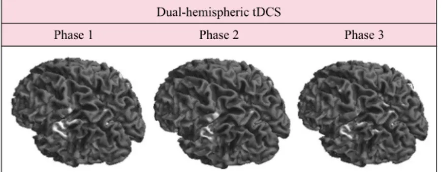
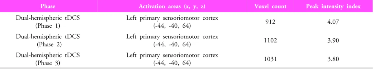
on both the Hemispheres: Single Case fMRI Study
The Journal of Korean Society of Physical Therapy
■
Jung-Won Kwon, PT, MS; Sung-Min Son, PT, MS; Chung-Sun Kim, PT, PhD, Professor
1; In-Sul Cho, PhD
2■
전체 글
The Journal of Korean Society of Physical Therapy
■
■
수치
관련 문서
첫째, UTAUT모형을 이용하여 스마트폰 어플리케이션의 구매에 대해 검정한 결과 독립변수들 (성과기대, 예상노력, 사회적 영향, 유희적 가치,
- 회로에 흐르는 전류의 방향과 크기가 일정한 전류를 직류(direct current) 라고 한다... 기전력이란 용어는 전기를 일으키는 힘이라는
• The supply and demand curves cross at the equilibrium price and quantity.. • You can read off approximate equilibrium values
1 John Owen, Justification by Faith Alone, in The Works of John Owen, ed. John Bolt, trans. Scott Clark, "Do This and Live: Christ's Active Obedience as the
The purpose of this study is to investigate the effect of online shopping mall characteristics on direct purchasing satisfaction of foreign consumers and purchase
Current study explores the effects of entrepreneurial orientation both on financial and innovative performance with 1,497 Korean small and medium-sized
This study analysed diverse data on colors and the current statues of actual education, and suggested an emotional coloring program which can motivate
Mean values of the distance from the most superior point of the mandibular condyle to the most inferior point of the articular eminence on transcranial and
