Hye-Sim Kang, MD1,3, Jeong Woo Park, MD1,2, Hee Sun Kim, MD4
1Department of Obstetrics and Gynecology, Jeju National University Hospital, Jeju,
2Department of Obstetrics and Gynecology, Seoul National University College of Medicine,
3Jeju National University College of Medicine and Graduate School of Medicine, Jeju, 4Department of Obstetrics and Gynecology, Ilsan- Paik Hospital, Inje University College of Medicine, Goyang, Korea
https://doi.org/10.14734/PN.2018.29.1.39 pISSN 2508-4887•eISSN 2508-4895
Objective: To characterize the hematologic profile of preterm fetuses delivered spontaneously with acute histologic chorioamnionitis (HCA).
Methods: This was a retrospective cohort study. The relationship between the presence of acute HCA and the change of hematologic profile was examined in 109 singleton pregnant women who were admitted and delivered between 24-32 weeks of gestation. Cases without results of placental histologic examination, cord blood cell count, and the differential count were excluded. From the cord blood, hemoglobin concentration, hematocrit, mean corpuscular volume, total leucocyte count and the differential count, platelet count, normoblast count, and umbilical arterial pH were obtain- ed. All the observed values were corrected for gestational age by calculating a ratio between the observed and mean expected value for gestational age.
Results: 1) The prevalence of acute HCA was 60.6% (66/109); 2) newborns with acute HCA had a higher median corrected leucocyte counts and corrected percentage of neutrophil in the differential count and a lower median corrected percentage of lymphocyte in the differential count than those without acute HCA; 3) neutrophilia was significantly frequent in newborns with acute HCA than in those without acute HCA; and 4) acute HCA was not associated with detectable changes in percentage of monocyte, eosinophil, basophil, and normoblast in the differential count, hemoglobin concentration, hematocrit, erythrocyte counts, mean corpuscular volume, platelet counts, or umbilical arterial pH.
Conclusion: The hematologic profile of preterm fetuses delivered spontaneously with acute HCA is characterized by significant changes in the total leucocyte count, neutrophil percentage, and lymphocyte percentage.
Key Words: Hematologic test , Preterm, Chorioamnionitis, Fetus
서론
급성 조직학적 융모양막염(acute histologic chorioamnionitis)은 태반의 급성 염증성 변 화를 의미하며 조기 분만된 태반에서 흔하게 관찰되는 소견이다.1-3 급성 조직학적 융모양막 염은 조산아에서 감염으로 인한 여러 주산기 합병증 및 뇌성마비의 위험인자로 알려져 있으 며 양수 내 백혈구 증가 및 여러 전염증성 시토카인(proinflammatory cytokine)의 증가와 밀 접한 관련이 있음이 밝혀져 있다.1,4-7 이러한 염증성 변화들은 결국 태아 염증반응을 유발하 게 되고, 태아 염증반응은 태아 염증반응증후군(fetal inflammatory response syndrome)으 로 명명되는 전신질환으로 설명되는데 이는 태아 혈액내 인터루킨(interleukin)-6 농도가
>11 pg/mL인 경우로 정의한다.8 자궁내 감염시, 태아에서 전염증성 시토카인의 합성이 촉 진되고 증가된 태아 혈액내 시토카인에 의해 성인에서의 전신 염증반응증후군(systemic inflammatory response syndrome)과 유사한 급성 염증반응이 일어나는 것으로 받아들여 지고 있다.
Received: 25 September 2017 Revised: 24 October 2017 Accepted: 8 December 2017 Correspondence to Jeong Woo Park, MD
Department of Obstetrics and Gynecology, Jeju National University Hospital, 15 Aran 13-gil, Jeju 63241, Korea
Tel: +82-64-717-1326 Fax: +82-64-717-1097 E-mail: [email protected] Copyright© 2018 by The Korean Society of Perinatology
This is an Open Access article distributed under the terms of the Creative Com- mons Attribution Non-Commercial License (http://creativecommons.org/
license/by-nc/4.0/), which permits unrestricted non-commercial use, distribution, and reproduction in any
The Effect of Acute Histologic Chorioam
nionitis on Hematologic Profile of Pre
term Fetuses
됨이 보고되었다.9-14 백혈구증가증(leucocytosis) 및 백혈구감 소증(leukopenia), 유핵적혈구(nucleated erythrocyte)의 출현 등은 좋지 않은 예후와도 관련이 있음이 알려져 있다.11,14 또한 신생아에서 중성구증가증(neutrophilia)이 호흡곤란증후군, 신 생아 패혈증, 뇌 손상과 관련되어 있다는 여러 저자의 보고가 있 었다.15-19
2011년 Romero 등은 태아 염증반응증후군을 가진 태아에서 신생아 및 성인에서 보이는 이러한 혈액학적 변화와 유사한 반 응이 일어남을 보고하였다.20,21 Romero 등20의 보고에 의하면, 태아 염증반응증후군으로 진단된 태아에서 염증반응증후군이 없는 태아에 비교해 전체 백혈구 수 및 중성구 수가 유의하게 증 가하고 유핵적혈구 수는 증가하는 경향성이 있다고 하였다. 한 편 다른 연구에서는 조기진통으로 조산한 태아에서 태아 염증 반응증후군 유무에 따른 제대동맥혈 내 pH값은 차이가 없었 다.21 또한 한 연구에서는 조직학적 융모양막염이 존재할 때 제 대혈 내 염증성 시토카인 및 대사물의 변화가 있다고 보고하였 다.22
그러나 급성 조직학적 융모양막염의 유무에 따른 신생아의 혈액계수 변화에 대해서는 부분적인 혈액계수 항목에 대한 연 구가 일부 있었고 모든 혈액계수에 대해 비교한 연구는 거의 없 었다. 2015년 Kim 등23은 34주 이전 조산한 태아 중, 제대염 (funisitis) 유무에 따른 모든 태아 혈액계수의 변화를 조사하여 보고하였다. 그러나 제대염은 태아 염증반응증후군의 태반 염 증성 변화의 대응물(counterpart)로 간주되고 있다.24 또한 조산 태아에서 제대염은 심한 태아 염증반응증후군과 관련이 깊다.25 따라서 제대염이 있을 때, 태아혈액계수가 태아 염증반응증후 군에서 보이는 변화와 비슷한 양상을 보이는 것은 당연하다고 할 수 있고 Kim 등26의 보고는 이를 확인한 것이라고 본다. 제대 염과 비교하였을 때, 급성 조직학적 융모양막염은 상대적으로 모체 부분의 태반 염증이며 경한 염증 단계로 받아들여진다. 급 성 조직학적 융모양막염이 있을 때 태아 혈액계수 항목의 전체 적인 변화를 조사한 연구는 지금까지는 없었다. 이에 본 연구의 저자들은 24-32주 사이에 조기진통 및 조기양막파열로 조산한 단태임신 태아에서 급성조직학적 융모양막염이 있으면서 혈액 계수의 변화를 보이는 태아에서 더 불량한 신생아 예후를 보일 것이라는 가설을 검증하기 위한 선행 연구로서 조직학적 융모 양막염과 태아 혈액계수의 변화 사이의 관련성을 조사하고자 하였다.
대상 및 방법
본 연구는 조산한 여성의 태반의 조직학적 소견과 출생시 제대 혈 내의 혈액계수의 변화와의 연관성을 알아보기 위하여 1998년 1월부터 2006년 12월까지 서울대학교병원에서 임신 24주에서 32주 사이에 조산한 임신부들 및 그 조산아를 대상으로 하였다.
본 연구대상의 선정 조건은 다음과 같다: 1) 단태임신; 2) 임신 24 주에서 32주 사이에 조산한 경우; 3) 의학적 적응증으로 조산하 지 않은 경우; 4) 분만 후 태반의 조직검사가 시행된 경우; 5) 분 만 직후 제대혈에서 전체혈구계산(complete blood cell count)을 시행하여 결과가 있는 경우; 6) 주요 선천기형(major con genital anomalies)이 없고 자궁내 태아사망(intrauterine fetal death)이 아닌 경우; 7) Rh 동종면역(isoimmunization)이 없는 경우; 8) 태 아수혈(fetal transfusion)을 시행하지 않은 경우로 하였다. 연구 대상은 태반의 조직학적 검사에 의한 급성 조직학적 융모양막염 의 존재 유무에 따라 두 군으로 나누었다. 혈구계산 및 감별계산 (differential count) 결과가 둘 다 없는 경우는 연구대상에서 제외 시켰다. 연구 수행 과정에서 모든 임상 자료와 검체는 충분한 설 명에 근거한 동의(informed consent)를 환자로부터 받고, 서울 대학교병원 생명윤리심의위원회의 승인(IRB No. 9712-038- 002) 하에 수집하였다.
임신부와 조산아의 인구학적 및 임상적 특성은 의무기록의 검토를 통해 확인하였다. 저자들은 임신부의 나이, 미분만부 (nulliparity) 여부, 분만 주수, 출생 체중, 조산의 원인(자연 조산 인지 의학적 적응증에 의한 조산인지 여부), 제대동맥혈 pH, 제 왕절개 유무, 분만 전 코르티코스테로이드(corticosteroid) 사용 유무, 분만 전 항생제 투여, 자궁수축억제제 사용, 양수 내 감염 (intra-amniotic infection) 유무, 중성구증가증 및 중성구감소증 의 빈도, 분만시 제대혈 내 백혈구계산(total leucocyte count) 및 감별계산(differential count), 적혈모세포(normoblast), 적혈구 계산(erythrocyte count), 혈색소 농도(hemoglobin concentra- tion), 적혈구용적률(hematocrit), 평균 적혈구용적(mean corpu- scular volume), 혈소판계산(platelet count)을 조사하였다. 혈구 계산 수치는 임신 주수에 따라 변하기 때문에 측정치는 측정치와 임신 주수에 따라 기대되는 평균치의 비(ratio)를 계산하여 분만 주수에 따라 교정하였다. 그러나 제대혈 내 호산구(eosino phil) 및 호염기구(basophil) 수는 임신 주수에 따라 변하지 않는다고 알려져 있으므로 교정값을 구하지 않았다.27 제대혈 내 백혈구계 산 및 감별계산, 적혈모세포계산, 적혈구계산, 혈색소 농도, 적혈 구용적률, 평균 적혈구용적, 혈소판계산, 제대동맥 pH의 임신 주수에 따른 참고치는 이전 연구들을 통해 얻었다.27-30 중성구 증가증은 재태 주수 대비 95백분위수 초과로, 중성구감소증은
연속형 변수의 비교는 student t-test 또는 Mann-Whitney U-test를 사용하였고, 범주형 변수의 비교는 chi-square test 또 는 Fisher’s exact test로 분석하였다. P값이 0.05 미만인 경우를 통계적으로 유의하다고 판정하였다. 통계프로그램은 SPSS version 19.0 (SPSS Inc., Chicago, IL, USA)를 사용하였다.
결과
연구 기간 동안 임신 24주에서 32주 사이의 생존아를 분만한 단태임신은 총 366예였다. 이 중에서 자발적인 조산이 아닌 모 체 및 태아의 적응증을 이유로 조산한 경우, 태반의 조직학적 검 사 결과가 없는 경우, 제대혈검사 결과가 없는 경우, 주요 선천기 형, 자궁내 태아사망, Rh 동종면역, 태아수혈을 시행한 경우를 제외한 109명이 포함기준을 충족하였다. 연구대상 가운데 급성 조직학적 융모양막염의 유병률은 60.6% (66/109)였고 양수 내 감염률은 20.3% (15/74)였다. 조산의 원인 중 조기 진통으로 분 만한 임신부는 56% (61/109)였으며, 조기 양막파열은 44%를 차지하였다. 급성 조직학적 융모양막염의 빈도는 조기 양막파열 군에서 조기 진통군에 비해 약간 더 높게 관찰되었으나 통계적 유의성은 없었다(68.8% [33/48] vs. 54.1% [33/61], P=0.120).
재태 주수 대비 5백분위수 미만으로 정의하였다.27
태반 조직 표본에서 제대(umbilical cord), 융모판(chorionic plate), 태반막(placental membranes)에서 조직을 얻어 10% 포 르말린에 고정시킨 후 파라핀에 포매(embedding)시켰다. 준비 된 조직블록의 절편을 헤마톡실린-에오신(hematoxylin-eosin) 으로 염색하였다. 급성 조직학적 융모양막염의 진단기준은 1995년 Yoon 등1이 보고한 바와 같이, 상기 조직 샘플 중 적어도 어느 하나에서 급성 염증성 병변이 존재하는 경우로 하였다. 조 기 진통의 진단은 적어도 20분 동안 4회 또는 1시간 동안 8회의 규칙적인 자궁수축이 있으면서 자궁경부의 변화가 있는 경우로 하였고, 조기 양막파열은 분만진통 이전에 양막이 파열되었다 는 병력과 무균 소독된 질경검사를 통한 질강 내 양수 확인, 니트 라진(nitrazine) 및 양치상화(ferning) 검사 등으로 양막파열 여 부를 확인하여 진단하였다. 임상적 융모양막염(clinical chorio- amnionitis)의 진단은 임신부의 체온이 37.8°C 이상이면서 자궁 압통, 악취 나는 질 분비물, 태아 빈맥, 모체 빈맥, 모체 백혈구증 가증 중 두 가지 이상의 소견을 보이는 경우로 하였다.
양수 내 감염 유무는 모든 대상 환자에서 사전 동의을 얻은 후, 복부 양수천자를 통하여 얻은 양수를 즉시 혈액배양병에 담아 마개를 씌워 차단하고 호기성, 혐기성 및 마이코플라즈마(my- coplasma)에 대하여 이미 보고된 방법에 의해 배양하였다.1
Table 1. Demographic and Clinical Characteristics of the Study Population according to the Presence or Absence of Acute Histologic Chorioamnio- nitis
Acute histologic chorioamnionitis
P-value
Absent (n=43) Present (n=66)
Maternal age (yrs) 30.2 (22.0-45.0) 31.5 (25.0-43.0) 0.314
Nulliparity 24/43 (55.8) 28/66 (42.4) 0.171
Etiology of preterm birth 0.167
Preterm labor 28/43 (65.1) 33/66 (50.0)
PROM 15/43 (34.9) 33/66 (50.0)
Cesarean delivery 26/43 (60.5) 28/66 (42.4) 0.066
Antenatal corticosteroids use 32/43 (74.4) 52/66 (78.8) 0.596
Antenatal antibiotics use 22/43 (51.2) 53/66 (80.3) 0.001
Tocolytics use 34/43 (79.1) 46/66 (69.7) 0.279
Clinical chorioamnionitis 1/43 (2.3) 4/66 (6.1) 0.646
Gestational age at delivery (wks) 29.7 (24.0-32.0) 28.2 (24.1-32.0) 0.001
Birthweight (g) 1,410.0 (710.0-2,610.0) 1,150.0 (530.0-2,020.0) 0.008
AF WBC count ≥20 cells/mm3* 8/23 (34.8) 32/51 (62.7) 0.025
Positive AF culture* 3/23 (13.0) 12/51 (23.5) 0.365
Values are presented as median (range) or number (%).
Abbreviations: PROM, premature rupture of membranes; AF, amniotic fluid; WBC, white blood cell.
*Transabdominal amniocentesis was performed only in 40 women from whom we received a written consent. In women without acute histologic chorioamnionitis, 61%
(14/23) were diagnosed as having preterm labor. Fifty three percent (27/51) had preterm labor in women with acute histologic chorioamnionitis.
급성 조직학적 융모양막염이 있었던 군에서 분만 주수 및 출생 체중의 중앙값은 더 낮고 임상적 융모양막염, 양수 내 백혈구 수 입방 밀리미터당 20개 이상 및 양수 내 감염의 빈도는 태반 염증 이 있는 군에 비해 더 높았다. 이 중 분만 주수, 출생 체중, 양수 내 백혈구 수는 통계적으로 유의한 차이가 있었다(Table 1).
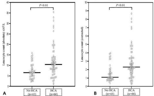
급성 조직학적 융모양막염의 존재 유무에 따른 제대혈 내 각 혈액계수의 절대값과 교정값, 백혈구 감별계산에서 백혈구 종 류에 따른 각 혈액계수의 백분위수와 교정 백분위수를 비교하 였다(Table 2). 급성 조직학적 융모양막염이 있었던 태아에서 제대혈 내 백혈구 수의 절대값 및 교정 백혈구 수의 중앙값은 통 계적으로 유의하게 높았다(Fig. 1). 급성 조직학적 융모양막염 이 있을 때 중성구증가증이 더 빈번하게 관찰되었으나 중성구 감소증은 차이가 없었다.
제대혈 내 백혈구 감별계산에서 중성구 백분위수 및 교정 중 성구 백분위수는 급성 조직학적 융모양막염이 존재할 때 증가하 였으나(Fig. 2), 림프구 백분위수 및 교정 림프구 백분위수는 모 두 감소하였다(Fig. 3). 한편, 백혈구 감별계산 중, 단핵구, 호산 구, 호염기구 백분위수는 두 군 간에 차이가 없었다(Table 2). 제 대혈 내 림프구 수, 단핵구 수, 호산구 수 및 호염기구 수의 절대 값은 급성 조직학적 융모양막염과 무관하였다(data not shown).
적혈구와 관련된 혈액계수 중에서 적혈구수, 혈색소 농도, 적
혈구용적률은 급성 조직학적 융모양막염이 있었던 태아에서 태 반 염증이 없었던 군과 비교하였을 때 감소하는 경향이었으나 모두 통계적으로 유의한 차이는 없었다. 평균적혈구용적, 혈소 판 수, 제대동맥 pH값도 두 군 간에 차이가 없었다. 적혈구 관련 모든 혈액계수, 혈소판 수, 제대동맥 pH값을 주수를 보정하여 계산하였을 때에도 두 군 간에 유의한 차이를 보이지 않았다 (Table 2).
고찰
본 연구에서는 임신 24-32주 사이에 자연 조산을 한 단태임 신 태아에서 태반의 급성 조직학적 융모양막염이 있을 때, 제대 혈 내 혈액계수의 분명한 변화가 있음을 확인하였다. 급성 조직 학적 융모양막염이 있는 태아에서 제대혈 내 백혈구 수, 중성구 수 및 백분위값이 유의하게 증가하였다. 반면 림프구 백분위수 는 유의한 감소를 보였다. 급성 조직학적 융모양막염이 존재할 때 적혈구 수, 혈색소 농도, 적혈구용적률이 통계적으로 유의하 지는 않지만 감소하는 경향을 보였다. 그 외 백혈구 감별계산에 서 단핵구, 호산구 및 호염기구 백분율, 혈소판 수, 제대동맥혈 pH값은 두 군 간에 유의한 차이가 없었다.
Fig. 1. Comparison of the fetal blood absolute and corrected leucocyte counts. (A) Fetuses with acute histologic chorioamnionitis had higher median absolute leucocyte counts than those without acute histologic chorioamnionitis (absolute leucocyte count median 10.5 [range, 1.1-35.4] vs. 6.6 [range, 2.4- 15.9] ×109/L, P<0.001). (B) Corrected leuco cyte counts by gestational age was also higher in fetuses with acute histologic chorioamnionitis (corrected leucocyte count median 2.31 [range, 0.27-8.68] vs. 1.11 [range, 0.41-3.97], P<0.001). HCA, histologic chorioamnionitis.
P<0.01 P<0.01
A B
P<0.01 P<0.01
A B
P<0.01 P<0.01
A B
Fig. 2. Comparison of the fetal blood percentage of neutrophil in the differential count and corrected neutrophil counts. (A) Median percentage of neutrophil was higher in fetuses with acute histologic chorio- amnionitis than in those without acute histologic chorioamnionitis (median percentage of neutrophil in the differential count 53.5% [range, 5.0-85.1] vs. 32.5% [range, 5.0-71.9], P<0.001). (B) Corrected median percentage of neutrophil counts was also higher in fetuses with acute histologic chorioamnionitis (corrected median percentage of neutrophil counts 5.51 [range, 0.43-11.2] vs. 2.20, [range 0.26-5.75], P<0.001). HCA, histologic chorioamnionitis.
Fig. 3. Comparison of the fetal blood percentage of lymphocyte in the differential count. (A) Median percentage of lymphocyte was lower in fetuses with acute histologic chorioamnionitis than those without acute histologic chorioamnionitis (median percentage of lymphocyte in the differential count 32.4%
[range, 8.3-86.0] vs. 53.0% [range, 18.1-85.0], P<0.001). (B) Corrected percentage of lymphocyte counts was also lower in fetuses with acute histologic chorioamnionitis (corrected median percentage of lympho- cyte counts 0.41 [range, 0.12-1.26] vs. 0.69 [range, 0.26-1.24], P<0.001). HCA, histologic chorioamnionitis.
Kim 등26의 보고에 의한 양수 내 상행감염의 과정을 살펴볼 때, 우선 양막 내 미생물이 출현하게 되면, 양수 내 전염증성 시 토카인 및 화학주성(chemotaxis) 인자의 농도가 증가한다. 다 음 과정으로 감염에 대한 일차 면역 방어 체계인 중성구의 출현 이 증가하게 된다. 따라서 본 연구에서 급성 조직학적 융모양막 염이 존재할 때, 출생시 제대혈 내 백혈구 수 및 중성구 수의 증 가는 상기 태반 연구와 Romero 등20의 태반 염증반응증후군에 서와 Kim 등23의 제대염에서 제대혈 내 태아혈액계수 변화 연구 결과와 일치하는 결과를 보여 주었다. 중성구증가증은 급성 조
직학적 융모양막염이 있을 때에는 거의 90% 이상에서 관찰되었 다. 중성구는 감염에 대한 급성 염증성 반응과 숙주 방어기제에 서 중심적인 역할을 담당하고 있다. 조산 태아의 혈액 내 백혈구 중에서는 림프구가 가장 지배적인 형태이다.27,29 임신 32주 이후 부터 중성구 수가 증가하기 시작하여 만삭이 되었을 때에는 중 성구가 가장 지배적인 형태가 되는 것으로 알려져 있다.27,31 또한 급성 조직학적 융모양막염이 있을 때 출생시 채취한 신생아 혈 액 내 백혈구 및 중성구가 증가한다는 관찰 결과는 자궁 내 염증 및 감염에 노출되었거나 세균혈증 소견을 보이는 조산한 태아 Table 2. Hematologic Profile of the Fetuses according to the Presence or Absence of Acute Histologic Chorioamnionitis
Acute histologic chorioamnionitis
P-value
Absent (n=43) Present (n=66)
Leucocyte (×109/L) 6.6 (2.4-15.9) 10.5 (1.1-35.4) <0.001
Corrected leucocyte 1.11 (0.41-3.97) 2.31 (0.27-8.68) <0.001
Neutrophilia 25/36 (69.4)† 52/57 (91.2)‡ 0.007
Neutropenia 1/36 (92.8)† 0/57 (0.0)‡ 0.387
Percentage of neutrophil (%)* 32.5 (5.0-71.9) 53.5 (5.0-85.1) <0.001
Corrected percentage of neutrophil* 2.20 (0.26-5.75) 5.51 (0.43-11.2) <0.001
Percentage of lymphocyte (%)* 53.0 (18.1-85.0) 32.4 (8.3-86.0) <0.001
Corrected percentage of lymphocyte* 0.69 (0.26-1.24) 0.41 (0.12-1.26) <0.001
Percentage of monocyte (%)* 7.2 (2.0-19.0)† 7.1 (0.0-24.0)‡ 0.981
Corrected percentage of monocyte* 2.38 (0.67-6.33)† 2.37 (0.0-8.0)‡ 0.981
Percentage of eosinophil (%)* 2.6 (0.0-11.0)† 1.6 (0.0-9.0)‡ 0.057
Percentage of basophil (%)* 0.1 (0.0-3.4)† 0.17 (0.0-6.2)‡ 0.976
Nucleated RBC (/100WBCs) 10.5 (0.0-237.0)† 0.9 (0.0-175.0)‡ 0.114
Corrected nucleated RBC 0.58 (0.0-12.2)† 0.04 (0.0-8.33)‡ 0.098
Erythrocyte (×1012/L) 3.60 (2.25-4.56) 3.42 (1.60-5.37) 0.059
Corrected erythrocyte 0.99 (0.65-1.31) 0.97 (0.48-1.41) 0.217
Hemoglobin (g/dL) 13.6 (2.4-18.1) 12.8 (6.0-20.6) 0.085
Corrected hemoglobin 1.04 (0.19-1.33) 0.98 (0.46-1.51) 0.176
Hematocrit (%) 41.5 (23.9-54.0) 39.8 (16.2-61.5) 0.229
Corrected hematocrit 1.00 (0.58-1.30) 0.95 (0.40-1.41) 0.401
MCV (×10-15/L) 111.9 (92.1-127.3) 114.9 (96.4-146.4) 0.173
Corrected MCV 0.96 (0.78-1.11) 0.97 (0.10-1.21) 0.507
Platelet (×109/L) 206.0 (16.0-380.0) 232.5 (7.0-491.0) 0.190
Corrected platelet 0.88 (0.06-1.57) 0.98 (0.03-2.03) 0.224
Umbilical arterial pH 7.29 (6.85-7.43)§ 7.30 (6.91-7.53)II 0.593
Corrected umbilical arterial pH 0.99 (0.93-1.00)§ 0.99 (0.92-1.00)II 0.480
Values are presented as median (range) or number (%).
Abbreviations: RBC, red blood cell; WBC, white blood cell; MCV, mean corpuscular volume.
*Percentage in the differential cell count.
†Available in 36.
‡Available in 57.
§Available in 40.
IIAvailable in 65.
및 신생아의 혈액에서 백혈구 및 중성구 수가 증가하였다는 여 러 연구의 결과와 일치한다. 2011년 Wirbelauer 등19은 급성 조 직학적 융모양막염을 보이는 1,500 g 미만으로 출생한 극소 저 체중 출생아(very low birthweight infant)에서 출생 후 1시간 이내에 얻은 혈액 내 백혈구 수와 중성구 수가 조직학적 융모양 막염이 없었던 군에 비해 유의하게 증가하였음을 확인하였다.
그러나 유핵적혈구 수는 두 군 사이에 차이가 없었다고 보고하 였다.19 2009년 Kim 등32은 태아 염증반응증후군의 조직학적 대 응부(counterpart)이자 급성 조직학적 융모양막염의 심한 형태 라고 할 수 있는 제대염이 존재할 때 임신부가 아닌 태아 제대혈 내 단핵구 및 과립구의 세포 표면 항원의 면역표현형이 변한다 는 사실을 증명하였다. 특히 과립구의 CD14, CD64, CD66b 그 리고 단핵구의 CD64의 평균통로밝기(mean channel bright- ness)의 중앙값이 유의하게 증가하였다는 것을 증명하였다.32 이 연구 결과의 의의는 본 연구 결과와 비교하였을 때, 비록 급 성 조직학적 융모양막염이 있을 때, 단핵구와 같이 그 절대적인 숫자의 변화가 관찰되지 않은 혈구 세포라고 할지라도 기능적 인 측면에서 여러 변화가 있음을 증명한 것이라고 할 수 있다.
1997년 Leikin 등33은 조기 진통 및 조기 양막파열로 조산하고 급성 조직학적 융모양막염이 확인된 신생아에서 출생 후 24시 간 이내에 채혈하여 시행한 혈액검사에서 유핵적혈구 수가 유 의하게 증가하였음을 보고하였다. 본 연구에서는 급성 조직학 적 융모양막염이 있었던 군에서 출생 직후 채취한 제대혈 내 백 혈구 수와 중성구는 유의하게 증가하였고 림프구 백분위수는 감소하였으나 유핵적혈구 수에서의 의미 있는 변화는 없었다.
앞서 말한 Wirbelauer 등19의 연구 결과와는 일치하였으나 Leikin 등33의 보고와는 다른 관찰 결과였다.
자궁 내 염증 및 감염의 결과라고 할 수 있는 태아 염증반응증 후군 또는 태반 염증의 존재 유무에 따른 유핵적혈구 수의 차이 에 있어서 성인에서의 전신 염증반응증후군에서와는 달리 일관 되지 않은 결과가 나오는 이유는 태아에 있어서 시토카인이나 염증 매개성 물질들이 적혈구 조혈 과정에 영향을 미치는 정도 가 성인에 비해 미약하기 때문으로 생각된다. 또한 적혈구형성 인자(erythropoietin)는 태반을 넘어가지 않으므로 태아는 태아 의 간 또는 신장에서 생성하는 적혈구형성인자의 영향만을 받 게 되는데 이는 태아가 성숙함에 따라 생산량이 증가하는 것으 로 알려져 있다.34 본 연구가 임신 24-32주 사이의 조산아를 연 구대상으로 하였기 때문에 유핵적혈구 수에 대한 영향이 보다 덜 반영되었을 것으로 보인다. 또 다른 설명으로는, 본 연구대상 군에서 유핵적혈구 수가 이미 알려진 임신 주수에 따른 유핵적 혈구 수 기준치와는 많은 차이를 보였다.28,29 특히 Table 2에서 유핵적혈구 교정값을 보았을 때, 급성 조직학적 융모양막염 유
무에 무관하게 양 군 모두 다른 선행 연구에서 알려진 평균값 기 준치 절반 또는 훨씬 못 미치는 수치를 보여주었으며 오히려 급 성 조직학적 융모양막염이 있는 군에서 더 낮은 수치를 보였던 점은 본 연구 결과와 다른 연구들과 차이가 있는 부분이었다.
본 연구는 임신 24-32주 사이의 자연 조산아에서 급성 조직 학적 융모양막염 유무에 따른 백혈구 수 및 감별계수, 적혈모세 포, 적혈구, 혈색소 농도, 혈소판, 적혈구용적률, 평균 적혈구용 적, 제대동맥 pH 등 거의 모든 혈액계수를 동시에 비교한 첫 번 째 연구이다. 이에 앞서 Kim 등23이 보고한 제대염이 있을 때 태 아 혈액계수의 변화와 거의 유사한 결과를 보여 준다. 이러한 연 구 결과의 의미는 태아 염증반응증후군의 대응부라고 할 수 있 는 제대염 즉 비교적 심한 태반 염증 형태를 보일 때 태아 혈액 계수의 분명한 변화를 보여준다는 기존 연구 결과와 비교하였 을 때, 비교적 초기 단계의 경한 태반 염증 형태까지 포함하는 조직학적 융모양막염이 존재할 때에도 이러한 태아 혈액계수의 변화는 이미 시작된다는 점을 보여주었다고 할 수 있다.
본 연구의 약점으로는 많은 연구대상 환자에서 채혈을 하기 전에 항생제, 스테로이드, 자궁수축억제제를 포함한 약물을 사 용하였다는 점이다. 특히 세팔로스포린계 항생제의 사용은 드 물지만 호산구증가증, 백혈구감소증 그리고 1% 미만에서 무과 립구증이 발생할 수 있는 것으로 알려져 있다.35,36 본 연구대상 군에서 급성 조직학적 융모양막염이 있는 군에서 조기 양막파 열 빈도가 더 높았으며, 따라서 항생제의 사용빈도가 더 높았다.
그러나 호산구 수 백분율에는 통계적으로 유의한 차이를 보이 지 않았으며 조직학적 융모양막염이 없었던 군에서 약간 더 높 은 경향을 보여 주었다. 또한 중성구감소증도 조직학적 융모양 막염 군에서는 관찰되지 않았다.
또한 본 연구대상군에서 급성 조직학적 융모양막염의 유병률 은 60.6% (66/109)로 높았다. 1979년 Russell37의 보고에 의하 면 분만 주수 32주 미만에서 급성 조직학적 융모양막염의 빈도 는 43.2% (70/162)였다. 또한 이 연구에 의하면, 분만 주수가 낮 을수록 태반 염증의 빈도는 증가하였고 구체적으로 21-24주에 는 94%, 25-28주에는 40%, 29-32주에는 약 35%에의 빈도를 보였다. 그리고 이 연구에서는 조기 진통 및 조기 양막파열 뿐만 아니라 다른 의학적 적응증에 의한 조산을 포함하는 모든 조산 아의 태반 염증 유무를 확인하여 보고한 연구이며 태반의 염증 은 임신성 고혈압 질환과 같은 의학적 적응증에 의한 조산보다 조기 진통 및 조기 양막파열과 같은 자연 조산에서 태반 염증의 빈도가 높음이 잘 알려져 있다.38 본 연구에서는 32주 이하의 자 연 조산한 경우만 연구대상으로 하였으므로 문헌에 알려진 43%보다는 약간 더 높은 빈도로 본 연구의 대상군에서 태반의 조직학적 융모양막염이 있을 것으로 예상할 수 있다. 따라서 본
연구에서 대상으로 한 분만 주수에서 약 60%의 태반 염증 유병 률은 기존 문헌의 알려진 유병률에 비하면 많이 높다고 할 수는 없을 것으로 사료된다.
결론적으로, 급성 조직학적 융모양막염이 있는 자연 조산아 의 혈액계수 상 특징으로는 백혈구 수의 증가, 중성구 백분위수 의 증가와 림프구 백분위수의 감소이다. 그리고 적혈구 생성 및 동맥혈 pH값에 미치는 영향은 미미하다는 점이다. 급성 조직학 적 융모양막염이 있을 때 거의 대부분에서 중성구증가증이 확 인되었다. 본 연구는 급성 조직학적 융모양막염이 있는 조산아 에서 모든 혈액계수의 변화를 동시에 보여준 첫 번째 연구라는 점에서 의의가 있겠다.
감사의 글
이 연구는 2017년도 제주대학교병원 연구비로 수행되었다.
References
1) Yoon BH, Romero R, Kim CJ, Jun JK, Gomez R, Choi JH, et al. Amniotic fluid interleukin-6: a sensitive test for antenatal diagnosis of acute in- flammatory lesions of preterm placenta and prediction of perinatal mor bidity. Am J Obstet Gynecol 1995;172:960-70.
2) Chang JW, Yoon BH, Syn HC. The relationship between the presence, severity and pattern of acute placental inflammation and amniotic fluid interleukin-8 in preterm labor. Korean J Obstet Gynecol 1999;42:
2669-74.
3) Kim JC, Yoon BH. The relationship between amniotic fluid white blood cell count and the presence and severity of acute placental inflamma- tion in preterm premature rupture of membrane. Korean J Obstet Gyne col 2000;43:885-90.
4) Murphy DJ, Sellers S, MacKenzie IZ, Yudkin PL, Johnson AM. Case-con- trol study of antenatal and intrapartum risk factors for cerebral palsy in very preterm singleton babies. Lancet 1995;346:1449-54.
5) Watterberg KL, Demers LM, Scott SM, Murphy S. Chorioamnionitis and early lung inflammation in infants in whom bronchopulmonary dys- plasia develops. Pediatrics 1996;97:210-5.
6) Lee JS, Choi SJ, Moon SO, Yang SH, Lee KS. The association of histologic chorioamnionitis and bronchopulmonary dysplasia in prematurity.
Korean J Obstet Gynecol 2002;45:1478-84.
7) Park KH, Yoon BH, Jun JK, Park JS, Kim GJ, Lee HK et al. The relationship between amniotic fluid tumor necrosis factor-, histologic chorioamnio- nitis, and congenital sepsis in preterm labor. Korean J Obstet Gynecol 2001;44:946-56.
8) Gomez R, Romero R, Ghezzi F, Yoon BH, Mazor M, Berry SM. The fetal inflammatory response syndrome. Am J Obstet Gynecol 1998;179:194-
202.
9) Fung YL, Fraser JF, Wood P, Minchinton RM, Silliman CC. The systemic inflammatory response syndrome induces functional changes and relative hyporesponsiveness in neutrophils. J Crit Care 2008;23:542-9.
10) Groselj-Grenc M, Ihan A, Pavcnik-Arnol M, Kopitar AN, Gmeiner-Stopar T, Derganc M. Neutrophil and monocyte CD64 indexes, lipopolysaccha- ride-binding protein, procalcitonin and C-reactive protein in sepsis of critically ill neonates and children. Intensive Care Med 2009;35:1950-8.
11) Grimm RH Jr, Neaton JD, Ludwig W. Prognostic importance of the white blood cell count for coronary, cancer, and all-cause mortality. JAMA 1985;254:1932-7.
12) Mammen EF. The haematological manifestations of sepsis. J Antimicrob Chemother 1998;41 Suppl A:17-24.
13) Koussoulas V, Tzivras M, Karagianni V, Spyridaki E, Plachouras D, Giama- rellou H, et al. Monocytes in systematic inflammatory response syn- drome: differences between sepsis and acute pancreatitis. World J Gastroenterol 2006;12:6711-4.
14) Stachon A, Segbers E, Holland-Letz T, Kempf R, Hering S, Krieg M.
Nucleat ed red blood cells in the blood of medical intensive care pati- ents indicate increased mortality risk: a prospective cohort study. Crit Care 2007;11: R62.
15) Nupponen I, Pesonen E, Andersson S, Mäkelä A, Turunen R, Kautiainen H, et al. Neutrophil activation in preterm infants who have respiratory distress syndrome. Pediatrics 2002;110(1 Pt 1):36-41.
16) Papoff P, Christensen RD, Calhoun DA, Juul SE. Granulocyte colony- stimulating factor, granulocyte macrophage colony-stimulating factor and neutrophils in the bronchoalveolar lavage fluid of premature in- fants with respiratory distress syndrome. Biol Neonate 2001;80:133-41.
17) Schrama AJ, de Beaufort AJ, Poorthuis BJ, Berger HM, Walther FJ. Se- cretory phospholipase A(2) in newborn infants with sepsis. J Peri natol 2008;28:291-6.
18) Selimović A, Skokić F, Selimović Z, Bazardzanović M. The predictive values of total white blood count and differential count in the diagnosis of early-onset neonatal sepsis. Med Arh 2008;62:205-10.
19) Wirbelauer J, Thomas W, Speer CP. Response of leukocytes and nucleat- ed red blood cells in very low-birth weight preterm infants after expo- sure to intrauterine inflammation. J Matern Fetal Neonatal Med 2011;
24:348-53.
20) Romero R, Savasan ZA, Chaiworapongsa T, Berry SM, Kusanovic JP, Hassan SS, et al. Hematologic profile of the fetus with systemic inflam- matory response syndrome. J Perinat Med 2011;40:19-32.
21) Romero R, Soto E, Chaiworapongsa T, Berry SM, Hassan SS, Kusanovic JP, et al. Blood pH and gases in fetuses in preterm labor with and without a systemic inflammatory response syndrome. J Matern Fetal Neonatal Med 2012;25:1160-70.
22) Kang MS, Kim YH, Kim CH, Kim KM, Cho MK, Kim JW, et al. Changes of interleukin-6, C-reactive protein, and lipid peroxide levels in the umbili- cal venous plasma of preterm birth with or without chorioamnionitis.
Korean J Perinatol 2007;18:352-61.
23) Kim EN, Kim CJ, Park JW, Yoon BH. Acute funisitis is associated with distinct changes in fetal hematologic profile. J Matern Fetal Neonatal
Med 2015;28:588-93.
24) Pacora P, Chaiworapongsa T, Maymon E, Kim YM, Gomez R, Yoon BH, et al. Funisitis and chorionic vasculitis: the histological counterpart of the fetal inflammatory response syndrome. J Matern Fetal Neonatal Med 2002;11:18-25.
25) Kim CJ, Yoon BH, Park SS, Kim MH, Chi JG. Acute funisitis of preterm but not term placentas is associated with severe fetal inflammatory res- ponse. Hum Pathol 2001;32:623-9.
26) Kim CJ, Romero R, Chaemsaithong P, Chaiyasit N, Yoon BH, Kim YM.
Acute chorioamnionitis and funisitis: definition, pathologic features, and clinical significance. Am J Obstet Gynecol 2015;213(4 Suppl):S29- 52.
27) Davies NP, Buggins AG, Snijders RJ, Jenkins E, Layton DM, Nicolaides KH.
Blood leucocyte count in the human fetus. Arch Dis Child 1992;67(4 Spec No):399-403.
28) Nicolaides KH, Thilaganathan B, Mibashan RS. Cordocentesis in the inve- stigation of fetal erythropoiesis. Am J Obstet Gynecol 1989;161:1197-200.
29) Forestier F, Daffos F, Catherine N, Renard M, Andreux JP. Developmental hematopoiesis in normal human fetal blood. Blood 1991;77:2360-3.
30) Nicolaides KH, Economides DL, Soothill PW. Blood gases, pH, and lac- tate in appropriate- and small-for-gestational-age fetuses. Am J Obstet Gynecol 1989;161:996-1001.
31) De Waele M, Foulon W, Renmans W, Segers E, Smet L, Jochmans K, et al.
Hematologic values and lymphocyte subsets in fetal blood. Am J Clin
Pathol 1988;89:742-6.
32) Kim SK, Romero R, Chaiworapongsa T, Kusanovic JP, Mazaki-Tovi S, Mittal P, et al. Evidence of changes in the immunophenotype and metabolic characteristics (intracellular reactive oxygen radicals) of fetal, but not maternal, monocytes and granulocytes in the fetal inflamma- tory response syndrome. J Perinat Med 2009;37:543-52.
33) Leikin E, Garry D, Visintainer P, Verma U, Tejani N. Correlation of neonatal nucleated red blood cell counts in preterm infants with histologic chorioamnionitis. Am J Obstet Gynecol 1997;177:27-30.
34) de Alarcón PA, Johnson MC, Werner EJ. Neonatal hematology, in Ery- thropoiesis, red cells, and the approach to anemia, edited by de Alarcón PA, Werner EJ, eds. 4th ed. New York, Cambridge University Press, 2005, p40-57.
35) Oakes M, MacDonald H, Wilson D. Abnormal laboratory test values during ceftriaxone therapy. Am J Med 1984;77:89-96.
36) Moskovitz BL. Clinical adverse effects during ceftriaxone therapy. Am J Med 1984;77:84-8.
37) Russell P. Inflammatory lesions of the human placenta. I. clinical signi- ficance of acute chorioamnionitis. Am J Diagn Gynecol Obstet 1979;
1:127-37.
38) Lee JH, Seong HS, Kim BJ, Jun JK, Romero R, Yoon BH. Evidence to sup- port that spontaneous preterm labor is adaptive in nature: neonatal RDS is more common in “indicated” than in “spontaneous” preterm birth. J Perinat Med 2009;37:53-8.