PGHN
Original Article
Acanthosis Nigricans as a Clinical Predictor of Insulin Resistance in Obese Children
Young Kwon Koh, Jae Hee Lee, Eun Young Kim, and Kyung Rye Moon
Department of Pediatrics, Chosun University School of Medicine, Gwangju, Korea
Purpose: This study aimed to evaluate the utility of acanthosis nigricans (AN) severity as an index for predicting insulin resistance in obese children.
Methods: The subjects comprised 74 obese pediatric patients who attended the Department of Pediatrics at Chosun University Hospital between January 2013 and March 2016. Waist circumference; body mass index; blood pressure;
fasting glucose and fasting insulin levels; lipid profile; aspartate transaminase, alanine transaminase, glycated hemo- globin, C-peptide, and uric acid levels; and homeostatic model assessment insulin resistance (HOMA-IR) and quanti- tative insulin check sensitivity index (QUICKI) scores were compared between subjects with AN and those without AN. Receiver operating characteristic curves were used to investigate the utility of the AN score in predicting insulin resistance. HOMA-IR and QUICKI were compared according to AN severity.
Results: The With AN group had higher fasting insulin levels (24.1±21.0 mU/L vs. 9.8±3.6 mU/L, p<0.001) and HOMA-IR score (5.74±4.71 vs. 2.14±0.86, p<0.001) than the Without AN group. The AN score used to predict insulin resistance was 3 points or more (sensitivity 56.8%, specificity 83.9%). HOMA-IR scores increased with AN severity, from the Without AN group (mean, 2.15; 95% confidence interval [CI], 1.72-2.57) to the Mild AN (mean, 4.15; 95%
CI, 3.04-5.25) and Severe AN groups (mean, 7.22; 95% CI, 5.08-9.35; p<0.001).
Conclusion: Insulin resistance worsens with increasing AN severity, and patients with Severe AN (AN score ≥3) are at increased risk of insulin resistance.
Key Words: Acanthosis nigricans, Insulin resistance, Hyperinsulinism, Pediatric obesity
Received:September 5, 2016, Revised:September 21, 2016, Accepted:October 24, 2016
Corresponding author: Kyung Rye Moon, Department of Pediatrics, Chosun University School of Medicine, 309 Pilmun-daero, Dong-gu, Gwangju 61452, Korea. Tel: +82-62-220-3052, Fax: +82-62-227-2904, E-mail: [email protected]
Copyright ⓒ 2016 by The Korean Society of Pediatric Gastroenterology, Hepatology and Nutrition
This is an openaccess article distributed under the terms of the Creative Commons Attribution NonCommercial License (http://creativecommons.org/licenses/by-nc/4.0/) which permits unrestricted noncommercial use, distribution, and reproduction in any medium, provided the original work is properly cited.
INTRODUCTION
With the adoption of Western dietary habits and a general decline in physical activity, the prevalence of childhood obesity in South Korea is increasing
gradually. The prevalence of overweight and obese children has increased to more than 1.5-fold be- tween 1998 and 2007, from 13.0% to 19.0% and 5.8%
to 9.7% respectively [1]. As such, there is an urgent need for preventive education and the establishment
of appropriate control measures.
Moderate or severe childhood obesity can result in metabolic complications such as insulin resistance, glucose intolerance, type 2 diabetes, and non- alcoholic fatty liver disease. In particular, insulin re- sistance is the most important mechanism under- lying metabolic abnormalities in obesity, and often leads to other metabolic complications and car- diovascular disease [2]. Insulin resistance causes dyslipidemia, glucose intolerance, cardiovascular disease, and type 2 diabetes. This phenomenon is known as “metabolic syndrome” [3].
Acanthosis nigricans (AN) is a relatively common skin lesion observed in obese patients. AN is charac- terized by thickened and rough skin, irregular wrin- kles, and brown pigmentation. It can develop in vari- ous parts of the body, including the neck, armpits, knees, elbows, and the curved surface of the inguinal region [4]. There is close correlation between AN and insulin resistance [5]. Insulin resistance increases with increased body fat, and may appear months to years before the onset of type 2 diabetes. It plays a central role in metabolic syndrome, and is an in- dependent factor affecting lipid metabolism and car- diovascular disease in adults [6]. Many studies have investigated the utility of AN as a screening test for insulin resistance [4]. However, there were few stud- ies have attempted a quantitative comparison of in- sulin resistance in relation to AN [7,8].
The present study quantified AN severity in obese children, with the aim of examining its utility as an index for predicting insulin resistance.
MATERIALS AND METHODS
Study subjects
Subjects comprised 74 children under 19 years of age who attended the Department of Pediatrics at Chosun University Hospital between January 2013 and March 2016, and who had an obesity index ([ac- tual body weight–standard body weight]/standard body weight×100) of 20% or greater. The standard body weight was taken as the median body weight by height used in the 2007 Korean National Growth
Chart [9], published by the Korea Centers for Disease Control and Prevention, and the Korean Journal of Pediatrics.
Physical measurements
Body mass index (BMI=body weight [kg]/height [m2]) was calculated from each subject’s height and weight. Z-scores were obtained by age and gender using data from the 2007 Korean National Growth Chart [9]. Waist circumference (cm) was measured at the highest point of the iliac crest, and was used to calculate waist-to-height ratio (WtHR=waist cir- cumference [cm]/height [cm]). Blood pressure (BP) was measured using an oscillometric sphygmoman- ometer after at least 10 minutes of rest. The width of the cuff used had to cover approximately 2/3 of the upper arm length. Hypertension was defined as sys- tolic and/or diastolic BP over the 95th percentile, based on the 2007 Korean National Growth Chart [9].
Blood serum tests
Subjects fasted for 9-12 hours (from 10 PM to 7-10 AM) prior to blood collection for the laboratory tests.
Fasting glucose, fasting insulin, total cholesterol, tri- glyceride, high-density lipoprotein (HDL), low-den- sity lipoprotein (LDL), aspartate aminotransferase (AST), alanine aminotransferase (ALT), glycated he- moglobin (HbA1C), C-peptide, and uric acid levels were measured.
Homeostatic model assessment insulin resistance (HOMA-IR) was used as an index of insulin resist- ance, and was calculated as follows: HOMA-IR=
fasting insulin (μU/mL)×fasting glucose (mg/dL)/405.
A HOMA-IR of 3.16 or higher was interpreted as in- sulin resistance [10]. The quantitative insulin sensi- tivity check index (QUICKI) was used as an index of insulin sensitivity, and was calculated as follows:
QUICKI=1/{log[fasting insulin (μU/mL)]+log[fast- ing glucose (mg/dL)]}.
Acanthosis nigricans
AN was clinically diagnosed when thick, rough, ir- regular wrinkles and brown pigmentation in the
Table 1. The Neck Severity of Acanthosis Nigricans
Neck severity Description
0 Absent: not detectable on close inspection.
1 Present: clearly present on close visual inspection, not visible to the casual observer, extent not measurable.
2 Mild: limited to the base of the skull, does not extend to the lateral margins of the neck (usually <7.62 cm in breadth).
3 Moderate: extending to the lateral margins of the neck (posterior border of the sternocleidomastoid) (usually 7.62‐
15.24 cm), should not be visible when the participant is viewed from the front.
4 Severe: extending anteriorly (>15.24 cm), visible when the participant is viewed from the front.
Adapted from Burke et al. (Diabetes Care 1999;22:1655-9) [11].
skin around the neck and armpits were observed. AN severity was evaluated based on the neck severity scale (Table 1) designed by Burke et al. [11], which had shown relatively good reproducibility in pre- vious studies.
Metabolic complications
Type 2 diabetes, which may present with symp- toms of polyuria, polydipsia, polyphagia, and weight loss, was diagnosed when fasting glucose was 110 mg/dL or higher, 2-hour glucose was 200 mg/dL or higher in an oral glucose tolerance test, or when HbA1C was 6.5% or higher [12]. No established diag- nostic criteria for metabolic syndrome in pediatric patients exist; as such, we used a modified version of the diagnostic criteria proposed by the Third Report of the National Cholesterol Education Program Expert Panel on Detection, Evaluation, and Treatment of High Blood Cholesterol in Adults (Adult Treatment Panel III) [13]. Metabolic syn- drome was diagnosed when 3 of the following 5 in- dices exceeded the cut-off value specific for the pa- tient’s gender, age, and height: 1) waist circum- ference ≥90th percentile, 2) triglycerides >110 mg/dL, 3) HDL <40 mg/dL, 4) fasting glucose >110 mg/dL, and 5) BP: systolic pressure or diastolic pres- sure ≥90th percentile.
Statistical processing
All statistical values were presented as mean±
standard deviation. For continuous variables, the means of the two independent groups were com- pared using the Student’s t-test and Mann-Whitney
non-parametric test. For categorical variables, the frequencies and percentages were compared using the Fisher exact test. A binary logistic regression model was used to investigate risk factors related to the presence of AN, and receiver operating charac- teristic (ROC) curves were used to find a test to pre- dict insulin resistance with high sensitivity and specificity. Comparison of means according to AN se- verity was performed using the one-way analysis of variance (ANOVA) and Kruskal-Wallis test. All anal- yses were performed with PASW Statistics ver. 18.0 (IBM Co., Armonk, NY, USA). Only p-values of less than 0.05 were considered to be statistically significant.
Ethics statement
This study was approved by the insititutional re- view board (IRB) at Chosun University Hospital (IRB no. CHOSUN 2016-04-011).
RESULTS
Subject characteristics
The mean age of the 74 subjects was 12.3±3.4 years. Thirty-four of the subjects were male and 40 were female. The mean obesity index was 40.7±71.2%, and the BMI Z-score was 2.11±0.51.
AN was observed in 56 subjects (75.7%). Three sub- jects (4.1%) were newly diagnosed with type 2 diabetes. Hypertension was observed in 13 patients (17.6%), and metabolic syndrome was diagnosed in 25 (36.8%) (Table 2).
Table 2. Clinical Characteristics of the Study Subjects
Characteristic Male (n=34) Female (n=40) Total (n=74)
Age (y) 12.3±3.8 12.4±3.1 12.3±3.4
Obesity index (%) 43.5±19.8 38.4±14.4 40.7±17.2
Body mass index Z‐score 2.07±0.54 2.14±0.48 2.11±0.51
Type 2 diabetes 2/34 (5.9) 1/40 (2.5) 3/74 (4.1)
Hypertension 6/34 (17.6) 7/40 (17.5) 13/74 (17.6)
Metabolic syndrome 13/33 (39.4) 12/35 (34.3) 25/68 (36.8)
Values are presented as mean±standard deviation or number (%).
Table 3. Clinical Characteristics and Biochemical Parameters according to the Absence or Presence of Acanthosis Nigricans
Characteristic Without AN (n=18) With AN (n=56) p‐value
Age (y) 11.9±3.2 12.5±3.5 NS
Gender, male* 4/18 (22.2) 30/56 (53.6) 0.029
Body weight (kg) 59.9±17.5 66.9±18.7 NS
Height (cm) 150.6±16.3 153.6±15.0 NS
Obesity index (%)* 33.9±12.1 42.9±18.0 0.048
Body mass index Z‐score 1.98±0.42 2.15±0.53 NS
Waist‐to‐height ratio* 0.548±0.058 0.586±0.049 0.011
Fasting insulin (mU/L)* 9.8±3.6 24.1±21.0 <0.001
Fasting glucose (mg/dL) 88.1±8.3 99.0±31.9 NS
Total cholesterol (mg/dL) 167.2±35.8 171.3±36.6 NS
Triglyceride (mg/dL)* 78.7±27.4 122.2±59.6 0.002
High‐density lipoprotein (mg/dL) 45.8±8.8 43.3±10.6 NS
Low‐density lipoprotein (mg/dL) 104.6±38.0 107.4±27.1 NS
Aaspartate aminotransferase (U/L)* 20.9±6.7 32.2±18.4 0.004
Alanine aminotransferase (U/L)* 22.2±20.1 45.6±40.3 0.005
HbA1C (%) 5.1±1.1 5.6±1.0 NS
C‐peptide (ng/mL)* 2.1±0.6 3.8±2.1 <0.001
Uric acid (mg/dL) 5.2±0.9 5.9±1.3 NS
HOMA‐IR* 2.14±0.86 5.74±4.71 <0.001
QUICKI* 0.346±0.025 0.310±0.028 <0.001
HOMA‐IR >3.16* 3/18 (16.7) 38/56 (67.9) <0.001
Type 2 diabetes 0/18 3/56 (5.4) NS
Hypertension* 6/18 (33.3) 7/56 (12.5) 0.043
Metabolic syndrome* 2/16 (12.5) 23/52 (44.2) 0.036
Values are presented as mean±standard deviation or number (%).
AN: acanthosis nigricans, HbA1C: glycated hemoglobin, HOMA‐IR: homeostatic model assessment insulin resistance, QUICKI:
quantitative insulin sensitivity check index, NS: not significant.
*p<0.05.
Clinical characteristics and biochemical results according to acanthosis nigricans status
There were no statistically significant differences between the Without AN and the With AN groups with regard to age, weight, height, or BMI Z-score.
There were significantly more males in the With AN group compared to the Without AN group (53.6% vs.
22.2%, p=0.029). Obesity index was higher in the
With AN group than the Without AN group (p=0.048), as was WtHR (p=0.011). Fasting glu- cose, total cholesterol, HDL, LDL, HbA1C, and uric acid levels were not significantly different between the two groups. However, fasting insulin (9.8±3.6 mU/L vs. 24.1±21.0 mU/L, p<0.001), triglyceride (78.7±27.4 mg/dL vs. 122.2±59.6 mg/dL, p=0.002), AST (20.9±6.7 U/L vs. 32.2±18.4 U/L, p=0.004), ALT
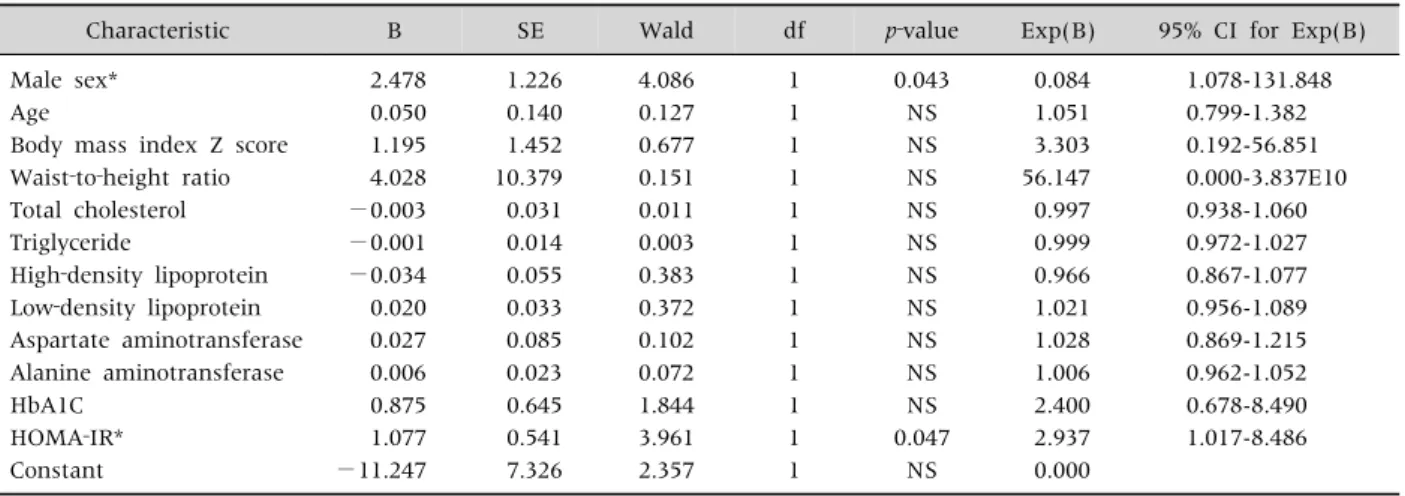
Table 4. The Interaction between Acanthosis Nigricans and Variables Using the Binary Logistic Regression Model
Characteristic B SE Wald df p‐value Exp(B) 95% CI for Exp(B)
Male sex* 2.478 1.226 4.086 1 0.043 0.084 1.078-131.848
Age 0.050 0.140 0.127 1 NS 1.051 0.799-1.382
Body mass index Z score 1.195 1.452 0.677 1 NS 3.303 0.192-56.851
Waist‐to‐height ratio 4.028 10.379 0.151 1 NS 56.147 0.000-3.837E10
Total cholesterol −0.003 0.031 0.011 1 NS 0.997 0.938-1.060
Triglyceride −0.001 0.014 0.003 1 NS 0.999 0.972-1.027
High‐density lipoprotein −0.034 0.055 0.383 1 NS 0.966 0.867-1.077
Low‐density lipoprotein 0.020 0.033 0.372 1 NS 1.021 0.956-1.089
Aspartate aminotransferase 0.027 0.085 0.102 1 NS 1.028 0.869-1.215
Alanine aminotransferase 0.006 0.023 0.072 1 NS 1.006 0.962-1.052
HbA1C 0.875 0.645 1.844 1 NS 2.400 0.678-8.490
HOMA‐IR* 1.077 0.541 3.961 1 0.047 2.937 1.017-8.486
Constant −11.247 7.326 2.357 1 NS 0.000
B: values for the logistic regression equation, SE: standard error, df: degrees of freedom, EXP(B): exponentiation of the B coefficient, CI: confidence interval, HbA1C: glycated hemoglobin, HOMA‐IR: homeostatic model assessment insulin resistance, NS: not significant.
*p<0.05.
(22.2±20.1 U/L vs. 45.6±40.3 U/L, p=0.005), and C-peptide (2.1±0.6 ng/mL vs. 3.8±2.1 ng/mL, p<0.001) levels were higher in the With AN group than the Without AN group. HOMA-IR was sig- nificantly higher in the With AN group (2.14±0.86 vs. 5.74±4.71, p<0.001), and QUICKI was sig- nificantly lower (0.346±0.025 vs. 0.310±0.028, p<0.001) in this group. In addition, the With AN group had a significantly higher rate of insulin resist- ance (p<0.001), hypertension (p=0.043), and meta- bolic syndrome (p=0.036; Table 3).
Factors related to the development of acanthosis nigricans
In order to evaluate risk factors for AN in obese pa- tients, a binary logistic regression analysis was per- formed on gender, age, BMI Z-score, WtHR, total cholesterol, triglyceride, HDL, LDL, AST, ALT, HbA1C, and HOMA-IR. The results showed that the male gender and a higher HOMA-IR score increased the risk of developing AN (Table 4).
Acanthosis nigricans severity in the prediction of insulin resistance: cut-off value and comparison with other indices
ROC curves were used to compare the predictive
power of AN severity, BMI Z-score, obesity index, and WtHR in the diagnosis of insulin resistance (HOMA-IR >3.16). Of these variables, only AN se- verity (p<0.001) and obesity index (p=0.041) showed a predictive power of at least 0.5 within the 95% confidence interval (CI). The area under the curve (AUC) for AN was 0.765, which was higher than the AUC of 0.645 for obesity index (Fig. 1). The ROC curve was used to find a cut-off value of AN se- verity that was suitably sensitive and specific. At an AN severity of 3 or greater, the sensitivity for predict- ing insulin resistance was 56.8%, and the specificity was 83.9%, providing relatively high specificity.
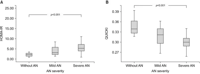
The relationship between insulin resistance and insulin sensitivity according to acanthosis nigricans severity
Subjects were divided into three groups: Without AN, Mild AN (AN severity score of 1-2), and Severe AN (AN severity score of 3-4). HOMA-IR and QUICKI were examined in these groups. HOMA-IR, which represents insulin resistance, increased with increasing AN severity (Without AN group: mean, 2.15; 95% CI, 1.72-2.57; Mild AN group: mean, 4.15;
95% CI, 3.04-5.25; Severe AN group: mean, 7.22; 95%
CI, 5.08-9.35; p<0.001) (Fig. 2A). QUICKI, which
Fig. 1. Receiver operating characteristic curves for prediction of insulin resistance. ROC: receiver operating characteristic, AUC:
area under curve, CI: confidence interval, AN: acanthosis nig- ricans, BMI: body mass index, WHtR: waist-to-height ratio.
*p<0.05.
Fig. 2. Comparison of acanthosis nigricans (AN) severity in terms of homeostatic model assessment insulin resistance (HOMA-IR) and quantitative insulin sensitivity check index (QUICKI). The p-value from (A) one-way ANOVA and (B) Kruskal-Wallis test is given. AN: acanthosis nigricans.
drepresents insulin sensitivity, decreased with in- creasing AN severity (Without AN group: mean, 0.346; 95% CI, 0.334-0.359; Mild AN group: mean 0.319; 95% CI, 0.310-0.329; Severe AN group: mean
0.300; 95% CI, 0.289-0.311; p<0.001) (Fig. 2B).
DISCUSSION
AN is a dermatological disorder characterized by symmetrical plaques with hyperpigmentation and hyperkeratosis. It is commonly seen in obese patients.
It is usually observed on the posterior neck, axilla, and groin, but may also be observed on the elbows, knuckles, and knees [14]. AN is known to occur when the concentration of insulin-like growth factor receptors in the skin is too low relative to the amount of insulin present, causing accumulation of insulin in the skin, proliferation of epidermal cells, and thickening of keratocytes [15]. AN is associated with obesity and endocrine diseases caused by severe in- sulin resistance, and may rarely be seen in patients with genetic diseases or malignant tumors [16,17].
The development of insulin resistance in obese pa- tients can cause several metabolic abnormalities to occur simultaneously. This phenomenon was first named “syndrome X” by Reaven [18]. Since then, various studies have demonstrated that insulin re- sistance can cause dyslipidemia, fatty liver disease, hypertension, and type 2 diabetes, and this phenom- enon is now more commonly referred to as “meta- bolic syndrome” [3]. Insulin sensitivity typically de-
clines during puberty, which results in physiological insulin resistance [19]. However, when obesity caus- es insulin resistance to develop before obesity, this pathological condition can become further ag- gravated in adulthood. Therefore, when insulin re- sistance is detected in obese children, it is important that appropriate treatment should be commence dearly.
Kahn et al. [5] first suggested an association be- tween AN and insulin resistance in obese children in 1976. Since then, many studies have been conducted with the aim of utilizing AN as an indicator for in- sulin resistance [4,11,20]. However, due to the lack of unified guidelines for classifying AN severity, these attempts have met with some difficulty. Stuart et al. [21] attempted to develop a quantitative scale for AN by observing its presentation in the neck, axil- la, elbow, and groin, and then assigning a score ac- cording to the number of body parts affected.
However, since they did not suggest criteria for measuring differences in the extent of AN in any giv- en area, the accuracy of any assessment was limited.
Following this, Burke et al. [11] conducted a study in adult Mexican American patients, in which they evaluated AN in 5 areas: the neck, axilla, knuckles, elbow, and knee. Of these, neck severity showed a relatively small inter-examiner error and good reproducibility. In addition, increasing neck severity of AN was found to be associated with high fasting insulin levels and BMI. Several studies have used this scale to compare AN severity quantitatively quantitatively [22,23]; however, there remains a dearth of such research in pediatric patients.
Using the scale proposed by Burke et al. [11], this study investigated the utility of AN severity in the neck as a predictor of insulin resistance in obese Korean children. We verified that increased AN se- verity was associated with an increase in insulin re- sistance and a decrease in insulin sensitivity. In addi- tion, we found that BMI did not have a separate ef- fect on AN, and that insulin resistance independently increased the risk of AN. Many studies have already demonstrated the key role that insulin resistance plays in metabolic syndrome, and these studies rec-
ommended the use of AN as a tool for the early de- tection of insulin resistance and metabolic syndrome [24,25]. We have shown that patients with severe AN, with a neck severity score of 3 or higher, have an increased risk of insulin resistance, and as such, re- quire thorough examination and treatment.
This study was conducted on obese patients with an obesity index of 20% or higher who visited Chosun University Hospital. The prevalence of AN was higher in male patients than in female patients.
This may reflect selection bias, in which only more severe obese male patients were likely to be included in the subject group. Other limitations include a rela- tively small sample size (74 subjects); use of sub- jective criteria for evaluating AN severity; and the lack of follow-up examinations, which made it im- possible to observe the rate of subsequent compli- cations. Therefore, there is a need for randomized controlled trials to investigate the utility of AN as an index for the early detection of metabolic syndrome risk. In addition, the index will need to be made more objective, by perhaps using an official index and comparing the assessments of 2 or more exa- miners. Follow-up examinations of pediatric pa- tients with AN will also be required to investigate the risk of developing metabolic syndrome.
ACKNOWLEDGEMENTS
This study was supported by research funds from Chosun University, 2013.
REFERENCES
1. Seo JW. Obesity in children and adolescents. Korean J Pediatr 2009;52:1311-20.
2. Weiss R, Kaufman FR. Metabolic complications of childhood obesity: identifying and mitigating the risk.
Diabetes Care 2008;31 Suppl 2:S310-6.
3. Hong YM. Metabolic syndrome in children and adolescents. Korean J Pediatr 2009;52:737-44.
4. Chueh HW, Cho GR, Yoo JH. Clinical significance of acanthosis nigricans in children and adolescents with obesity induced metabolic complications. Korean J Pediatr 2007;50:987-94.
5. Kahn CR, Flier JS, Bar RS, Archer JA, Gorden P, Martin MM, et al. The syndromes of insulin resistance and acanthosis nigricans. Insulin-receptor disorders in man. N Engl J Med 1976;294:739-45.
6. Dietz WH. Overweight in childhood and adolescence. N Engl J Med 2004;350:855-7.
7. Slyper AH, Kashmer L, Huang WM, Re'em Y.
Acanthosis nigricans, vitamin D, and insulin resistance in obese children and adolescents. J Pediatr Endocrinol Metab 2014;27:1107-11.
8. Patidar PP, Ramachandra P, Philip R, Saran S, Agarwal P, Gutch M, et al. Correlation of acanthosis nigricans with insulin resistance, anthropometric, and other metabolic parameters in diabetic Indians. Indian J Endocrinol Metab 2012;16:S436-7.
9. Centers for Disease Control and Prevention. 2007 Korean National Growth Charts [Internet]. Osong:
2008 [cited 2008 Aug 21]. Available from: http://cdc.go.kr/
CDC/notice/CdcKrInfo0201.jsp?menuIds=HOME001- MNU1154-MNU0005-MNU1889&cid=1235.
10. Keskin M, Kurtoglu S, Kendirci M, Atabek ME, Yazici C. Homeostasis model assessment is more reliable than the fasting glucose/insulin ratio and quantitative in- sulin sensitivity check index for assessing insulin re- sistance among obese children and adolescents. Pediat- rics 2005;115:e500-3.
11. Burke JP, Hale DE, Hazuda HP, Stern MP. A quantita- tive scale of acanthosis nigricans. Diabetes Care 1999;
22:1655-9.
12. American Diabetes Association. Diagnosis and classi- fication of diabetes mellitus. Diabetes Care 2014;37 Suppl 1:S81-90.
13. National Cholesterol Education Program (NCEP) Expert Panel on Detection, Evaluation, and Treatment of High Blood Cholesterol in Adults (Adult Treatment Panel III). Third report of the National Cholesterol Education Program (NCEP) expert panel on detection, evaluation, and treatment of high blood cholesterol in adults (Adult Treatment Panel III) final report. Circu- lation 2002;106:3143-421.
14. Yosipovitch G, DeVore A, Dawn A. Obesity and the skin:
skin physiology and skin manifestations of obesity. J
Am Acad Dermatol 2007;56:901-16.
15. Hermanns-Lê T, Scheen A, Piérard GE. Acanthosis nig- ricans associated with insulin resistance: pathophysi- ology and management. Am J Clin Dermatol 2004;5:
199-203.
16. Dunaif A, Green G, Phelps RG, Lebwohl M, Futterweit W, Lewy L. Acanthosis nigricans, insulin action, and hyperandrogenism: clinical, histological, and bio- chemical findings. J Clin Endocrinol Metab 1991;73:
590-5.
17. Schwartz RA. Acanthosis nigricans. J Am Acad Dermatol 1994;31:1-19.
18. Reaven GM. Role of insulin resistance in human disease (syndrome X): an expanded definition. Annu Rev Med 1993;44:121-31.
19. Caprio S, Plewe G, Diamond MP, Simonson DC, Boulware SD, Sherwin RS, et al. Increased insulin se- cretion in puberty: a compensatory response to reduc- tions in insulin sensitivity. J Pediatr 1989;114:963-7.
20. Stuart CA, Smith MM, Gilkison CR, Shaheb S, Stahn RM. Acanthosis nigricans among native Americans: an indicator of high diabetes risk. Am J Public Health 1994;84:1839-42.
21. Stuart CA, Peters EJ, Prince MJ, Richards G, Cavallo A, Meyer WJ 3rd. Insulin resistance with acanthosis nigricans: the roles of obesity and androgen excess.
Metabolism 1986;35:197-205.
22. Venkatswami S, Anandam S. Acanthosis nigricans: a flag for insulin resistance. J Endocrinol Metab Diabetes South Afr 2014;19:68-74.
23. Noviarti D, Rini EA, Oenzil F. Association of resistin level with acanthosis nigricans in obese adolescents.
Paediatr Indones 2016;56:32-6.
24. Kong AS, Williams RL, Rhyne R, Urias-Sandoval V, Cardinali G, Weller NF, et al. Acanthosis nigricans:
high prevalence and association with diabetes in a prac- tice-based research network consortium--a PRImary care Multi-Ethnic network (PRIME Net) study. J Am Board Fam Med 2010;23:476-85.
25. Yamazaki H, Ito S, Yoshida H. Acanthosis nigricans is a reliable cutaneous marker of insulin resistance in obese Japanese children. Pediatr Int 2003;45:701-5.