Paresthesia diagnosed using cone-beam computed tomography: a case report
Umesh Kumar
1, Charan Kamal Kaur
2, Ruchi Vashisht
1, Vidya Rattan
31Unit of Conservative Dentistry & Endodontics, Oral Health Sciences Centre, Post Graduate Institute of Medical Education & Research (PGIMER), Chandigarh, India
2Medical Officer (Dental), Government Multispecialty Hospital, Chandigarh, India
3Unit of Oral and Maxillofacial Surgery, Oral Health Sciences Centre, Post Graduate Institute of Medical Education & Research (PGIMER), Chandigarh, India
Various dental procedures, such as injection administration, surgical treatment, and endodontic treatment, can cause injury to the nerves. The most commonly injured nerves are the inferior alveolar and lingual nerves.
This can manifest as altered sensation to the area of innervation of the injured nerve, such as the lower lip, chin, teeth, tongue, and mucosa. Altered sensations or loss of sensation are relatively infrequent complications in daily dental practice. Here, we report an uncommon case of altered sensation in the midfacial region caused by an endodontic procedure and discuss the need to consider local dental causes in the differential diagnosis of numbness in the facial region.
Keywords: Altered Sensation; Cone-Beam Computed Tomography; Delayed Paresthesia.
This is an Open Access article distributed under the terms of the Creative Commons Attribution Non-Commercial License (http://creativecommons.org/licenses/by-nc/4.0/) which permits unrestricted non-commercial use, distribution, and reproduction in any medium, provided the original work is properly cited.
Received: February 4, 2019•Revised: March 13, 2020•Accepted: March 29, 2020
Corresponding Author:Umesh Kumar, Unit of Conservative Dentistry & Endodontics, Oral Health Sciences Centre, Post Graduate Institute of Medical Education
& Research (PGIMER) Chandigarh - 160012, India
Tel: +919862969792,+917087003365 Fax: +01722756899 E-mail: [email protected] Copyrightⓒ 2020 Journal of Dental Anesthesia and Pain Medicine
INTRODUCTION
Paresthesia is defined as a temporary or permanent unpleasant alteration in sensations or loss of sensation caused by nerve lesions [1]. The etiology is known in 83% of all cases of facial paresthesia, and dental procedures account for 48% of the cases [2]. The inferior alveolar and lingual nerves are the most commonly involved nerves in paresthesia resulting from dental procedure. Various dental procedures, such as injection administration, surgical treatment, and endodontic treat- ment, can injure the inferior alveolar nerve and manifest as altered sensation in the area of innervation, such as the lower lip, chin, teeth, tongue, and mucosa [1,3]. The
patient usually notes altered sensation or paresthesia on the day of the dental procedure after recovery from local anesthesia. However, rarely, delayed paresthesia can occur a few days to months after the procedure [4]. In a retrospective study of 1377 third molar surgeries by Kipp DP et al., only 5% of the 60 cases of delayed paresthesia were reported [5]. Doh et al. reported a case of delayed paresthesia induced by inflammatory edema after third molar surgery involving the inferior alveolar nerve [6]. Delayed paresthesia occurs a few days to months later than classic paresthesia, which occurs immediately after the procedure [3]. Considering the limitations (superimposition, distortion, etc.) of two- dimensional radiography, the introduction of cone-beam computed tomography (CBCT) devices improved the
Fig. 1. An extraoral preoperative photograph shows the area with altered sensations.
Fig. 2. An intraoral preoperative photograph shows gingival recession on the palatal side with sloughing in relation to 13.
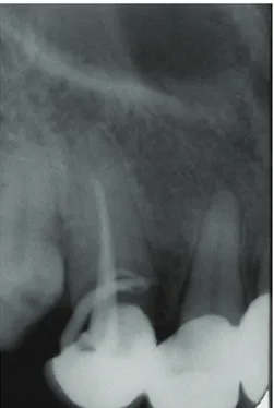
Fig. 4. Sagittal cone-beam computed tomography shows inadequate root canal filling and a large periapical radiolucency in relation to 13.
Fig. 3. An intraoral preoperative radiograph shows inadequate root canal filling with a large periapical radiolucency in relation to 13.
endodontic evaluation of the morphology of teeth, including the location and number of canals, pulp chamber size and degree of calcification, root structure, direction and curvature, fractures, iatrogenic defects, and the extent of dental caries. CBCT, which provides a small field of view, has become the standard of care for endodontic diagnosis, treatment planning, and follow-up [7].
Here, we report an uncommon case of altered sensation in the midfacial region caused by an endodontic procedure diagnosed using CBCT and discuss the need to consider local dental causes in the differential diagnosis of numbness in the facial region.
CASE REPORT
A 53-year-old woman was referred to the Unit of Endodontics at the Oral Health Science Centre, PGIMER, Chandigarh, India, for the management of numbness of
the right side of the face. She had undergone emergency root canal treatment for toothache in the upper right canine and had subsequently developed numbness on the right side of the face from the upper lip to the infraorbital
Fig. 5. Cross-sectional cone-beam computed tomography shows inadequate root canal filling in the periodontal ligament space and a large periapical radiolucency in relation to 13.
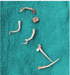
Fig. 6. A photograph shows gutta percha retrieved from the periodontal ligament space in relation to 13.
area. She had no relevant medical or family history.
Physical extraoral and intraoral examinations were unremarkable, except for paresthesia. Extraoral exami- nation of the affected area showed altered sensation in a 15–20-mm region from the upper lip to the infraorbital area (Fig. 1). Intraoral examination revealed a dressing through a porcelain-fused-to-metal (PFM) crown on the maxillary right canine. On the palatal side, gingival recession along with sloughing was noted through which gutta percha was seen (Fig. 2). The patient reported of tingling sensation of the gums in the first quadrant, particularly in the canine and premolar regions. The tooth was slightly tender to percussion. No swelling could be seen at the buccal sulcus. Radiographic examination revealed inadequate root canal filling and a large periapical radiolucency at the maxillary right canine with loss of the periodontal ligament (PDL) space and a wide lamina dura (Fig. 3). However, an intraoral periapical radiograph could neither reveal whether the material was inside or outside the canal nor precisely confirm which tooth was responsible for the symptoms and signs. CBCT confirmed the presence of radiopaque filling materials adjacent to the root canal in the PDL space and revealed empty root canals (Figs. 4 and 5). No extruded filling material was found beyond the canine apex. The patient was diagnosed with classic paresthesia from symptomatic apical periodontitis due to mechanical pressure and ischemia related to inflammatory processes (edema) or due to toxic metabolic or inflammatory products of bacteria. Retreatment was planned, and informed consent was obtained from the patient. When the dressing was removed, gutta percha was found outside of the canal
(Fig. 6). Irrigation was performed thoroughly with normal saline and povidone-iodine solutions. At the 1-week follow-up, the patient reported of rapid improvement of numbness. The root canal treatment was completed by her dentist a few weeks later, and the lost sensation recovered.
DISCUSSION
The basic principle of endodontic treatment is elimi- nation of the diseased pulp and dentin with adequate shaping and cleaning and three-dimensional sealing of the root canal space using biocompatible filling materials [1,8]. The filling materials should be limited within the root canal space without overextending to the periapical
tissue or neighboring structures; otherwise, serious neuro- toxic complications, such as anesthesia, hypoesthesia, paresthesia, and dysesthesia, can occur [9,10,11]. Neura- praxia is the mildest form of nerve injury, as classified by Seddon. It results from temporary loss of sensation due to blunt injuries or pressure from a local infection [12]. Altered sensations or loss of sensation are relatively infrequent complications in daily dental practice that are caused by improper treatment, particularly endodontic treatment. Instrumentation and overfilling beyond the root canal or in the PDL space while performing an endo- dontic procedure with hand or rotary files can produce serious clinical manifestations, such as pain, swelling, and, occasionally, altered sensations [3,8].
In our patient, although the root did not seem overfilled, the master cone along with other accessory gutta percha was introduced into the PDL space from access preparation through the PFM crown. Based on the rate of recovery of sensory sensations, the most probable cause was pressure from an acute infection of the periradicular tissue because chemical effects from filling materials on the nerve usually take longer to recover. The typical feature of such types of nerve injury is the fast recovery of sensory function after elimination of pressure from the local infection. In the present case, after removal of obturated materials from the canal prepared in the periradicular region, followed by irrigation with normal saline and povidone-iodine solutions, immediately relieved the pressure, leading to recovery within a short period of 2–3 weeks. Although the area of altered sensation in this case was supplied by the anterior and middle superior alveolar nerves along with the infraorbital nerve, branches of the maxillary nerve could have sustained trauma during instrumentation of the PDL space, or local infection could have blocked the accessory nerve branches or alveolar nerve plexus [11,13].
This case highlights the need to exercise great care during non-surgical endodontic procedures, particularly when performing through a crown. The requisites of endodontic procedures are straight-line access pre- paration, instrumentation within the canal, passive
irrigation, and proper sealing. Positioning the file inside the root canal space during instrumentation should be ensured with an electronic apex locator and radiograph;
otherwise, over instrumentation or instrumentation within the PDL space may cause damage to the nerves, leading to paresthesia.
AUTHOR ORCIDs
Umesh Kumar: https://orcid.org/0000-0001-7267-6446 Charan Kamal Kaur: https://orcid.org/0000-0001-6484-7620 Ruchi Vashisht: https://orcid.org/0000-0001-9299-9330 Vidya Rattan: https://orcid.org/0000-0002-6313-5098
AUTHOR CONTRIBUTIONS
Umesh Kumar: Investigation, Methodology, Writing – original draft Charan Kamal Kaur: Writing – review & editing.
Ruchi Vashisht: Writing – original draft, Writing – review & editing Vidya Rattan: Supervision, Writing – review & editing
CONFLICT OF INTEREST: All authors declare that they have no competing interests.
REFERENCES
1. Ahmad M. The anatomical nature of dental paresthesia:
a quick review. Open Dent J 2018; 12: 155-9.
2. Shadmehr E, Shekarchizade N. Endodontic periapical lesion-induced mental nerve paresthesia. Dent Res J 2015;
12: 192-6.
3. Smith MH, Lung KE. Nerve injuries after dental injection:
a review of the literature. J Can Dent Assoc 2006; 72:
559-64.
4. Devine M, Hirani M, Durham J, Nixdorf DR, Renton T. Identifying criteria for diagnosis of post-traumatic pain and altered sensation of the maxillary and mandibular branches of the trigeminal nerve: a systematic review. Oral Surg Oral Med Oral Pathol Oral Radiol 2018; 125: 526-40.
5. Kipp DP, Goldstein BH, Weiss WW Jr. Dysesthesia after mandibular third molar surgery: a retrospective study and
analysis of 1,377 surgical procedures. J Am Dent Assoc 1980; 100: 185-92.
6. Di Lenarda R, Cadenaro M, Stacchi C. Paresthesia of the mental nerve induced by periapical infection: a case report.
Oral Surg Oral Med Oral Pathol Oral Radiol Endod 2000;
90: 746-9.
7. Borgonovo A, Bianchi A, Marchetti A, Censi R, Maiorana C. An uncommon clinical feature of IAN injury after third molar removal: a delayed paresthesia case series and literature review. Quintessence Int 2012; 43: 353-9.
8. Morse DR. Endodontic-related inferior alveolar nerve and mental foramen paresthesia. Compend Contin Educ Dent 1997; 18: 963-8, 970-3, 976-8.
9. Ozkan BT, Celik S, Durmus E. Paresthesia of the mental nerve stem from periapical infection of mandibular canine
tooth: a case report. Oral Surg Oral Med Oral Pathol Oral Radiol Endod 2008; 105: e28-31.
10. Di Lenarda R, Cadenaro M, Stacchi C. Paresthesia of the mental nerve induced by periapical infection: a case report.
Oral Surg Oral Med Oral Pathol Oral Radiol Endod 2000;
90: 746-9.
11. Donoff RB. Nerve regeneration: basic and applied aspects.
Crit Rev Oral Biol Med 1995; 6: 18-24.
12. Seddon HJ. Three types of nerve injury. Brain 1943; 66:
237-88.
13. Campbell WW. Evaluation and management of peripheral nerve injury. Clin Neurophysiol 2008; 119: 1951-65.
14. Dahlin LB, Danielsen N, Ehira T, Lundborg G, Rydevik B. Mechanical effects of compression of peripheral nerves.
J Biomech Eng 1986; 108: 120-2.