ABSTRACT
C2–3 disc herniation is rare and a definitive treatment of choice has not been established.
The purpose of this case report is to suggest posterior approach as one of the best options.
A 49-year-old man visited our clinic with a 7-year history of neck pain and occipital headache and a 2-month history of right arm pain. C2–3 intervertebral disc herniation of the central type was diagnosed on magnetic resonance imaging (MRI), and surgery was performed, including C1 laminectomy, C2–3 laminoplasty, and C2–3 posterior fixation. The posterior approach was used because the patient's neck was difficult to operate anteriorly. After 3 months postoperatively, MRI showed widened cerebrospinal fluid space at the C2–3 level.
The visual analogue scale score for pain improved in the occipital area and right arm.
However, the untouched protruded central disc, subjective weakness in right hand grasping, and numbness persisted. In conclusion, this case highlights posterior decompression and fixation as a good treatment of choice for decompression at the C2–3 level disc herniation, from where it is difficult to remove compressive lesions directly via the anterior corridor.
Keywords: Herniated disc; Laminectomy; Laminoplasty; Cervical spine
INTRODUCTION
High cervical degenerative disc disease is rare, and the overall incidence of disc herniation between C2 and C3 is less than 1%.1) Upper cervical disc herniations are usually seen in elderly patients, and their mechanisms are related to spondylotic changes. Degenerative changes and loss of soft tissues, such as the intervertebral discs, in the middle or lower cervical regions lead to excessive movement of the upper cervical region and excessive axial load.6,13) It can make the high cervical disc more degenerative and protruded. Although there are several approaches to resolve spinal cord compression due to C2–3 disc herniation, a definitive treatment of choice has not been established. In addition, reports on the treatment are limited.16) Therefore, we herein report a case of C2–3 intervertebral disc herniation treated with the posterior approach and share the risks and benefits of this approach.
Case Report
Received: Jul 5, 2020 Revised: Sep 23, 2020 Accepted: Oct 7, 2020 Address for correspondence:
Sung Hwan Hwang
Department of Neurosurgery, The Armed Forces Capital Hospital, PO Box #99, 81 Saemaeul-ro 177beon-gil, Bundang-gu, Seongnam 13574, Korea.
E-mail: [email protected]
Copyright © 2021 Korean Neurotraumatology Society
This is an Open Access article distributed under the terms of the Creative Commons Attribution Non-Commercial License (https://
creativecommons.org/licenses/by-nc/4.0/) which permits unrestricted non-commercial use, distribution, and reproduction in any medium, provided the original work is properly cited.
ORCID iDs Sunho Kim
https://orcid.org/0000-0002-5597-7404 Sung Hwan Hwang
https://orcid.org/0000-0003-4232-4719 Byung-Kyu Cho
https://orcid.org/0000-0002-0578-6597 Sang Hoon Yoon
https://orcid.org/0000-0003-0212-4819 Joonho Yoon
https://orcid.org/0000-0001-7406-3459 Conflict of Interest
The authors have no financial conflicts of interest.
Sunho Kim , Sung Hwan Hwang , Byung-Kyu Cho , Sang Hoon Yoon , and Joonho Yoon
Department of Neurosurgery, The Armed Forces Capital Hospital, Seongnam, Korea
Posterior Approach in C2–3 Disc Herniation: C1 Laminectomy, C2–3
Laminoplasty and Posterior Fixation in
C2–3 Disc Herniation
CASE REPORT
After approval from the Institutional Review Board (No. AFCH-20-IRB-009), we retrospectively reviewed medical records of patient who underwent surgery for C2–3 intervertebral disc herniation.
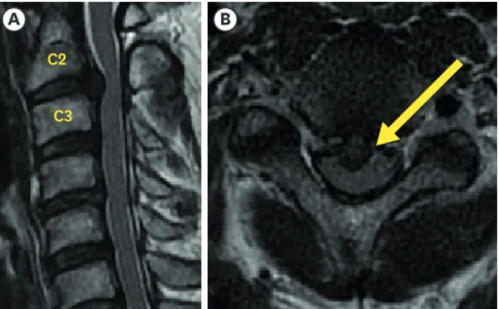
A 49-year-old man presented with neck pain, occipital headache, and right arm pain. Neck pain and occipital headache had persisted for 7 years despite conservative treatment. Two months before, he had developed right arm pain and numbness on the lateral side. In addition, he experienced right arm weakness and motor grade IV. To obtain the diagnosis, we performed three-dimensional cervical spine computed tomography (CT) and magnetic resonance imaging (MRI), which revealed C2–3 disc herniation of the central type
compressing the spinal cord (FIGURE 1). The patient was obese and had a short neck, so an anterior approach was difficult.9) And according to other research, anterior approach can cause some complications such as dysphonia, dysphagia, carotid vessel injury, esophageal and tracheal injury.7,9,11,15) In the published papers, there were surgical methods showing successful results for the anterior approach, but these were also mass invasive surgical methods (TABLE 1).16) This cannot be denied that the stress for postoperative complications and the risk during surgery are clearly high even for experienced surgeons. Therefore, considering the C2–3 posterior approach, we performed vertebral artery CT angiography. It showed left vertebral artery dominant feature and high-riding vertebral artery on the left side and a narrow pedicle (FIGURE 2).
There are various treatment approaches for C2–3 disc herniation. However, there is no definite evidence of what approach is more beneficial.16) In our case, we chose the posterior midline approach to avoid injury of the anterior cervical structures because of the short neck.
In the prone position, surgery was performed under general anesthesia, and we used the Mayfield fixator to obtain the head-fixed state and slightly flexed neck position for the patient. In addition, intraoperative neurophysiological monitoring was performed to avoid spinal cord and nerve root injuries. A midline incision was made at the C1–3 level posterior neck, and we performed blunt dissection until we reached the posterior tubercle and arch of C1 and the spinous process and lamina of C2–3. After checking the cervical spine level, C1 laminectomy and C2 and C3 laminoplasty were performed for decompression. The reason for
A B
C2 C3
FIGURE 1. Sagittal (A) and axial (B) magnetic resonance images acquired before operation show central disc protrusion and spinal cord compression.
C1 laminectomy was spinal cord kinking at the C1 posterior arch when the spinal cord shifted to the posterior side after C2–3 decompression. In addition, we performed C2–3 laminoplasty instead of C2–3 laminectomy because recent studies have reported various disadvantages of laminectomy, such as instability of the vertebra, acceleration of deformity, epidural soft- tissue scar change and adhesion with the dura mater, and insufficient protection of the spinal cord due to the absence of posterior elements, such as the lamina.12,18) Therefore, we performed laminoplasty and added posterior instrumented fixation to stabilize the vertebra. In case of laminoplasty alone, if motion remains, the possibility of disc protrusion progression remains and postoperative cervical kyphosis was reported up to 10.8% in retrospective study.3) Because of that reason, we thought posterior instrumented fixation can be benefit to stabilize disc progression and prevent cervical kyphosis. In this step, we performed posterior screw fixation at the C2–3 level, in which the pars screw was used on the left side of C2 because of the high-riding vertebral artery, and pedicle screw was used for the right side C2 and lateral mass screws were used for both sides of the C3.
TABLE 1. Review of cases published in literature
Study Year Number of
cases Presentation Location of the disc
material Surgical technique Outcome
Espersen et al. 1984 1 C2–3 level Cloward's technique
Jomin et al. 1986 2 C2–3 level ACDF
Rosemberg et al. 1991 2 Myelopathy Retro-odontoïd Posterior transdural 1 improved, 1 stable
Nishizawa et al. 1996 1 Myelopathy Retro-odontoïd Posterior transdural
Chen and Luis 1997 1 Myelopathy Retro-odontoïd Transoral odontoidectomy+C1–2 fusion Improved
Nishizawa et al. 1999 3 Myelopathy 2 Retro-odontoïd Posterior transdural All improved
1 C2–3 level
Antich et al. 1999 1 Myelopathy C2–3 level ACDF Improved
Campbell 2000 1 Myelopathy Retro-odontoïd Transoral odontoidectomy
Chen 2000 8 Myelopathy C2–3 level ACDF 6 improved, 2 stable
Matsutano et al. 2004 1 Myelopathy Retro-odontoïd Far lateral
Deshmukh et al. 2004 1 C2 Radiculopathy Retro-odontoïd Posterior extradural Improved
Türe et al. 2007 1 C3 radiculopathy C2–3 level Anterolateral extradural approach Improved
Chan et al. 2009 1 Myelopathy C2–3 level C3 transcorporeal
Kotil and Sengoz 2011 5 Myelo-radiculopathy C2–3 C2–3 level Discectomie antérieure+greffe 3 improved, 2 stable
N'Dri et al. 2014 1 Myelo-radiculopathy C2–3 level Anterolateral extradural approach Improved
Current case 2020 1 Myelopathy C2–3 level Posterior approach (decompression & fixation) Improved This table is sourced from Oka et al.16)
ACDF: anterior cervical discectomy and fusion.
A B C
FIGURE 2. Vertebral artery CT angiography shows left high-riding vertebral artery (B) compared to the right vertebral artery (A). The axial image (C) shows a narrow pedicle on the left side (red arrow). A narrow pedicle is defined by a pedicle width of 4 mm or less, which is measured in axial CT scans.
CT: computed tomography.
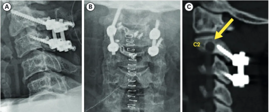
At the end of the procedure, a C-arm radiography image was acquired to confirm the accuracy of the C2–3 level screw fixation state.
Postoperative radiography and vertebral artery CT angiography showed no injury of the left high- riding vertebral artery at the C2 level and safe insertion of the pars and pedicle screws (FIGURE 3).
In addition, postoperative radiography showed some cervical parameters4,5,20) such as cervical lordosis (CL: Cobb angle between the lower endplates of C2 and C7) of 6.4°, a C2–3 angle (angle between the lower endplates of C2 and C3) of 3.5°, and a C2–7 sagittal vertical axis (SVA: distance from the posterosuperior corner of C7 to a vertical line from the center of the C2 vertebra) of 30.1 mm compared to the pre-operative measured values of 7.7°, 1.5°, and 51.63 mm, respectively (FIGURE 4). Normal ranges of CL and C2–7 SVA in adults are 19.2±11.9° and 12.0±9.6 mm, respectively.20) Initially, our patient's cervical parameter values
A B C
C2
FIGURE 3. Radiography shows safe insertion of the C2–3 pars and pedicle screw (A, B), and postoperative computed tomography angiography shows no injury of the left vertebral artery (arrow, C).
A B
3.5° 1.5°
6.4° 7.7°
51.63 mm 30.1 mm
FIGURE 4. Lateral preoperative cervical spine radiography (B) shows C2–7 CL (yellow) of 7.7°, a C2–3 angle (skyblue) of 1.5°, and a C2–7 SVA (red) of 51.63 mm. Postoperative radiography (A) shows CL of 6.4°, a C2–3 angle of 3.5°, and a C2–7 SVA of 30.1 mm. There is no significant change in CL or C2–3 angle, but C2–7 SVA has improved, indicating the improvement of sagittal imbalance.
CL: cervical lordosis, SVA: sagittal vertical axis.
showed decreased cervical lordosis and increased C2–7 SVA, indicating an increase in sagittal imbalance. Postoperatively, there were no significant changes in CL, and sagittal imbalance had improved. Moreover, the C2–3 angle showed no significant change from preoperative 1.5°
to postoperative 3.5°.
MRI showed posterior decompression at the C1–3 level; however, due to the epidural hematoma, the spinal cord was still mildly compressed (FIGURE 5A & B).
After 3 months postoperatively, the visual analogue scale score for pain improved from 7 to 2 points in the occipital area and from 7 to 3 points in the right arm. MRI showed that the posterior epidural collection had resolved, and posterior cerebrospinal fluid (CSF) space in the spinal cord had expanded compared to preoperative MRI (FIGURE 5C & D). However, the right hand grasping subjective weakness and numbness persisted.
DISCUSSION
C2–3 disc herniation is very rare, with an incidence rate of less than 1% of all cases of cervical disc herniation.1) In addition, there is no definite choice of treatment. Approximately, literature review shows this as the 31st case published in the literature on C2–3 disc herniation and 8th case of using the posterior approach (TABLE 1).16) However, all cases reported so far included surgical approaches for discectomy of the herniated discs. To the best of our knowledge, this is the first case without a direct protrusion disc removal.
Outcomes of previously reported cases were almost good and improved.16) However, there was no consensus on the most appropriate approach. In our case, the posterior approach was considered to avoid other risk factors. Anterior approaches are extremely difficult in patients with a short and thick neck, and there is a possibility of injury to the anterior
A
D B C
C2 C3
C2 C3
FIGURE 5. After 3 months, magnetic resonance imaging shows expansion of the posterior space (yellow arrow) of the C2–3 spinal cord (C, D) compared to the preoperative MRI (A, B).
MRI: magnetic resonance imaging.
cervical structures, such as carotid vessels and branches and cranial nerves.9) In a review of 85 patients who underwent this approach to the cervico-thoracic spine (C3–T1), 9 (11%) had postoperative voice changes while 3 (3.5%) had permanent vocal cord paralysis.7,11,15) The strict lateral approach is rarely used and is therefore limited to the lateral foraminal hernia.13) We decided to choose the posterior approach for the aforementioned reasons. When planning the posterior approach, the risk of vertebral artery injury should be considered in case of a high-riding vertebral artery.8,10,17,19) This can occur when posterior screw insertion occurs.
A high-riding vertebral artery was defined as an isthmus height of 5 mm or less and/or internal height of 2 mm or less on a sagittal image, which is 3 mm lateral to the cortical margin of the spinal canal wall at C2.2,14) Our case fulfilled the criteria for the diagnosis of the high-riding vertebral artery; therefore, we used a pars screw on the C2 left side to avoid vertebral artery injury.
For 12 months follow-up period, the patient's symptoms improved significantly, and MRI revealed that the subarachnoid space through which CSF flow was secured using sufficient decompression. In addition, there was no deformity of cervical spine so far, no injury of the high-riding vertebral artery after pars screw fixation.
Limitations of our case report are; 1) short follow up to evaluate the long-term outcome, 2) unexplainable symptoms and signs corresponding to the C2–3 spinal level lesion, 3) the uncertainty of C1 laminectomy in the delayed swan neck deformity, 4) persisting untouched protruded central disc's fate.
CONCLUSION
There are various surgical approach methods for C2–3 disc herniation, but we concluded that in case of a short neck, decompression and stabilization through the posterior approach can also be a good treatment option. In addition, if the diagnosis of a high-riding vertebral artery can be made using vertebral artery CT angiography, safe insertion of the posterior screw at the C2 level is possible using another screw insertion method, such as the pars screw insertion method.
REFERENCES
1. Antich PA, Sanjuan AC, Girvent FM, Simó JD. High cervical disc herniation and Brown-Sequard syndrome. A case report and review of the literature. J Bone Joint Surg Br 81:462-463, 1999 PUBMED | CROSSREF
2. Bloch O, Holly LT, Park J, Obasi C, Kim K, Johnson JP. Effect of frameless stereotaxy on the accuracy of C1–2 transarticular screw placement. J Neurosurg 95:74-79, 2001
PUBMED | CROSSREF
3. Cao J, Zhang J, Yang D, Yang L, Shen Y. Multivariate analysis of factors associated with kyphotic deformity after laminoplasty in cervical spondylotic myelopathy patients without preoperative kyphotic alignment.
Sci Rep 7:43443, 2017 PUBMED | CROSSREF
4. Cobb JR. Outline for the study of scoliosis. Instr Course Lect 5:261-275, 1948
5. Glassman SD, Bridwell K, Dimar JR, Horton W, Berven S, Schwab F. The impact of positive sagittal balance in adult spinal deformity. Spine 30:2024-2029, 2005
PUBMED | CROSSREF
6. Haid RW Jr. Natural history and management in Cooper P (ed): Degenerative Diseases of the Cervical Spine, Chicago, IL: AANS Publications, pp113-124, 1993
7. Heeneman H. Vocal cord paralysis following approaches to the anterior cervical spine. Laryngoscope 83:17-21, 1973
PUBMED | CROSSREF
8. Jung YG, Lee S, Jeong SK, Kim M, Park JH. Subaxial cervical pedicle screw in traumatic spinal surgery.
Korean J Neurotrauma 16:18-27, 2020 PUBMED | CROSSREF
9. Kotil K, Sengoz A. The management in the C2–C3 disc herniations: a clinical study. Turk Neurosurg 21:15-21, 2011
PUBMED
10. Madawi AA, Casey AT, Solanki GA, Tuite G, Veres R, Crockard HA. Radiological and anatomical evaluation of the atlantoaxial transarticular screw fixation technique. J Neurosurg 86:961-968, 1997 PUBMED | CROSSREF
11. McAfee PC, Bohlman HH, Riley LH Jr, Robinson RA, Southwick WO, Nachlas NE. The anterior retropharyngeal approach to the upper part of the cervical spine. J Bone Joint Surg Am 69:1371-1383, 1987 PUBMED | CROSSREF
12. Menku A, Koc RK, Oktem IS, Tucer B, Kurtsoy A. Laminoplasty with miniplates for posterior approach in thoracic and lumbar intraspinal surgery. Turk Neurosurg 20:27-32, 2010
PUBMED
13. Murphey F, Simmons JC, Brunson B. Surgical treatment of laterally ruptured cervical disc. Review of 648 cases, 1939 to 1972. J Neurosurg 38:679-683, 1973
PUBMED | CROSSREF
14. Neo M, Matsushita M, Iwashita Y, Yasuda T, Sakamoto T, Nakamura T. Atlantoaxial transarticular screw fixation for a high-riding vertebral artery. Spine 28:666-670, 2003
PUBMED | CROSSREF
15. Nishizawa S, Ryu H, Yokoyama T, Uemura K. Myelopathy caused by retro-odontoid disc hernia: case report. Neurosurgery 39:1256-1259, 1996
PUBMED | CROSSREF
16. Oka DN, Kouakou F, Haro Y, Sarki S. Cervical spine disc herniation at C2–C3 level: study of a clinical observation and literature review. Rom Neurosurg 29:459-464, 2015
CROSSREF
17. Peng CW, Chou BT, Bendo JA, Spivak JM. Vertebral artery injury in cervical spine surgery: anatomical considerations, management, and preventive measures. Spine J 9:70-76, 2009
PUBMED | CROSSREF
18. Sun S, Li Y, Wang X, Lu G, She L, Yan Z, et al. Safety and efficacy of laminoplasty versus laminectomy in the treatment of spinal cord tumors: a systematic review and meta-analysis. World Neurosurg 125:136- 145, 2019
PUBMED | CROSSREF
19. Wright NM, Lauryssen CAmerican Association of Neurological Surgeons/Congress of Neurological Surgeons. Vertebral artery injury in C1-2 transarticular screw fixation: results of a survey of the AANS/CNS section on disorders of the spine and peripheral nerves. J Neurosurg 88:634-640, 1998
PUBMED | CROSSREF
20. Xing R, Liu W, Li X, Jiang L, Yishakea M, Dong J. Characteristics of cervical sagittal parameters in healthy cervical spine adults and patients with cervical disc degeneration. BMC Musculoskelet Disord 19:37, 2018 PUBMED | CROSSREF