Lab Anim Res 2016: 32(4), 267-271 https://doi.org/10.5625/lar.2016.32.4.267
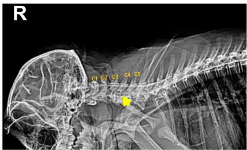
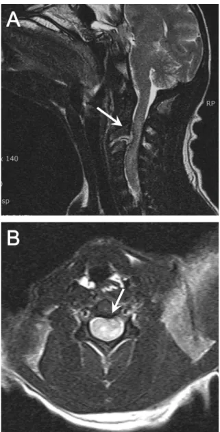
Clinical and magnetic resonance imaging features of compressive cervical myelopathy with traumatic intervertebral disc herniation in
cynomolgus macaque (Macaca fascicularis)
Yun-Jung Choi
1,2, Hye-Jin Park
1, Chul-Ho Sohn
4, Kyeong Cheon Jung
1,3, Seong Hoe Park
1,2, Jae-Il Lee
1,2,*
1
Transplantation Research Institute, Medical Research Center, Department of Medicine, Seoul National University College of Medicine, Seoul, Korea
2
Department of Medicine, Seoul National University College of Medicine, Seoul, Korea
3
Department of Pathology, Seoul National University College of Medicine, Seoul, Korea
4