Ⅰ. 서 론
치아 결손 후 기능적, 심미적 문제점을 해결하는 보철치료에서 임플랜트의 사용빈도가 증가되고 그 성공률도 높아지고 있다. 임플랜트 시술의 성공률은 적절한 임플랜트의 모양 및 재질, 수술기법, 환자 해 당부위 해부학적 구조의 이해, 시술상태 등이 결정 하는 것으로 알려져 있다.1-3)
Ulm 등4)은 치아가 결손된 상태가 지속되면 상악골 에서 골흡수가 증가하고 상악동이 팽창하여 치조돌 기의 폭과 높이가 감소하고, 이때 나타나는 해면골 의 감소는 치아가 결손된 기간과 골다공증이 영향을 미치지만 치조돌기의 형태와 골량은 직접적인 상관 관계가 없다고 하였다. Toffler5)와 Ulm 등6)은 상악 대 구치의 결손으로 골흡수가 증가되고, 치조돌기는 치 아가 주던 골자극이 감소하여 골위축이 일어나며, 이때 보철물을 사용하여도 치조돌기에 압력을 주지 못해 골흡수는 막지 못한다고 하였다. Kraut7), 그리 고 Chanavaz8)는 치아결손 후 골흡수와 함께 상악동 점막인 Schneiderian 막에서 파골능력(osteoclastic activity)이 증가하여 상악동 하연이 아래쪽으로 신장 되면서 치아와 상악동의 경계는 얇은 골판(osseous plate)으로 이루어진다고 하였다.
Razavi 등2)은 상악골 치조돌기에서 치밀골의 두께 가 4 mm 이상인 부위의 높이가 가장 낮은 곳은 상 악동저 최저점이고, 골질이 가장 나쁜 곳은 상악결 절 주변이라고 하였으며 Boss와 Taylor9)는 상악에서
발치 후 치조돌기가 흡수되고 상악동이 내려와 수직 골이 부족해지고 골질이 불량해져 임플랜트 지지 보 철물의 설계가 어렵고, 시술시 골유착이 지연된다고 하였다. 한편 Tidwell 등10)은 상악동저에 대한 해부학 구조의 폭넓은 이해가 상악동의 augmentation 수술 시 매우 중요하다고 하였고, Fredholm 등11)은 상악의 임플랜트 시술시 방사선 촬영을 통해 골높이, 골량, 치조융선의 기울기와 같은 주변 구조물에 대한 정보 를 많이 획득하는 것이 시술의 성공률을 높일 수 있 다고 하였으며, Kwak 등12)도 임플랜트 시술시 상악 동저를 구성하는 벽과 치근첨 사이 거리, 주변 치밀 골의 두께를 아는 것이 시술의 계획을 세우고 시술 할 때 매우 중요하다고 하였다.
이에 저자들은 상악에 임플랜트 식립을 위한 한국 인의 계측치를 제공하기 위하여 두개골 중 치조돌기 의 단면에서 치밀골의 두께, 상악동저와 치조능선사 이 거리, 치조돌기의 크기와 뼈두께 등을 계측하 였다.
Ⅱ. 연구 재료 및 방법 1. 대 상
이 연구는 한양대학교 의과대학 해부∙세포생물학 교실에 보관 중인 방부처리 시신 19구(남자 10구, 여 자 9구)의 두개골을 대상으로 하였으며, 손상되었거 나 치아의 위치가 불분명한 경우는 계측에서 제외하 대한치과보철학회지:Vol. 45, No. 2, 2007
임플랜트 시술을 위한 치조돌기와 상악동 주변 구조물의 형태계측적 연구
한양대학교 의과대학 치과학교실, *해부∙세포생물학교실 박주진∙이영수∙백두진*∙박원희∙유동엽
였다. 시신의 사망시 연령은 평균 69.9세(44~88 세)였다(Fig. 1).
2. 방 법
1) 계측 방법
상악골 주변의 연조직을 제거하고 70% alcohol로 닦은 후, 탈회용액인 Calci-clear Rapid�(national diagnostics. USA)에 1주일 동안 담가 탈회시키고 물 렁해진 것을 확인한 다음 치과용드릴(Escort-III, Korea)을 이용하여 상악골의 중앙에 구멍을 낸 후, 상악동의 전벽 각 모서리를 따라 삼각형모양으로 절 개하여 떼어내고 Digimatic Caliper (CD-15cp Mitutoyo, Japan)로 계측하였다. 이때 안와하공 (infraorbital foramen) 위치의 뼈조각은 파손되었다.
2) 계측항목
(1) 치조연에서 상악동하연까지의 높이
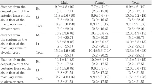
상악동 내측연과 하외측연 사이에 위치하는 상악 동하연의 중간에서 중격(septum)과 중격의 전∙후 에서 함요가 관찰되었다. 상악동하연과 치조연(alve- olar crest) 사이에서 다음과 같은 항목을 계측하 였다.
∙치조연에서 상악동하연 중격 전 함요 최심점까 지의 거리
∙치조연에서 상악동하연 중격까지의 거리
∙치조연에서 상악동하연 중격 후 함요 최심점 깊 은 점까지의 거리
∙치조연에서 상악동하연까지의 거리 중 최단점의 치아 위치
∙상악동하연에서 중격이 있는 치아 위치
(2) 치조궁과 상악동하연의 상관관계
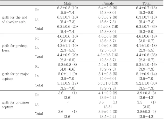
상악동하연의 중격과 최심점에서 내린 수직선이 치조돌기 외측연과 만나는 지점을 치조궁 정중점에 서 치조궁 둘레의 길이로 계측하였다(Fig. 4).
∙치조궁정중점(pr)에서 치조돌기끝 지점까지의 둘레
∙치조궁정중점에서 상악동 최심점까지의 둘레
∙치조궁정중점에서 상악동하연의 중격까지 둘레
(3) 상악동 기저부와 치조돌기 사이의 길이와 치 밀골의 두께
치조돌기에서 견치와 제1소구치, 제1소구치와 제2 소구치, 제2소구치와 제1대구치, 제1대구치와 제2대 구치, 그리고 제2대구치와 제3대구치 사이의 치조골 을 상악동까지 절단하고 다음과 같은 항목을 계측하 였다(Fig. 2).
∙치조돌기 절단면의 외판(outer table)의 길이
∙치조돌기 절단면의 내판(inner table)의 길이
∙치조돌기 외판 중앙 피질골의 두께
∙치조돌기 내판 중앙 피질골의 두께
∙상악동 하벽 중앙 피질골의 두께
Ⅲ. 결 과
1. 치조연에서 상악동하연 중격(septum)까지의 높이
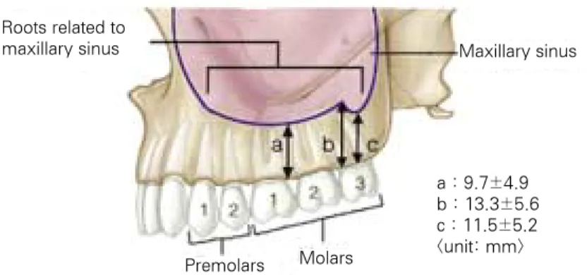
치조연이 손상되지 않은 37쪽 중, 중격은 28쪽 (76%)에서 관찰되었다. 치조연에서 상악동하연 중 격 전 함요 최심점까지의 거리는 9.7 ± 4.9 mm, 치 조연에서 상악동하연 중격까지의 거리는 13.3 ± 5.6 mm, 그리고 치조연에서 중격 후 최심점까지의 거리는 11.5 ± 5.2 mm이었다(Table I, Fig. 3).
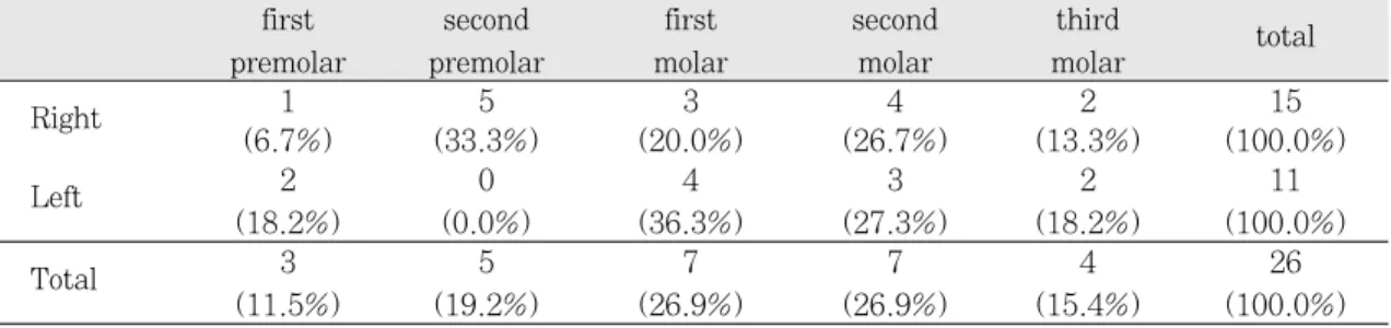
치아가 존재하는 26쪽 중 치조연에서 상악동하연 최심점의 치아위치는 우측에서 제2소구치 5예 Fig. 1. Ages distribution of the samples.
Fig. 2. Vertical section of interdental alveolar process.
1; buccal length from alveolar crest to sinus floor, 2; lingual length from alveolar crest to sinus floor, 3; thick- ness of buccal wall of alveolar compact bone, 4; thickness of lingual wall of alveolar compact bone, 5; thick- ness of superior wall of alveolar compact bone.
Table I. Distances from inferior border of the maxillary sinus to the alveolar crest
Male Female Total
distance from the Rt 9.9±4.5 (10) 7.7±4.7 (9) 8.9±4.6 (19)
deepest point of the [3.0~17.1] [2.5~15.8] [2.5~17.1]
anterior fossa on the Lt 11.8±5.6 (10) 8.9±4.6 (8) 10.5±5.2 (18)
sinus floor of the [3.5~22.0] [3.9~16.6] [3.5~22.0]
maxillary sinus to Total 10.9±5.0 (20) 8.3±4.5 (17) 9.7±4.9 (37)
alveolar crest [3.0~22.0] [2.5~16.6] [2.5~22.0]
distance from Rt 13.9±3.6 (8) 10.7±5.8 (7) 12.8±4.9 (15)
the septum on the [9.6~20.7] [5.2~20.2] [5.2~20.7]
sinus floor of the Lt 16.5±5.9 (8) 10.0±6.0 (5) 14.0±6.5 (13)
maxillary sinus to [9.6~25.1] [5.2~20.1] [5.2~25.1]
alveolar crest Total 15.2±4.9 (16) 10.4±5.6 (12)* 13.3±5.6 (28)
[9.6~25.1] [5.2~20.2] [5.2~25.1]
distance from the Rt 12.1±4.1 (8) 10.0±6.1 (7) 11.1±5.1 (15)
deepest point of the [5.5~17.5] [2.2~17.5] [2.2~17.5]
posterior fossa on the Lt 13.4±5.6 (8) 9.6±5.3 (5) 12.0±5.6 (13)
sinus floor of the [3.8~21.5] [2.5~17.3] [2.5~21.5]
maxillary sinus Total 12.7±4.8 (16) 9.8±5.6 (12) 11.5±5.2 (28)
to alveolar crest [3.8~21.5] [2.2~17.5] [2.2~21.5]
Note: Mean (mm)±S.D. [minimum~maximum]
maxillary sinus maxillary sinus
(19.2%)와 좌측에서 제1대구치 4예(36.3%)로 나타 났으며 전체적으로는 제1대구치와 제2대구치 각각 7예씩(각 26.9%)이었다(Table II). 상악동하연에 서 주중격이 있고 치아의 위치가 확인되는 19예에서 중격의 위치는 좌, 우측 모두 제3대구치 각 5예(합계 10예, 52.6%)로 가장 많이 나타났으며(Table III), 상 악동하연에서 중격 외에 부중격이 있는 경우가 4예 로 제2소구치(2예)와 제1대구치(2예)에서 관찰되 었다.
2. 치조궁과 상악골하연의 상관관계
치조궁정중점에서 치조궁의 끝 지점까지의 둘레는 6.3 ± 0.7 cm, 치조궁정중점에서 상악동하연 최심 점까지의 둘레는 4.4 ± 0.8 cm, 치조궁정중점에서 상악동하연 중격까지의 둘레는 5.2 ± 1.0 cm이었 다. 치조궁의 일부가 파손된 2쪽은 제외하였다 (Table IV, Fig. 4).
3. 상악동 기저부와 치조골 사이에서 피질골의 길 이와 두께
1) 견치와 제1소구치 사이 치조골 절단면
견치와 제1소구치 사이의 절단면에서 치조돌기 외 판의 길이는 21.1 ± 4.3 mm, 치조골 내판의 길이는 19.7±3.3 mm, 치조돌기 외판 피질골의 두께는
1.5 ± 0.3 mm, 치조돌기 내판 피질골의 두께는 1.6
± 0.5 mm, 그리고 상악동 하벽 피질골의 두께는 2.1 ± 0.7 mm이었다. 치조돌기 외판의 길이와 상악 동 하벽 피질골의 두께에서는 우측(21.0 ± 2.7 mm, 1.8 ± 0.4 mm)과 좌측(18.3 ± 3.5 mm, 2.4
± 0.7 mm)에서 통계적으로 유의한 차이(P<0.05)를 보였다(Table V, Fig. 2).
2) 제1소구치와 제2소구치 사이 치조골 절단면 제1소구치와 제2소구치 사이의 절단면에서 치조 돌기 외판의 길이는 19.2 ± 5.9 mm, 치조돌기 내판 의 길이는 17.0 ± 4.9 mm, 치조돌기 외판 피질골의 두께는 1.9 ± 0.7 mm, 치조돌기 내판 피질골의 두 께는 1.9 ± 0.6 mm, 그리고 상악동 하벽 피질골의 두께는 2.0 ± 0.9 mm이었다(Table V, Fig. 2).
3) 제2소구치와 제1대구치 사이 치조골 절단면 제2소구치와 제1대구치 사이의 절단면에서 치조 돌기 외판의 길이는 15.1 ± 5.0 mm, 치조돌기 내판 의 길이는 14.1 ± 4.3 mm, 치조돌기 외판 피질골의 두께는 1.8 ± 0.7 mm, 치조돌기 내판 피질골의 두 께는 2.1 ± 0.8 mm, 그리고 상악동 하벽 피질골의 두께는 1.9 ± 0.5 mm이었다. 치조돌기 내판 피질골 의 두께에서는 우측(1.7 ± 0.6 mm)과 좌측(2.5 ± 0.9 mm)에서 통계적으로 유의한 차이(P<0.05)를 보 였다(Table V, Fig. 2).
Fig. 3. Measurement items and results for the lateral view of the maxillary sinus.
a; height from the anterior fossa of the sinus lower border to alveolar crest, b; height from the septum of the sinus lower border to alveolar crest, c; height from the posterior fossa of the sinus lower border to alveolar crest.
Roots related to maxillary sinus
Premolars Molars
Maxillary sinus
a : 9.7±4.9 b : 13.3±5.6 c : 11.5±5.2
<unit: mm>
Fig. 4. Tooth position for the floor of the maxillary sinus.
Table II. Tooth position for the lowest point on the floor of the maxillary sinus
first second first second third
total
premolar premolar molar molar molar
Right 1 5 3 4 2 15
(6.7%) (33.3%) (20.0%) (26.7%) (13.3%) (100.0%)
Left 2 0 4 3 2 11
(18.2%) (0.0%) (36.3%) (27.3%) (18.2%) (100.0%)
Total 3 5 7 7 4 26
(11.5%) (19.2%) (26.9%) (26.9%) (15.4%) (100.0%)
unit: number (%)
Table III. Tooth position of the Underwood’s septum on the floor of the maxillary sinus
second first second third outer
total
premolar molar molar molar molar
Right 0 1 3 5 0 9
(0.0%) (11.1%) (33.3%) (55.6%) (0.0%) (100.0%)
Left 1 1 2 5 1 10
(10.0%) (10.0%) (20.0%) (50.0%) (10.0%) (100.0%)
Total 1 2 5 10 1 19
(5.3%) (10.5%) (26.3%) (52.6%) (5.3%) (100.0%)
unit: number (%)
(unit : mm)
4) 제1대구치와 제2대구치 사이 치조골 절단면 제1대구치와 제2대구치 사이의 절단면에서 치조 돌기 외판의 길이는 12.7 ± 3.9 mm, 치조돌기 내판 의 길이는 13.3 ± 3.8 mm, 치조돌기 외판 협측 피 질골의 두께는 1.7 ± 0.8 mm, 치조돌기 내판 피질 골의 두께는 1.7 ± 0.7 mm, 그리고 상악동 하벽 피 질골의 두께는 1.7 ± 0.4 mm이었다(Table V, Fig.
2).
5) 제2대구치와 제3대구치 사이 치조골 절단면 제2대구치와 제3대구치 사이의 절단면에서 치조 돌기 외판의 길이는 14.2 ± 4.4 mm, 치조돌기 내판 의 길이는 13.8 ± 4.1 mm, 치조돌기 외판 피질골의 두께는 1.8 ± 0.5 mm, 치조돌기 내판 피질골의 두 께는 1.7 ± 0.4 mm, 그리고 상악동 하벽 피질골의 두께는 1.7 ± 0.5 mm이었다(Table V, Fig. 2).
Table IV. Measurement of buccal circumference of alveolar process from the prosthion to the each points on the floor of the maxillary sinus
Male Female Total
Rt 6.3±0.5 (10) 6.4±0.9 (8) 6.4±0.7 (18)
[5.5~7.4] [5.3~8.0] [5.3~8.0]
girth for the end
Lt 6.3±0.7 (10) 6.3±0.7 (8) 6.3±0.7 (18)
of alveolar arch [5.4~7.3] [5.6~7.3] [5.4~7.3]
Total 6.3±0.6 (20) 6.4±0.8 (16) 6.3±0.7 (36)
[5.4~7.4] [5.3~8.0] [5.3~8.0]
Rt 4.6±0.6 (10) 4.6±0.8 (8) 4.6±0.6 (18)
[3.5~5.4] [3.6~5.7] [3.5~5.7]
girth for pr-deep Lt 4.2±1.1 (10) 4.0±0.8 (8) 4.1±1.0 (18)
fossa [2.3~5.5] [2.5~5.0] [2.3~5.5]
Total 4.4±0.9 (20) 4.3±0.8 (16) 4.4±0.8 (36)
[2.3~5.5] [2.5~5.7] [2.3~5.7]
Rt 5.2±0.8 (8) 5.4±1.2 (8) 5.3±1.0 (16)
[4.0~6.6] [3.9~7.3] [3.9~7.3]
girth for pr-major
Lt 5.0±1.1 (9) 5.1±0.8 (5) 5.1±0.9 (14)
septum [3.5~7.0] [4.0~6.0] [3.5~7.0]
Total 5.1±0.9 (17) 5.3±1.0 (13) 5.2±1.0 (30)
[3.5~7.0] [3.9~7.3] [3.5~7.3]
Rt 3.6 (1) 4.1±0.2 (2) 3.9±0.3 (3)
[3.6] [3.9~4.2] [3.6~4.2]
girth for pr-minor Lt - 3.5 (1) 3.5 (1)
septum [3.5] [3.5]
Total 3.6 (1) 3.9±0.4 (3) 3.8±0.3 (4)
[3.6] [3.5~4.2] [3.5~4.2]
Note: Mean (cm)±S.D. [minimum~maximum]
pr-deep fossa; the length of circumference between pr and the deepest fossa of the maxillary sinus, pr-Major septum; the length of circumference between pr and the major crest of the maxillary sinus, pr-minor septum;
the length of circumference between pr and the minor crest of the maxillary sinus.
Ⅳ. 고 찰
치아가 결손된 상악 치조돌기에서 비가역적인 골 흡수가 일어나 수직골(vertical bone)의 양이 감소하 고, 상악동의 함기화가 증가하여 치조돌기의 높이가 감소하면 임플랜트 시술에 장애요인이 된다. 이러한 장애요인은 상악동거상술, 자가골이식 혹은 대체골 이식을 이용한 augmentation 시술로 임플랜트 보철
의 안정성을 확보하여 해결할 수 있다. 이러한 과정 을 성공적으로 마치기 위해서 상악골에서 치조돌기, 상악동하연, 치아와 연관된 구조물의 해부학적 특성 을 이해하는 것은 매우 중요하다.13-17)
상악동에서 발견되는 중격이 상악동을 완전하게 나누기도 하지만,18-20) 대부분은 상악동하연에 위치 하면서 상악동을 불완전하게 나누는 Underwood 중 격21)이다. 이 중격은 치아의 결손으로 골흡수가 일어 Table V. Measurement of the vertical sections between teeth from canine to the 3rd molar
canine- 1st- 2nd premolar 1st- 2nd-
1st premolar 2nd premolar -1st molar 2nd molar 3rd molar Right 21.5±2.9 (10) 19.0±5.8 (10) 14.0±4.3 (10) 12.4±4.6 (10) 15.0±5.0 (8) buccal length from [18.5~27.1] [11.5~30.5] [8.3~21.2] [4.9~20.2] [6.7~22.8]
alveolar crest to Left 20.7±5.5 (10) 19.3±6.3 (10) 16.2±5.7 (10) 13.2±2.8 (8) 13.3±3.6 (7) sinus floor [11.9~28.6] [12.1~32.4] [9.4~28.2] [9.5~17.9] [9.5~18.2]
Total 21.1±4.3 (20) 19.2±5.9 (20) 15.1±5.0 (20) 12.7±3.9 (18) 14.2±4.4 (15) [11.9~28.6] [11.5~32.4] [8.3~28.2] [4.9~20.2] [6.7~22.8]
Right 21.0±2.7 (10) 17.4±4.6 (10) 13.5±4.0 (10) 13.2±4.3 (10) 13.9±4.5 (8) lingual length from [17.1~25.8] [9.6~24.0] [9.3~20.5] [7.4~20.3] [8.1~19.6]
alveolar crest to Left 18.3±3.5 (10) 16.4±5.5 (9) 14.7±4.7 (10) 13.4±3.2 (8) 13.6±3.8 (7) sinus floor [13.4~24.0] [9.4~24.4] [9.2~24.3] [10.3~19.4] [9.0~20.2]
Total 19.7±3.3 (20) 17.0±4.9 (19) 14.1±4.3 (20) 13.3±3.8 (18) 13.8±4.1 (15 [13.4~25.8]* [9.4~24.4] [9.2~24.3] [7.4~20.3] [8.1~20.2]
Right 1.5±0.4 (9) 2.0±0.7 (10) 2.0±0.7 (10) 1.5±0.5 (10) 1.7±0.4 (7) thickness of buccal [0.9~2.1] [1.0~2.9] [1.1~3.2] [0.9~2.4] [1.2~2.4]
wall of alveolar Left 1.6±0.3 (9) 1.8±0.8 (9) 1.7±0.7 (9) 2.1±1.0 (8) 1.9±0.7 (7)
compact bone [1.0~1.9] [0.9~3.0] [1.1~3.1] [0.7~3.3] [1.0~2.6]
Total 1.5±0.3 (18) 1.9±0.7 (19) 1.8±0.7 (19) 1.7±0.8 (18) 1.8±0.5 (14 [0.9~2.1] [0.9~3.0] [1.1~3.2] [0.7~3.3] [1.0~2.6]
Right 1.7±0.6 (9) 1.7±0.5 (10) 1.7±0.6 (10) 1.5±0.5 (10) 1.5±0.2 (7) thickness of lingual [0.9~2.9] [1.0~2.8] [0.6~2.5] [0.8~2.3] [1.1~1.8]
wall of alveolar Left 1.5±0.4 (10) 2.1±0.5 (9) 2.5±0.9 (9) 2.0±0.8 (8) 1.9±0.4 (7)
compact bone [1.1~2.2] [1.4~3.1] [1.3~4.6] [1.2~3.5] [1.3~2.6]
Total 1.6±0.5 (19) 1.9±0.6 (19) 2.1±0.8 (19) 1.7±0.7 (18) 1.7±0.4 (14) [0.9~2.9] [1.0~3.1] [0.6~4.6]* [0.8~3.5] [1.1~2.6]
Right 1.8±0.4 (9) 2.0±0.5 (10) 1.8±0.5 (10) 1.7±0.5 (9) 1.9±0.6 (7) thickness of superior [1.0~2.4] [1.3~2.9] [1.0~2.4] [1.1~2.7] [1.2~2.7]
wall of alveolar Left 2.4±0.8 (10) 2.0±1.2 (9) 2.1±0.4 (8) 1.8±0.4 (7) 1.5±0.4 (7)
compact bone [1.4~4.1] [1.2~5.1] [1.5~2.8] [1.3~2.2] [1.1~2.2]
Total 2.1±0.7 (19) 2.0±0.9 (19) 1.9±0.5 (18) 1.7±0.4 (16) 1.7±0.5 (14) [1.0~4.1]* [1.2~5.1] [1.0~2.8] [1.1~2.7] [1.1~2.7]
Note: Mean (mm)±S.D. [minimum~maximum]
* significant difference between right and left (P<0.05)
나는 두 곳 사이이고 상악동의 함기화가 다양하게 일어난 흔적이며 저작력을 전달받는 골주이다.6,15,22)
이 실험에서 상악동을 완전히 나누는 중격은 관찰 되지 않았다. Underwood 중격이 나타나는 빈도는 약 76%로, 오스트리아인 41예에서 관찰한 Ulm 등6) 의 37%, 치아가 결손된 환자 200예 중 치아가 결손 되어 치조돌기의 위축이 일어난 41예의 CT에서 관 찰한 Krennmair 등23)의 26.8%보다 현저하게 높았 다. 이는 시신기증을 해주신 분의 건강상태, 영양상 태, 치아건강상태 등과 연관이 있을 것으로 생각 된다.
이 실험에서 중격은 제3대구치(52%)와 제2대구치 (26.3%)에 주로 위치하여 대부분 소구치가 있는 부 분에 위치한다는 Krennmair 등24)과 Ulm 등4)의 보고 와 큰 차이가 있었다. 이 중격의 높이를 치조연에서 중격까지의 거리와 치조연에서 깊은 점까지 거리의 차이라고 했을 때 4.5 ± 3.2 mm로, Ulm 등4)의 7.9 mm나 Krennmair 등23)의 8.1 ± 2.5 mm보다 낮았 으며, 중격 전후에 위치하는 함요의 깊이가 서로 차 이가 난다는 것은 일치하였다. 또 상악동하연에서 가장 깊은 곳은 제1 혹은 제2대구치 위치이었으며 Kwak 등12), Chanavaz8)그리고 고광준과 이상래39)의 보고와 유사하였다.
1910년 Underwood가 기술보고한 Underwood 중 격은 상악동거상술시 장애요인으로 상악동 외측벽 에 창(window) 형성을 방해하고 상악동점막이 찢 어지게도 한다. 이 중격은 치아가 결손된 경우 잘 생기며 치아가 있는 경우에는 위치를 예측하기 어 렵다. 이 중격을 확인하는 방법은 파노라마 촬영시 보다 CT 촬영시 확인하기 쉬우며, 중격의 위치, 형 태, 높이 등에 대한 해부학 지식이 깊을수록 상악동 하연과 연관된 수술에서 합병증을 감소시킬 수 있다.6,23,24,26)
치아 사이에서 상악골 치조돌기의 외판(outer table)을 구성하는 치밀골의 길이는 4.9 ~ 28.2 mm이었다. 견치와 제1소구치 사이부터 짧아져 제1 대구치와 제2대구치 사이가 가장 짧았으며 제2와 제 3대구치 사이는 다소 길어졌다. 내판(inner table)을 구성하는 치밀골의 길이는 7.4 ~ 25.8 mm이었으며 제 1대구치와 제2대구치 사이가 가장 짧았다. 일반 적으로 임플랜트시술에는 잔존 치조돌기의 높이가 4
mm 이상인 경우 임플랜트의 일차적 안정성이 유지 될 수 있다고 하였으며,27-29)Misch (1987)는 잔존 치 조골의 높이가 4 ~ 5 mm, 너비는 6 ~ 7 mm이상 이어야 임플랜트의 초기고정이 가능하며 높이가 5 mm 이하인 경우는 골이식술이 선행되어야 한다고 하였고, 이용찬30)은 잔존 치조돌기가 10 mm 미만인 경우 골이식을 겸한 상악동절단술, 치조골증대술, 상악동점막거상술 등의 외과적 수술이 필요하였으 며, Smiler 등26)은 잔존골의 높이가 충분하지 않은 경 우 임플랜트 시술시 합병증의 발현빈도가 증가된다 고 하였다.
각 치아 사이에서 관찰되는 내, 외판의 길이는 4.9
~ 28.2 mm이어서 임플랜트 시술에 큰 문제가 없는 것처럼 보였으나 Table II에서 보면 치아 중앙에서 상악동하연 최심점까지의 거리가 4 mm 이하인 경 우가 많았다. Table II의 결과는 치아결손 기간이 길 어 치아위치를 확인할 수 없는 경우도 포함된 것이 므로 치아결손된 기간이 길수록 치조돌기의 높이가 낮아진다는 것을 간접적으로 알 수 있었다. 그리고 치아 사이보다는 치조연 중심에서의 거리가 실제 임 상에서 활용도가 높을 것으로 생각되었다.
치조돌기에서 각 부위별 외판 중앙부에서 치밀골 의 두께는 0.9 ~ 3.3 mm이었고 내판 중앙부의 두께 는 0.9 ~ 4.6 mm이었으며, 치밀골의 경계가 불분명 하여 두께의 계측에 어려움이 있었다. 오른쪽과 왼 쪽에서 의미있는 차이는 없었으며, 여자의 경우는 치아의 위치를 확인할 수 없는 경우가 많아 구체적 인 차이를 규명할 수 없었다.
한편, 상악동하벽의 두께에 대한 해부학 정보는 상 악치아의 외과적처치, 인공치아이식 등의 시술에 앞 서 진단과정에서 획득되며 임상적으로 매우 중요하 다고 보고한 곽현호 등31)은 한국인의 경우 치아 위치 에 따라 다르지만 대개 0.37 ~ 0.77 mm 사이에 있 으며 제2소구치 주변이 가장 두껍다고 하였고, Harrison32)은 상악동하벽의 두께는 대개 0.5 mm이 며, 치아위치에 따라 0.5 mm 이상되는 경우는 5 ~ 46%라고 하였다.
이 실험에서는 상악동하벽에서 치밀골의 두께가 1.0 ~ 5.1 mm이었고 견치에서 제3대구치쪽으로 가 면서 두께가 얇아지는 것은 치아결손에 의한 골흡수 와 관련 있을 것으로 생각되었다.
이 실험에서 일부 결과는 외국인과 뚜렷한 차이를 나타내기도 하였으나 시신수가 적고 일부 연령에 집 중되고 있어 한국인 전체를 대표한다고 보기 어렵 다. 또 더욱 의미있는 자료를 확보하기 위해서 치아 가 결손된 경우와 잔존하는 경우 보철물을 착용한 경우를 분리하여 결과를 획득하고 비교 분석하는 것 이 필요할 것으로 생각되었다. 일반적으로 CT 촬영 을 통해 획득하는 자료는 편리하게 대량 확보할 수 있으며 관찰하기 어려운 부분까지 확인할 수 있고, 부비동의 경계가 불분명한 파노라마 촬영보다는 정 확하여 많은 연구자들이 활용하고 있으나, 주변 연 조직의 밀도와 기계적 오차에 의해 실제보다 벽 두 께가 크게 나오는 단점이 있다. 건조골(마른 뼈)을 사용하는 경우는 연조직이 결손되어 부피가 실제보 다 크게 계측되나,33-35)이 실험에서는 해부실습이 종 료된 후 계측하여 비교적 실제적인 자료를 획득할 수 있었다.
이 실험의 결과들은 평균수명이 현저하게 증가하 는 한국인 고령자의 상악동 치조돌기 주변의 해부학 자료를 제공하여 임플랜트 시술의 계획이나 예후 판 단에 도움이 될 것으로 생각된다. 이후 지속적인 연 구를 통하여 연령별 자료의 확보, 병력이 확인된 경 우 치아결손에 따른 상악골 치조돌기의 흡수상태 확 인, 임플랜트 시술 후 치조돌기 골의 변화 등을 구명 하여 효과적인 임플랜트 시술에 필요한 정보의 제공 이 필요할 것으로 예상된다.
이 실험을 통하여 확보된 치조연과 상악동하연 사 이의 거리, Underwood 중격의 위치와 높이, 치조돌 기의 높이, 치조돌기 주변의 치밀골두께 등의 자료 는 임플랜트 수술에 유용한 자료로 이용될 수 있다 고 생각된다.
Ⅴ. 결 론
현재 치아결손으로 발생되는 문제점을 해결할 때 추천하고 있는 임플랜트 시술에서 시술부위의 해부 학 지식은 임플랜트 설계, 시술 및 성공률을 결정짓 는 중요한 요소이다.
이에 저자는 상악골 치조돌기와 상악동하연 사이 에서 임플랜트 시술에 필수적인 뼈의 길이와 두께, 그리고 위치에 대한 해부학 자료를 확보하기 위하여
시신 19구에서 치조연에서 상악동하연까지의 높이, 치조돌기의 높이와 단면에서 치밀골의 두께 등을 관 찰하여 다음과 같은 결과를 얻었다.
1. 상악동하연에서 중격은 28예(76%)에서 관찰되었 으며, 제3대구치(52.6%)와 제2대구치(26.3%) 에 해당되는 부위에 위치하였다. 상악동하연에서 가장 깊은 곳은 제1대구치 혹은 제2대구치 위치 이며 치조연에서 9.7 ± 4.9 mm 떨어진 곳이 었다.
2. 각 치아단면에서 치조돌기의 길이는 4.9 ~ 28.2 mm이었으며 제1대구치와 제2대구치 사이가 가 장 짧았고, 돌기 중앙의 두께는 0.9 ~ 3.2 mm이 었다. 치조돌기 내판의 길이는 7.4 ~ 25.8 mm이 었고 제1대구치와 제2대구치 사이가 가장 짧았으 며 중앙의 두께는 0.9 ~ 4.6 mm이었다.
이상의 결과는 임플랜트 수술에 유용하게 사용될 수 있는 상악골의 해부학 자료라고 생각된다.
참고문헌
1. Hong SB, Chai GJ, Jung UW, Kim CS.
Clinical evaluation of Branemak Ti-Unite implant and ITI SLA implant in post maxillary area with sinus elevation tech- nique. J Korean Acad Periodont 2005;
35:813-822.
2. Razavi R, Zena RB, Khan Z, Gould AR.
Anatomic site evaluation of edentulous max- illae for dental implant placement. J Prosthodont 1995;4:90-94.
3. Albrektsson T, Bra�nemark PI, Hansson HA, Lindstrom J. Osseointegrated titanium implants. Requirements for ensuring a long-lafting direct bone-to-implant anchor- age in man. Acta Orthop Scand 1981;
52:155-170.
4. Ulm CW, Solar P, Gsellmann B, Matejka M, Watzek G. The edentulous maxillary alveolar process in the region of the max- illary sinus - a study of physical dimension.
Int J Oral Maxillofac Surg 1995a;24:279-
282.
5. Toffler M. Minimally invasive sinus floor elevation procedures. N Y State Dent J 2004;70:38-44.
6. Ulm CW, Solar P, Krennmair G, Matejka M, Watzek G. Incidence and suggested sur- gical management of septa in sinus-lift pro- cedures. Int J Oral Maxillofac Implants 1995b;10:462-465.
7. Kraut R, Kessler H. Quantification of bone in dental implant sites after composite grafting of the mandible. Int J Oral Maxillofac Surg 1989;47:238-242.
8. Chanavaz M. Maxillary sinus: Anatomy, physiology, surgery, and bone grafting related to implantology-eleven years of surgical experience (1979-1990). J Oral Implantol 1990;16:199-209.
9. Boss LP, Taylor TD. Problems associated with implant rehabilitation of the edentulous maxilla. Dent Clin North Am 1998;42:117- 126.
10. Tidwell JK, Blijdorp PJ, Stoelinga PJW, Brouns JB, Hinderks F. Composite graft- ing of the maxillary sinus for placement of endosteal implants. A preliminary report of 48 patients. Int J Oral Maxillofac Surg 1992;21:204-209.
11. Fredholm U, Bolin A, Andersson L.
Preimplant radiographic assessment of available maxillary bone support. Compa- rison of tomographic and panoramic tech- nique. Swed Dent J 1993;17:103-109.
12. Kwak HH, Park HD, Yoon HR, Kang MK, Koh KS, Kim HJ. Topographic anato- my of the inferior wall of the maxillary sinus in Koreans. Int J Oral Maxillofac Surg 2004;33:382-388.
13. Boyne PJ, James RA. Grafting of the maxillary sinus floor with autogenous marrow and bone. J Oral Surg 1980;
38:613-616.
14. Misch CE. Density of bone: Effect on treatment plans, surgical approach, heal- ing and progressive bone loading. Int J Oral Implant 1990;6:23-31.
15. Vinter I, Krmpotic-Nemanic J, Jalsovec D.
Verschwindet der processus alveolaris des oberkiefers immer nach dem Zahnausfall.
Laryngorhinootologie 1993;72:605-607(in German).
16. Lustmann J, Lewinstein I. Interpositional bone grafting technique to widen narrow maxillary ridge. Int J Oral Maxillofac Implants 1995;10:568-577.
17. Barone A, Crespi RC, Aldini NN, Fini M, Giardino R, Covani U. Maxillary sinus augmentation: Histologic and histomor- phometric analysis. Int J Oral Maxillofac Implants 2005;20:519-525.
18. Shin KS, Kim YD. A case of mucopy- ocele of septated Maxillary sinus. J Korean Ophthalmological Society 1990;31:814-817.
19. Schatz CJ, Becker TS. Normal CT anato- my of the paranasal sinuses. Radiol Clin North Am 1984;22:107-118.
20. Bhatia PL, Gupta SK, Sasibabu K, Varanasi BS. Clinical significance of completely subdivided maxillary antrum. Oral Surg Oral Med Oral Pathol 1977;44:679-683.
21. Underwood AS. An inquiry into the anato- my and pathology of the maxillary sinus.
A Anat Physiol 44: 354-369, 1910 cited from Ulm et al. (1995b).
22. Poyton HG. Maxillary sinuses and the oral radiologist. Dent Radiogr Photogr 1972;45:43-50.
23. Krennmair G, Ulm C, Lugmayr H.
Maxillary sinus septa : incidence, mor- phology and clinical implications. J Craniomaxillofac Surg 1997;25:261-265.
24. Krennmair G, Ulm CW, Lugmayr H, Solar
P. The incidence, location, and Height of maxillary sinus septa in the edentulous and dentate maxilla. J Oral Maxillofac Surg 1999;57:667-671.
25. Ko KJ, Lee SR. A radiographic study on the morphology of maxillary sinus in ado- lescents and adults. J Korean Acad Dent Radiology 1984;14:17-26.
26. Smiler DG, Johnson PW, Lozada JL, Misch CE, Rosenlicht JL. Sinus lift grafts and endosseous implants. Treatment of the atrophic posterior maxilla. Dent Clin North Am 1992;36:151-186.
27. van den Bergh JP, ten Bruggenkate CM, Krekeler G, Tuinzing DB. Sinus floor ele- vation and grafting with autogenous bone from the iliac crest. Clin Oral Implants Res 1998;9:429-435.
28. Hurzeler MB, Kirsch A, Ackermann KL, Quinones CR. Reduction of the severely resorped maxilla with dental implants with augmentated maxillary sinus. 5 year clinical investigation. Int J Oral Maxillofac Implants 1996;11:466-475.
29. Misch CE. Maxillary sinus augmentation for endosteal implants: Organized alter-
native treatment plans. Int J Oral Implantol 1987;4:49-58.
30. Lee YC. A sinus floor elevation method using maxillary tuberosity bone. J Korea Assoc Maxillofac Plast Reconstr Surg 1996;18:236-244.
31. Kwak HH, Yoon HL, Hur KS, Kang MG, Park HD, Koh KS, Park CS, Kim KD, Kim HJ. Topography of the Inferior Wall of the Maxillary Sinus in Koreans. Korean J Physical Anthropology 2001;14:137-149.
32. Harrison DF. Oro-antral fistula. Br J Clin Pract 1961;15:169-174.
33. Kasabah S, Sleza′k R, S̆imu�nek A, Krug J, Lecaro MC. Evaluation of the accuracy of panoramic radiograph in the definition of maxillary sinus septa. Acta Medica 2002;45:173-175.
34. Kanda S. Quantitative analysis in max- illofacial regions using computed tomog- raphy. J Jpn Stomatol Soc 1992;4:793- 794(in Japanese).
35. Taguchi M. Development of maxillary sinus in childhood. J Iwate Med Ass 40:
363-372, 1988 cited from Ariji et al. (1994).
Reprint request to:
Young-Soo Lee, D.D.S., M.S., Ph.D.
Department of Dentistry, College of Medicine, Hanyang University
#17 Haeng Dang-Dong, Seong Dong-Gu, Seoul, 133-792, Korea [email protected]
Statement of problem: Following tooth loss, the edentulous alveolar process of maxilla is affected by irreversible reabsorption process, with progressive sinus pneumati- zation leads to leaving inadquate bone height for placement of endosseous implants. Grafting the floor of maxillary sinus by sinus lifting surgery and augmentation of autologous bone or alternative bone material is a method of attaining sufficient bone height for maxillary implants placement and has proven to be a highty successful.
Purpose: This study was undertaken to clarify the morphometric characteristics of infe- rior maxillary sinus and alveolar process for installation of implants.
Material and method: Nineteen skulls (37 sinuses, 10M / 9F) obtained from the col- lection of the department of anatomy and cell biology of Hanyang medical school were stud- ied. The mean age of the deceased was 69.9 years (range 44 to 88 years).
The distance between alveolar border and inferior sinus margin at each tooth, the height of alveolar process and the thickness of cortical bone of the outer and inner table of alveolar process and the inferior wall of maxillary sinus were measured.
Results and Conclusion:
1. The septum of inferior maxillary sinus were observe 28 sides (76.%) and located at the third molar (52.6%) and the second molar (26.3%). The deepest points of inferior bor- der of maxillary sinus were located the first or second molar. The distance between alve- olar margin and the deepest point of inferior maxillary sinus is 9.7 ± 4.9 ㎜.
2. The length of the outer table of alveolar process were 4.9 ~ 28.2 ㎜ and the shortest point was between the first and the second molors. The thickness of them were 0.9~3.2
㎜. The length of the inner table of alveolar process were 7.4~25.8 ㎜ and the shortest point was between the first and the second molars. The thickness of the were 0.9~4.6 ㎜.
The results of this study are useful anatomical data for installing of maxillary implants.
MORPHOMETRICS OF ALVEOLAR PROCESS AND ANATOMICAL STRUCTURES AROUND INFERIOR MAXILLARY SINUS FOR
MAXILLARY IMPLANTATION
Ju-Jin Park, D.D.S., Ph.D., Young-Soo Lee, D.D.S., Ph.D., Doo-Jin Paik, M.D., Ph.D.*, Won-Hee Park, D.D.S., Ph.D., Dong-Yeob Yoo, D.D.S., M.S.
Department of Dentistry, College of Medicine, Hanyang University
*Department of Anatomy and Cell Biology, College of Medicine, Hanyang University ABSTRACT
Key words : Maxillary sinus, Alveolar process, Skull, Endosseous implant