1)
Introduction
Salivary glands are composed of acinar cells, either serous or mucous, and ductal cells of several varieties. The primary saliva is formed in the lumen of the acinar cells, where the accumulation of ions generates a transepithelial osmotic gradient driving water movement. In the second stage of secretion, sodium is actively reabsorbed while the primary saliva passes through the duct. Therefore, altera- tions of sodium transporters and water channels may affect the formation and the ductal modification of the saliva.
Received February 25, 2008. Accepted March 27, 2008.
Corresponding author : Sun-Youl Ryu, D.D.S.
Department of Oral and Maxillofacial Surgery, Chonnam National University Professional Graduate School of Dentistry, 5 Hak-dong, Dong-gu, Gwangju, 501-746, Korea
Tel : +82-62-220-5430, Fax : +82-62-232-8126 E-mail: [email protected].
Immunofluorescence labelling and confocal microscopy revealed a polarized distribution of Na+,K+-ATPase, the secretory isoform of Na+/K+/2Cl cotransporter (NKCC2), and type 1 Na+/H+ exchanger (NHE1) in the basolateral membranes of acinar and intralobular duct cells1). Na+,K+- ATPase produces an inward-directed sodium chemical gradient in the acinar cell2). Although sodium influx is ne- gligible in resting cells, it is rapidly increased by acti- vation of NKCC2, NHE1, and nonselective cation channels in the acinar cell3). Blockade of NKCC2 causes significant reduction of acetylcholine-stimulated secretion4), in which the residual secretion is attributed to the operation of Na+/H+ and Cl /HCO3 antiport5, 6). The amiloride-sensi- tive epithelial sodium channels (ENaC) are localized to the surface epithelial cells of the secretory duct. Among their three homologous subunits, only -subunit can produce anα amiloride-sensitive current7), whileβ- and γ-subunits are
Altered Expression of Sodium Transporters and Water Channels in the Submandibular Gland of Rats
Treated with Nitric Oxide Synthesis Inhibitors
Il-Young Seo, D.D.S.1, Miwon Kim, Ph.D.2, JongUn Lee, M.D.3 and Sun-Youl Ryu, D.D.S.1
1Department of Oral and Maxillofacial Surgery, Chonnam National University Professional Graduate School of Dentistry
2Department of Nursing, Chonnam National University College of Nursing
3Department of Physiology, Chonnam National University Medical School, Gwangju, Korea
A role of nitric oxide (NO) in the regulation of sodium transporters and water channels in the salivary gland was investigated. Male Sprague-Dawley rats were treated with NG-nitro-L-arginine methyl ester (L- NAME, 100 mg/L drinking water) for 1 week. The control group was supplied with normal tap water. The expression of Na+,K+-ATPase, type 2 Na+/K+/2Cl- cotransporter (NKCC2), type 1 Na+/H+ exchanger (NHE1), α-subunit of epithelial sodium transporter (ENaC), and aquaporin-5 (AQP5) and aquaporin-1 (AQP1) proteins were determined in the submandibular gland by Western blot analysis. Following the treatment with L-NAME, the expression of Na+,K+-ATPase α1-subunit, NKCC2, NHE1, and ENaC α- subunit increased significantly. On the contrary, the expression of AQP5 was significantly decreased, while that of AQP1 was not significantly altered. These findings indicate that the sodium transporters and water channels may be under a tonic regulatory influence of NO in the salivary gland.
Key Words : nitric oxide; sodium transporters; aquaporins; submandibular gland
not functional on their own8).
On the other hand, there have been known multiple iso- forms of aquaporin (AQP) water channels in the salivary gland. Among them, AQP5 may be the only isoform of AQP playing a major role in the salivary secretion process.
Strong AQP5 labelling is located in the apical membrane of serous-type acinar cells from the rat submandibular gland9). The transepithelial water movement occurs through the apical AQP5 channels and paracellular pathways10), leading to the secretion of isotonic primary saliva. Although the presence of AQP1 and AQP8 has been also generally accepted, their role in the salivary secretion remains con- troversial11).
Over the last two decades, nitric oxide (NO) has been found to play a role in the regulation of various physiolo- gical functions. It has been also implicated in mediating the nerve-evoked vasodilator and secretory responses in the salivary gland. Parasympathetic nerve activity increases the submandibular blood flow, and elicits the flow of saliva and output of protein by mechanisms that involve in situ generation of NO in the ferret12). A previous study revealed that NO synthase (NOS) is not present in the acinar cells, but in neural terminals within the gland and in the apical membrane of the excretory and striated ducts, the cyto- plasm of granular convoluted tubules, and, to a lesser ex- tent, in the cytoplasm of excretory and striated ducts13). However, later investigators demonstrated that acinar cells also posses endogenous NOS activity14). Furthermore, the salivation induced by cholinergic agonists or that produced by the chorda stimulation was decreased by inhibition of NO synthesis by NG-nitro-L-arginine methyl ester (L- NAME) in the rat submandibular gland11, 15).
Nevertheless, the role of NO in the regulation of sodium transporters and AQP channels in the salivary gland has not been established. Therefore, the present study was aimed to explore the role of NO in the regulation of sodium transporters and AQP channels in the salivary gland. Rats were inhibited of endogenous generation of NO by treat- ment with L-NAME, and the expression of sodium trans- porters and water channels was determined in the sub- mandibular gland.
Materials and Methods 1. Animals
Male Sprague-Dawley rats (200-250 g) were used. The experimental group was treated with L-NAME (100 mg/L drinking water) for 1 week. The control group was suppli- ed with normal tap water. The experimental procedure conformed to the Institutional Guidelines for Experimental Animal Care and Use.
2. Western blot analysis
The submandibular glands were rapidly isolated under ketamine anesthesia. They were rapidly frozen and kept at -70 until analyzed. For protein preparation, they were thawed and homogenized with Polytron homogenizer at 3,000 rpm in a solution containing sucrose (250 mmol/L), EDTA (1 mmol/L), phenylmethylsulfonyl fluoride (0.1 mmol/L), and potassium phosphate buffer (20 mmol/L), at pH 7.6. The homogenate was centrifuged at 1,000 g for 15 min to remove the whole cells, nuclei, and mito- chondria. The supernatant was then ultracentrifuged at 100,000×g for 1 h to produce a pellet containing mem- brane fractions enriched for both plasma membranes and intracellular vesicles. The pellet was resuspended in homo- genizing solution for protein blotting.
Protein samples were loaded and electrophoretically size-separated with a discontinuous system consisting of 8-12.5% polyacrylamide resolving gel and 5% polyacry- lamide stacking gel. The proteins were then transferred to a nitrocellulose membrane at 40 V for 3 h. The membrane was washed in Tris-based saline buffer (pH 7.4) con- taining 0.1% Tween-20 (TBST), blocked with 5% nonfat milk in TBST for 1 h, and incubated with antibodies in 2% nonfat milk/TBST for 1 h at room temperature. The antibodies used were polyclonal anti-rabbit α-1 and β-1 subunits of Na+,K+-ATPase (1:2,500, Upstate Biotech- nology, Lake Placid, NY, USA), NKCC2 (1:1,000, Che- micon, Temecula, CA, USA), NHE1 (1:500, Alpha Diag- nostic, San Antonio, TX, USA), α-subunit of ENaC (1:
500, Alpha Diagnostic, San Antonio, TX, USA), AQP1 (1:1,000, Alomone, Jerusalem, Israel), AQP5 (1:1000,
Alpha Diagnostic, San Antonio, TX, USA), endothelial NOS (eNOS) and neuronal NOS (nNOS) (1:750, Transduc- tion; Lexington, KY, USA).β-Actin was used as internal control (1:1,000, Sigma, St. Louis, MO, USA). The mem- brane was then incubated with horseradish peroxidase- labeled goat anti-rabbit IgG (1:1,200) in 2% nonfat milk in TBST for 2 h. The bound secondary antibody was detected by enhanced chemiluminescence (Amersham, Buckinghamshire, England). The protein levels were de- termined, using the transmitter scanning videodensitometer (Biomed Instruments, Fullerton, CA, USA).
3. Drugs and statistical analysis
Drugs were purchased from Sigma, unless stated other- wise. Results are expressed as mean±SEM. The statistical significance of values between groups was determined by the unpaired t-test.
Results
In the L-NAME-treated group, the systolic blood pres- sure measured indirectly by the tail cuff method in a con- scious state was significantly higher in the experimental group than in the control (146±9 vs 121±6 mmHg, p<0.05, n=6 each). Accordingly, the expression of eNOS and nNOS in the submandibular gland was decreased significantly
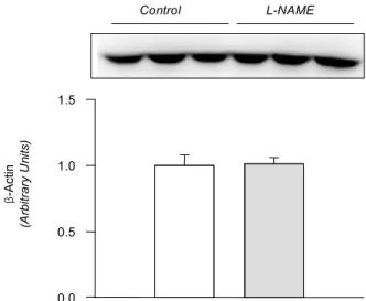
(Fig. 1). The amount ofβ-actin was not significantly alter- ed (Fig. 2). On the contrary, the expression of 1-subunitα of Na+,K+-ATPase was significantly increased, although that of β1-subunit remained unchanged (Fig. 3). The expression of NKCC2, NHE1, and -subunit of ENaC wasα increased significantly (Fig. 4). The expression of AQP5 was significantly decreased, while that of AQP1 was not significantly altered (Fig. 5).
eNOS (Arbitrary Units) nNOS (Arbitrary Units)
0 1 2
Control L-NAME
0 1 2
Control L-NAME
* ***
Fig. 1. Expression of endothelial NOS (eNOS) and neuronal NOS (nNOS) in the submandibular gland. Representative immunoblots and densitometric data are shown. The open column denotes control, and the hatched column depicts NG-nitro-L-arginine methyl ester (L-NAME)-treated group. Each column represents mean±SEM of 6 rats.
*p<0.05, ***p<0.001, compared with control.
β-Actin (Arbitrary Units)
0.0 0.5 1.0 1.5
Control L-NAME
Fig. 2. Expression of -actin in the submandibular gland. Legendsβ as in Fig. 1. Mean±SEM of 6 rats each.
0 1 2
Control L-NAME
Na+,K+-ATPaseβ1 (ArbitaryUnits) 0
1 2
Control L-NAME
Na+,K+-ATPaseα1 (ArbitaryUnits) **
Fig. 3. Expression of α1- and β1-subunits of Na+,K+-ATPase in the submandibular gland. Legends as in Fig. 1. Mean±SEM of 6 rats each.
**p<0.01, compared with control.
0 1 2 3
Control L-NAME
***
0 1 2 3
Control L-NAME
*
0 1 2 3
Control L-NAME
**
NKCC2 (ArbitaryUnits) ENaCα (ArbitaryUnits)
NHE1 (ArbitaryUnits)
Fig. 4. Expression of Na+/K+/2Cl-cotransporter (NKCC2), Type 1 Na+/H+exchange (NHE1), andα-subunit of epithelial sodium channel (ENaC ) in the submandibular gland. Legends as in Fig. 1. Mean±SEM of 6 rats each.α
*p<0.05, **p<0.01, ***p<0.001, compared with control.
0 1 2
Control L-NAME
AQP5 (Arbitrary Units)
0 1 2
Control L-NAME
AQP1 (Arbitrary Units)
*
Fig. 5. Expression of aquaporin-1 (AQP1) and aquaporin-5 (AQP5) proteins in the submandibular gland. Legends as in Fig. 1. Mean±SEM of 6 rats each.
*p<0.05, compared with control.
Discussion
NO has been implicated in mechanisms mediating nerve-evoked vasodilator and secretory responses in sali- vary glands. The salivation induced by cholinergic ago- nists or chorda stimulation was decreased by NO synthesis inhibition in the submandibular gland12, 13, 15)
. The for- mation of primary and secondary saliva involves transe- pithelial electrolyte and water movement. Therefore, it is plausible to hypothesize that NO plays a role in regulating the sodium transporters and AQP channels in the salivary gland.
Previous studies have shown that L-NAME treatment decreases the gene and protein expression of eNOS16, 17). The present study also demonstrated a decreased expres- sion of both eNOS and nNOS in the submandibular gland following the treatment with L-NAME, which may have resulted in a decreased formation of NO. The down-re- gulation of NOS was associated with an increased ex- pression of a1-subunit of Na+,K+-ATPase, NKCC2, NHE1, and a-subunit of ENaC, suggesting that these sodium transporters should be under tonic inhibitory influence of NO in the submandibular gland. The up-regulation of these transporters may then, at least in part, account for the decreased salivation following inhibition of NO synthesis in rat submandibular gland13, 15), possibly through an en- hancement of the ductal reabsorption of sodium.
On the other hand, the expression of AQP5 was signifi- cantly decreased by the treatment with L-NAME. This finding suggests that AQP5 is under tonic excitatory influ- ence of NO in the submandibular gland. A key role of AQP5 in saliva fluid secretion has been well known.
Transgenic mice lacking AQP5 display reduced pilocarpine- stimulated saliva secretion (60%), in which the secreted saliva is more hypertonic and viscous18). In Sjøgren's syn- drome, an abnormal distribution of AQP5 has been demon- strated in the acini, which is likely to contribute to the de- ficiency of fluid secretion19). A down-regulation of AQP5 may result in a decrease of water flux in the acinar cell and hence the formation of primary saliva. In this context, the L-NAME-induced reduction of secretory responses12, 13,
15) may be attributed in part to the down-regulation of AQP5. The expression of AQP1 has been demonstrated to be continuously distributed to microvasculature during embryonic and postnatal development in the rat submandi- bular gland20, 21). However, the expression of AQP1 proteins was not significantly altered by the L-NAME-treatment. In transgenic mice lacking AQP1, pilocarpine-induced salivary secretion showed no defects in volume or composition of saliva18, 22). It is unlikely that AQP1 should play a major role in salivary secretion.
Finally, it is worth to note that the systolic blood pres- sure was significantly increased, indicating that the ani- mals were in a state of systemic withdrawal of tonic in- fluences of endogenous NO. Furthermore, the alteration of sodium transporters and water channels cannot be attri- buted to the increased blood pressure, perse, since an attenuated salivation has not been documented in other models of hypertension.
In summary, these results indicate that sodium transpor- ters and water channels are under tonic regulatory influ- ence of NO in the submandibular gland. An altered NO synthesis may affect the expression of these transporters and channels, and hence the formation and secretion of saliva. Further studies will be needed to directly measure the sodium and water flux in the ductal epithelia to de- termine the actual contribution of each transporter.
References
1) Robertson MA, Woodside M, Foskett JK, Orlowski J, Grinstein S: Muscarinic agonists induce phosphorylation- independent activation of the NHE-1 isoform of the Na+/H+ antiporter in salivary acinar cells. J Biol Chem 272:287-294, 1997
2) Peagler FD, Redman RS: Enzyme histochemical localiza- tion of Na(+),K(+)-ATPase and NADH-DE in the de- veloping rat parotid gland. Anat Rec 256:72-77, 1999 3) Robertson MA, Foskett JK: Na+ transport pathways in
secretory acinar cells: membrane cross talk mediated by [Cl-]i. Am J Physiol 267:C146-156, 1994
4) Pirani D, Evans LA, Cook DI, Young JA: Intracellular pH in the rat mandibular salivary gland: the role of Na-H and Cl-HCO3 antiports in secretion. Pflugers Arch 408:178- 184, 1987
5) Case RM, Hunter M, Novak I, Young JA: The anionic basis of fluid secretion by the rabbit mandibular salivary
gland. J Physiol 349:619-630, 1984
6) Young JA, Cook DI, Evans LA, Pirani D: Effects of ion transport inhibition on rat mandibular gland secretion. J Dent Res 66:531-536, 1987
7) Awayda MS, Tousson A, Benos DJ: Regulation of a cloned epithelial Na+ channel by its beta- and gamma-subunits.
Am J Physiol 273:C1889-1899, 1997
8) McNicholas CM, Canessa CM: Diversity of channels gene- rated by different combinations of epithelial sodium channel subunits. J Gen Physiol 109:681-692, 1997 9) Nielsen S, King LS, Christensen BM, Agre P: Aquaporins
in complex tissues. II. Subcellular distribution in respira- tory and glandular tissues of rat. Am J Physiol 273:C1549- 1561, 1997
10) Koyama Y, Yamamoto T, Tani T, Nihei K, Kondo D, Funaki H, et al.: Expression and localization of aquaporins in rat gastrointestinal tract. Am J Physiol 276:C621-627, 1999
11) Delporte C, Steinfeld S: Distribution and roles of aqua- porins in salivary glands. Biochim Biophys Acta 1758:
1061-1070, 2006
12) Tobin G, Edwards AV, Bloom SR, Ekstrom J: Nitric oxide in the control of submandibular gland function in the anaesthetized ferret. Exp Physiol 82:825-836, 1997 13) Lomniczi A, Suburo AM, Elverdin JC, Mastronardi CA,
Diaz S, Rettori V, et al.: Role of nitric oxide in salivary secretion. Neuroimmunomodulation 5:226-233, 1998 14) Looms DK, Dissing S, Tritsaris K, Pedersen AM, Nauntofte
B: Adrenoceptor-activated nitric oxide synthesis in salivary acinar cells. Adv Dent Res 14:62-68, 2000
15) Takai N, Uchihashi K, Higuchi K, Yoshida Y, Yamaguchi M: Localization of neuronal-constitutive nitric oxide syn- thase and secretory regulation by nitric oxide in the rat submandibular and sublingual glands. Arch Oral Biol 44:
745-750, 1999
16) Toba H, Nakagawa Y, Miki S, Shimizu T, Yoshimura A, Inoue R, et al.: Calcium channel blockades exhibit anti- inflammatory and antioxidative effects by augmentation of endothelial nitric oxide synthase and the inhibition of angiotensin converting enzyme in the N(G)-nitro-L-arginine methyl ester-induced hypertensive rat aorta: vasoprotective effects beyond the blood pressure-lowering effects of amlodipine and manidipine. Hypertens Res 28:689-700, 2005
17) De Gennaro Colonna V, Rigamonti A, Fioretti S, Bonomo S, Manfredi B, Ferrario P, et al.: Angiotensin-converting enzyme inhibition and angiotensin AT1-receptor antago- nism equally improve endothelial vasodilator function in L-NAME-induced hypertensive rats. Eur J Pharmacol 516:
253-259, 2005
18) Ma T, Song Y, Gillespie A, Carlson EJ, Epstein CJ, Verk- man AS: Defective secretion of saliva in transgenic mice lacking aquaporin-5 water channels. J Biol Chem 274:
20071-20074, 1999
19) Steinfeld S, Cogan E, King LS, Agre P, Kiss R, Delporte C: Abnormal distribution of aquaporin-5 water channel protein in salivary glands from Sjogren's syndrome pa- tients. Lab Invest 81:143-148, 2001
20) King LS, Nielsen S, Agre P: Aquaporins in complex tissues. I. Developmental patterns in respiratory and glan- dular tissues of rat. Am J Physiol 273:C1541-1548, 1997 21) Akamatsu T, Parvin MN, Murdiastuti K, Kosugi-Tanaka C, Yao C, Miki O, et al.: Expression and localization of aquaporins, members of the water channel family, during development of the rat submandibular gland. Pflugers Arch 446:641-651, 2003
22) Verkman AS, Yang B, Song Y, Manley GT, Ma T: Role of water channels in fluid transport studied by phenotype analysis of aquaporin knockout mice. Exp Physiol 85 Spec No:S233-241, 2000