1)
Introduction
Protein kinase C (PKC) is a family of protein kinases that specifically phospholylate serine/threonine resi- dues. The family includes at least 11 isoforms ( , I, II,α β β , , , , , , , and ) in mammalian tissue. These γ δ ε ζ η θ λ µ
isoforms are divided into three subgroups based on their structure and mode of activation. The first group, the classical or conventional PKCs (cPKCs), including the
isoenzymes , I, II and , are dependent on activationα β β γ through diacylglycerol (DAG) and Ca2 . The second group, the new or novel PKCs (nPKCs), including , ,δ ε η and , are activated by DAG. The third group, the
θ µ
atypical PKCs (aPKCs), including ζ and λ, are not activated by DAG or Ca2 1-3).
PKC plays a central role in intracellular signal trans- duction pathways for hormones, neurotransmitters and growth factors, and significantly contributes to the control of various renal functions, including cellular proliferation, differentiation, exocytosis, and ion and water transport3-5).
In cultured cells, PKC inhibits activation of Na ,K -ATPase4, 6)and activates the Na /H -exchanger and
Immunolocalization of Protein Kinase C Isoenzymes , I, II and in Adult and Developing Rat Kidney
⍺ β β γ
Wan-Young Kim, Ph.D.1, Gye-Sil Lee, M.D.1, Young-Hee Kim, Ph.D.1, Eun-Young Park1 Jin-Sun Hwang1, Hyang Kim, M.D.2, and Jin Kim, M.D.1
1Department of Anatomy and MRC for Cell Death Disease Research Center, The Catholic University of Korea College of Medicine
2Department of Internal Medicine, Sungkyunkwan University, Kangbuk Samsung Hospital, Seoul, Korea
Protein kinase C (PKC) plays an important role not only in signal transduction mechanisms in various biological processes, but also in the regulation of growth and differentiation during development. We studied the classical PKC , I, II and , with regard to their expression in adult and developing rat kidney. PKCα β β γ α appeared in the ureteric bud at embryonic day (E) 16, and the proximal and distal anlage at E18. After birth, the immunoreactivity of PKC gradually decreased. In adult, PKC was expressed intensely in theα α connecting tubule (CNT), the collecting ducts (CD) and the renal corpuscle, and weakly in the proximal and distal tubules. PKC I appeared in the ureteric bud at E16, and the proximal anlage at E18. After birth,β the immunoreactivity of PKC I gradually disappeared from the CD and proximal tubule. In adult, PKC Iβ β was expressed in the intercalated cells of the CNT and cortical CD, the proximal straight tubule, and the renal corpuscle. PKC II appeared in distal anlage at E18, and increased markedly after birth. In the CD,β PKC II immunoreactivity appeared after birth. In adult, PKC II was expressed in the distal tubule, the CNTβ β and the CD. The immunoreactivity for PKC appeared only in the proximal anlage at E18, and increasedγ temporally around the time of birth. However, no immunoreactivity for PKC was observed in adult ratγ kidney. These results indicate that classical PKC isoforms appear to play a role in the regulation of various renal functions and differentiation within specific functional units of the uriniferous tubule in rat kidney.
Protein kinase C, Development, Kidney Original article
Na /HCO3-cotransporter7, 8). According to the findings, PKC may be involved in the modulation of intracellular transporters.
There are several studies showing that various PKC isoforms that are expressed in the rat kidney. Kosaka et al.9)and Ono et al.10)showed the PKC isoenzymes , , andα β
; Wetsel et al.
ζ 11), , II, , , and ; Caterina et al.α β δ ε ζ 12)and Aristimuno and Good13), , , , , and ; Ostlund et al.α β δ ε ζ 14) and Serlachius et al.15), , , , and ; and Pfaff et al.α δ ε λ 16), ,α I, and II. Although most of these studies identified the
β β
PKC isoenzymes using molecular biologic approaches, little is known about their localization along the nephron.
Hashimoto et al.17)and Hirataet al.18)detected PKC in brain tissue and Puceat et al.19) and Rybin et al.20) detected it in heart tissue. Serlachius et al.15)suggested a distinct and differential expression and distribution of PKC isoenzymes depending on embryonal develop- ment in the kidney. Moreover, they reported that inhi- bition of PKC activation enhances apoptosis and in- duces impairment of nephron formation. These findings support that PKC plays a role in growth and differentiation in development21-25).
To identify the function of PKC in the kidney, we studied the differential expression and localization of the PKC isoenzymes ,α β βI, II andγ in the developing rat kidney using immunohistochemistry.
Materials and Methods
1. Animals and preservation of kidneys
Male Sprague Dawley rats weighing approximately 250 to 300 g were used in all experiments. Prenatal kidneys were obtained from 16-, 18- and 20-day-old fetuses.
Postnatal kidneys were obtained from 1-, 3-, 7-, 14- and 21-day-old pups and adult. The animals were anesthetized with an intraperitoneal injection of urethane (16.5%) and perfused with periodate-lysine-parafor- maldehyde (PLP) solution for 3-5 minutes through the abdominal aorta. Kidneys were removed, and cut into 2-mm-thick slices, including the renal papilla. Slices were then immersed in PLP solution for 6-12 hours at 4 .
Tissues were embedded in wax or EPON 812. For immunohistochemistry using a pre-embedding method, PLP-fixed tissues were cut on a vibratome (Lancer Vibratomes Series 10 00; Technical Products Interna- tional, St. Louis, MO) to a thickness of 50 m.µ
2. Immunohistochemistry
The 50- m-thick wax sections were dewaxed inµ xylene and hydrated through an ethanol series and washed for 10 minutes. Sections were incubated with 1.4% methanolic H2O2 for 30 minutes and with 0.5%
Triton X-100 (0.01 M PBS, pH 7.4) for 15 minutes.
After rinsing three times in PBS, sections were in- cubated for 1 hour in PBS containing 10% normal goat serum (Vector Laboratories, Burlington, CA, USA).
Sections were immunostained with rabbit polyclonal IgGs (Santa Cruz technology, CA, USA) against PKCs ,α
I, II and as primary antibodies, using a Vec
β β γ tastain
ABC kit (Vector Laboratories) according to the manufacturer s instructions. The sections were in’ - cubated overnight in PBS solutions containing anti- bodies diluted 1:2000 for PKC , 1:2000 for PKC I,α β 1:1500 for PKC II and 1:5000 for PKC . Tissue secβ γ - tions were washed three times for 10 minutes and then incubated for 2 hours at room temperature with PBS containing biotin-conjugated goat anti-rabbit IgG (Vector Laboratories), diluted 1:500, as the secondary antibody. Avidin-biotin-peroxidase complex (Vector Laboratories) diluted 1:100 was used as the tertiary reagent. After the sections were washed three times for 10 minutes with 0.05 M Tris-HCl buffer (pH 7.6), the 0.05% diaminobenzidine and 0.0033% H2O2were used as chromogen. After the immunostaining, sections were counterstained with hematoxylin.
To identify the immunoreactivity of PKC in inter- calated cells in adult rat kidney, 1 mm semi-thin sec- tions, embedded in EPON-812, were cut into slices
displaying cortex, outer medulla and inner medulla. The EPON was removed using saturated sodium hydroxide.
Antibodies against PKC , I, II andα β β γwere used. H - ATPase (1:2,000) were used on adjacent sections.
Antibodies against aquaporin-1 (AQP-1; 1:2,000) were used to differentiate descending thin limbs of Henle from proximal convoluted tubules. Immuno- staining was performed with avidin-biotin-peroxidase complex (ABC), and then the sections were examined after staining with the blue-gray-colored Vector SG (Vector Laboratories).
Results
The PKC isoenzymes ,α βI and II, but not , wereβ γ
expressed in the adult rat kidney in the tubules, and the distribution in the tubules was variable (Table 1, 2). PKC isoenzymes α β, I, βII and γ were expressed in the developing kidney, and distinct and differential expres- sion patterns were shown during development.
2. PKCα
In the adult kidney, PKC immunostaining was geα - nerally observed in the entire tubule, but was most strongly observed in the connecting tubules and the cortical collecting ducts. On staining with H -ATPase, PKC -positive cells were strongly evident in the inα - tercalated cells. Type A intercalated cells were stained in the supranuclear portion of the cytoplasm and type B intercalated cells were stained throughout the entire cytoplasm. The connecting tubule cells and principal
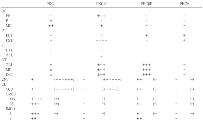
Table 1. Immunoreactivity of Classical Protein Kinase (PKC) Isoforms in Rat Kidney
PKCα PKC Iβ PKC IIβ PKCγ
RC PE P MC PT
PCT PST IT
DTL ATL DT
TAL MD DCT CNT CD
CCD OMCD
OS IS IMCD
i t
+±
++
+– ––
±±
+ ±(++~+++)
+ (++~+++)
+~++ ( )±
++~ ( )±
+++ ( )–
++
±~+–
+
+~++–
++
_
±~+±~+
– ±~+(++~+++)
– (+~+++)
– ( )–
– ( )–
– ( )–
–
–– –
+– ––
++++++
+++++( )–
++ ( )–
+ ( )–
+ ( )–
+ ( )–
++
–– –
++ ––
–– – – ( )–
– ( )– – ( )– – ( )– – ( )– –
Abbreviations : RC, renal corpuscle; PE, parietal epithelium; P, podocytes; MC, mesangial cells; PT, proximal tubules; PCT, proximal convoluted tubules; PST, proximal straight tubules; IT, intermediate tubules; DTL, descending thin limb; ATL, ascending thin limb; DT, distal tubules; TAL, thick ascending limb; MD, macular densa; DCT, distal convoluted tubules;
CNT, connecting tubules; CD, collecting ducts; CCD, cortical CD; OMCD, outer medullary CD; OS, outer stripe of the OMCD; IS, inner stripe of the OMCD; IMCD, inner medullary CD; i, initial part of the IMCD; t, terminal part of the IMCD.
Symbols designate not detectable ( ), faint ( ), weak ( ), moderate (– + + ++), and high (+++) levels of immunoreactivity.
Values in parentheses represent the levels of immunore activity in the intercalated cells.
cells were stained weakly at the basolateral plasma membrane. In the medullary collecting ducts, the inter- calated cells were PKC negative and the principal cellsα
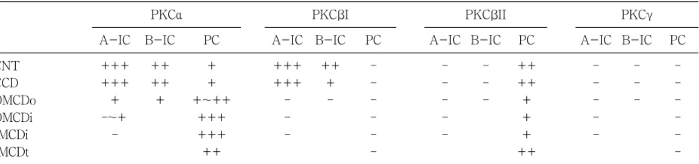
were PKCα positive at the basolateral plasma mem- brane. The inner stripe of the outer medulla and the initial part of the inner medulla showed strong immu- Table 2. Immunoreactivity of Conventional Proten Kinase C (PKC) Isoforms in the Intercalated Cells of Rat Kidney
PKCα PKC Iβ PKC IIβ PKCγ
A-IC B-IC PC A-IC B-IC PC A-IC B-IC PC A-IC B-IC PC
CNT CCD OMCDo OMCDi IMCDi IMCDt
++++++
–~++ –
++++
+
++
+~+++++
+++++
++++++
–– –
+++ –
–– –– ––
–– –– –
–– –
++++
++
+++
–– –– –
–– –
–– –– –– A-IC, type A intercalated cells; B-IC, type B intercalated cells; PC, principal cells; CNT, connecting tubules; CCD, cortical collecting duct; OMCDo, outer stripe part of outer medullary collecting duct; OMCDi; inner stripe part of OMCD;
IMCDi, initial part of the inner medullary collecting duct; IMCDt, terminal part of the IMCD. Symbols designate not detectable ( ), faint ( ), weak ( ), moderate (– + + ++), and high (++ ) levels of immunoreactivity.+
Fig. 1. Differential interference contrast (DIC) micrographs of wax sections illustrating immunostaining for protein kinase Cα PKC )( α in the cortical labyrinth (A), medullary ray (B), initial part of the inner medulla (C), and the terminal part of the inner medulla (D) of adult rat kidneys. PKC immuα - nostaining is observed in the cytoplasm of mesangial cells (arrows) and the parietal epithelium (arrowhead) of renal corpuscles, intercalated cells (open arrows) of the connecting tubule (CNT) and cortical collecting duct (CCD), and on the basolateral plasma membrane of the CNT cells and the principal cells of the collecting duct. Note the PKC -negative intercalatedα cells (open arrowheads) in the initial part of the inner medullary collecting duct (IMCDi). IMCDt, terminal part of the IMCD. Magnifications: A-D,
×400.
noreactivity. In the renal corpuscle, mesangial cells showed moderate immunoreactivity, and we observed weak immunoreactivity in parietal epithelial cells and podocytes. In the proximal tubule, the convoluted part showed weak immunoreactivity on the microvilli and
faint immunoreactivity was observed in the straight portion. The cytoplasm of distal tubule cells showed faint immunoreactivity. We did not observe any PKCα immunoreactivity in the descending or ascending limbs of the Loop of Henle (Fig. 1; Table 1, 2).
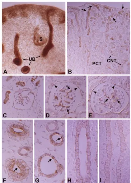
Fig. 2. DIC micrographs of wax sections illustrating immunostaining for PKC in 16- (A), 18- (C & F), and 20-day-old (G) fetal kidneys,α and 3- (D & H) and 7-day-old (B, E & I) pups. protein kinase Cα (PKC )α appeared in the ureteric buds (UB) at 16 days of gestation (A) and in the proximal and distal anlage (stars) at 18 days of gestation (C). B. Note that the PKCa-positive tubular profiles (arrows), which are newly formed proximal and distal tubules, are located only in the subcapsular region in 7-day-old pups, whereas PKCα immunoreacti- vity is decreased in the mature tubules such as the proximal convoluted tubule (PCT) and connecting tubule (CNT) cells located in the inner cortex. C-E, Note the PKCα immunoreactivity in the mesangial cells (arrows) and parietal epithelium (arrowheads) of the developing renal corpuscle. F-G. Immunoreactivity for PKCα in the basolateral plasma membrane of inner medullary collecting duct (IMCD) cells gradually decreased during development. Note the disappearance of apical PKC expression in the intercalated cells (arrows) of the medullary collecting duct (MCD) after birth. Magnifications: A,×200; B,×200; C-I,×528.
In the developing kidney, PKCα appeared in the ureteric bud at 16 days of gestation, but there was no staining of the renal vesicle and S-shaped body (Fig.
2A). The PKCα immunoreactivity of the collecting tubule gradually decreased during development and showed a mature pattern from 14 days after birth (Fig.
2F-I). PKC appeared strongly in the mesangial andα parietal cells of the developing renal corpuscle in stage III, proximal anlage and distal anlage of the 18- day-old pups, whereas immunoreactivity for PKCα gradually decreased in mature proximal convoluted and distal convoluted tubules (Fig. 2A-E). In the intercalated cells, immunoreactivity was shown in the connecting and
collecting tubules of 18-day-old pups.
3. PKCβI
In the adult kidney, there was strong positive PKC Iβ staining in the connecting segment and intercalated cells of the cortical collecting duct. Similar to PKC , type Aα intercalated cells were positive in the supranuclear area and type B cells were positive throughout the entire cytoplasm. PKC I staining was negative in principalβ cells. In the proximal tubule, the convoluted part was negative and the straight portion was moderately positive. In the renal corpuscle, the mesangial cells were weakly positive, parietal cells faintly positive, and the Fig. 3. DIC micrographs of wax sections illustrating protein kinase
C β (PKC II β ) immunostaining in the cortical labyrinth (A), medullary rays (B), outer stripe of the outer medulla (C), inner stripe of the outer medulla (D), and initial part of the inner medulla (E) of the adult rat kidney. PKC I immunoreactivity is strong in theβ intercalated cells (open arrows) of the connecting tubules (CNT) and cortical collecting ducts (CCD), moderate in the proximal straight tubules (PST), and weak in the mesangial cells (arrows) and parietal epithelium of the renal corpuscle. In the inner medulla, PKC I immuβ nostaining is observed only in the descending thin limb (stars) of the Loop of Henle. There is no immunoreactivity in the CNT cells, principal cells of the CCD, outer (OMCDo) and inner stripe of the outer medullary collecting duct (OMCDi), and initial inner medullary collecting duct (IMCDi). Note that there is no im- munoreactivity in the intercalated cells (open arrowheads) of the OMCD and IMCDi. Magnifications: A-E, ×400.
podocytes negative. The thick ascending limb of the Loop of Henle and the distal convoluted tubule were also negative. We did not observe immunoreactivity in the outer and inner medullary collecting tubules. However, we did detect moderately positive staining in the inner medulla, at the apical plasma membrane of the descending thin limb (Fig. 3; Table 1, 2).
In the developing kidney, PKC I immunoreactivityβ appeared from 16 days of gestation and was strongly positive in the ureteric bud (Fig. 4A). The immuno- reactivity of the collecting tubule was strong in the fetus, but decreased markedly after birth, and the principal
cells were negative 3 days after birth (Fig. 4E-G). In the renal corpuscle, mesangial cells, parietal cells, and the proximal anlage were strongly positive in 18-day-old pups (Fig. 4B, C). The renal vesicles and S-shaped bodies were negative (Fig. 4A). During development, the straight portion of the proximal anlage remained moderately immunopositive and the immunoreactivity in the convoluted portion disappeared after birth, being negative from 14 days after birth. In the distal nephron, the distal anlage was negative, with immunoreactivity only becoming positive from 21 days after birth. PKC Iβ immunoreactivity in the intercalated cells showed a Fig. 4. DIC micrographs of wax sections illustrating immu-
nostaining for protein kinase C I (PKC I) in 16- (A), 18- (B)β β and 20-day-old (E) fetal kidneys, and 3-(F) and 7-day-old (C, D & G) pups. PKC I appeared in the ureteric buds (UB) at 16 daysβ of gestation and in the proximal anlage (open arrows) at 18 days of gestation. PKCβI immunostaining appeared in the differ- entiating proximal tubules (open arrows) in the fetal stage and disappeared from the mature proximal tubule after birth. PKCβ I-positive proximal anlage are observed in the subcapsular region until 7 days after birth. Note the PKCβI immunostaining in the mes- angial cells (arrows) and parietal epithelium (arrowheads) of the developing renal corpuscle in D. Immunoreactivity for PKCβI in the medullary collecting duct (MCD) cells gradually disappeared after birth (E-G). IMCD, inner medullary collecting duct. Magnifica- tions: A, ×200; B, ×264; C & D-G,×528.
pattern similar to PKC in appearance and distribution.α
4. PKCβII
In the adult kidney, immunoreactivity for PKC II wasβ strongly positive in the thick ascending limb of the Loop of Henle, the macula densa, the distal convoluted tubule, and the basolateral membrane of the connecting tubule.
In the collecting tubule, the basolateral membrane of the principal cells was moderately positive, but no immunoreactivity was seen in the intercalated cells of the connecting and collecting tubules (Fig. 5B-D).
There was weak basolateral labeling in the proximal convoluted tubule. There was no immunoreactivity in the renal corpuscle or intermediate tubule (Fig. 5A;
Table 1, 2).
In the developing kidney, PKC II immunoreactivityβ appeared in the basolateral membrane of the distal anlage at 18 days of gestation. Immunoreactivity in- creased markedly in the distal tubule, including the thick ascending limb of the Loop of Henle, the distal convoluted tubule and the connecting tubule from 1 day after birth. PKC II immunoreactivity gradually deβ - Fig. 5. DIC micrographs of 1-mm-thick plastic sections illustrating immu-
nostaining for protein kinase C II (PKC II)β β in the cortex (A), medullary rays (B), the border between the outer and inner medulla (C), and the terminal part of the inner medulla (D) of adult rat kidney. Immunoreactivity for PKC II is well localized on the basolateral plasma membrane of distalβ convoluted tubule (DCT) cells, connecting tubule (CNT) cells, thick ascending limb (TAL) cells, and principal cells throughout the collecting duct. Note that there is no immunoreactivity for PKC II in both the typeβ A intercalated cells (arrow) and type B intercalated cells (arrowhead) in the cortical collecting duct (CCD). There is weak basolateral PKC IIβ labeling in the proximal convoluted tubule (PCT). The open arrow indicates a PKC II-negative interβ calated cell in the CNT. ATL, ascending thin limb;
G, glomerulus; OMCDi, inner stripe of the outer medullary collecting duct;
IMCDi, initial part of the inner medullary collecting duct; IMCDt, terminal part of the IMCD. Magnifications: A, ×368; B, ×640; C & D, ×320.
creased from 7 days after birth and had a similar pattern to the adult rat from 21 days. The intercalated cells in the connecting and collecting tubules were negative for PKC βII immunostaining. The principal cells were negative during the initial stages of development, but immunoreactivity gradually increased after birth and showed a similar pattern to adult rats from 14 days after birth (Fig. 6).
5. PKCγ
There was very weak immunoreactivity for PKC onlyγ in the proximal tubule, whereas no immunoreactivity in other uriniferous tubules (Fig. 7; Table 1, 2). In the developing kidney, PKC immunoreactivity was strong inγ the proximal tubule. Immunoreactivity appeared in the proximal anlage at 18 days of gestation. There was strong positive staining in the entire proximal tubule at 1, 3, and Fig. 6. DIC micrographs of wax sections illustrating immunostaining for protein
kinase C II (PKC II) in 18-day-old fetuses (A), and 1- (B), 3- (C), and 7-β β day-old (D) pups. A. PKCβII immunoreactivity appeared in the distal tubules (asterisks) at 18 days of gestation. B-C. Note a marked increase in PKCβII immunostaining in the distal tubules (asterisks) after birth. Insets demonstrat- ing PKCβII-negative medullary collecting ducts (MCD) from an 18-day-old fetus (A) and a 1-day-old pup (B). In the thick ascending limb (TAL), PKCβII immunoreactivity appeared around the time of birth. P, proximal tubule; S, S-shaped body; III, stage III renal corpuscle; V, renal vesicle. Magnifications:
A-D,×200; insets, 528.×
20 days after birth. Subsequently, immunoreactivity decreased and had a similar pattern to adult rats from day 21 after birth (Fig. 8).
Discussion
PKC plays a central role in intracellular signal
transduction. The various PKC isoforms are expressed in the rat kidney with distinct and differential ex- pression patterns (Fig. 9). As a member of the cPKC group, PKC expression was predominant in the adultα kidney. PKCβ was localized in the tubules. PKCγ is known to be detected in the central nervous system10). Our study, using immunohistochemistry and immuno- Fig. 8. Light micrographs of wax sections illustrating immunostaining for protein kinase
Cγ (PKCγ) in kidneys from 20-day-old fetuses (A & B), and 1- (C & D), 3- (E), and 21-day-old (F) pups. PKC immunoreactivity is highly expressed in the proximalγ tubules (PT) during development and markedly decreased in the PT from 21 days after birth. B and D are higer magnification area indicate by rectangular in A and C, re- spectively. DT, distal tubule; PCT, proximal convoluted tubule; PST, proximal straight tubule. Magnifications: A, C, E & F, ×83; B & D,×330.
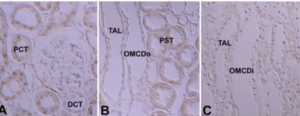
Fig. 7. Light micrographs of wax sections illustrating immunostaining for protein kinase Cγ(PKC ) in adult rat kidneys. There is very weak immunostaining for PKCγ γ only in the cytoplasm of proximal convoluted tubules (PCT) and proximal straight tubules (PST). Co, cortex; DCT, distal convoluted tubule; OMCDi, inner stripe part of outer medullary collecting duct; OMCDo, outer stripe part of OMCD; OSOM, outer stripe of outer medulla; ISOM, inner stripe of outer medulla; TAL, thick ascending limb. Magnifications: A-C, ×330.
blotting, demonstrates that the expression of PKC , Iα β and II, but not PKC , is evident in in the tubules of theβ γ rat kidney and PKC , I, II, andα β β γare expressed in the developing kidney.
Wetsel et al.11), La Porta et al.12), Dong et al.22), Pfaff et al.26), Saxena et al.27), Ostlund et al.14), Aristimuno and Good13), and Serlachius et al.15)have reported the ex- pression of PKCα in the kidney. In our study, the expression of PKC was detected in the cortex, outerα stripe of the outer medulla, inner stripe of the outer medulla and, using immunoblotting, in cytosolic and membrane fractions from the inner medulla. Using immunohistochemistry, Dong et al.28)and Fukuzaki et al.29)reported PKC expression in the renal corpuscle,α proximal straight tubule and collecting duct of the inner medulla of rat and human kidneys. However, our study demonstrates that PKC staining was diffusely posiα - tive, with the exception of the intermediate tubule.
Especially, strong positive staining was observed in the connecting tubule, intercalated cells of the cortical collecting tubule, mesangial cells of the renal corpuscle, outer and inner stripes of the outer medulla, and the
principal cells in the collecting duct of the inner me- dulla.
Wetsel et al.11), Ostlund et al.14)and Aristimuno and Good13)reported the expression of PKC I in the kidβ - ney, but not PKC II. La Porta et al.β 12)identified PKC inβ renal corpuscles using immunohistochemistry. Our study demonstrates the expression of PKC I and II. Onβ β immunoblotting, PKC I was faintly detected in theβ cortex and outer stripe of the outer medulla in the cytosolic fractions. Expression was generally weakly detected in the membrane fraction. Using immunohis- tochemistry, PKC I immunoreactivity was detected inβ the connecting segment, cortical collecting tubules, proximal straight tubules, mesangial cells of the renal corpuscle and the parietal epithelium. The connecting tubules and intercalated cells of the cortical collecting tubules were strongly positive. The apical plasma membrane of the descending thin limb of Henle was positive. These results were consistent with the im- munoblotting findings.
A PKC II band was observed in the membraneβ fractions. On immunohistochemistry, PKC II was exβ - Fig. 9. Changes in immunoreactivity of classical protein kinase C
isoforms in uriniferous tubules during renal development in the rat. RC, renal corpuscle; PT, proximal tubule; DT, distal tubule; CD, collecting duct.
pressed in the proximal convoluted tubules, distal con- voluted tubules, and the basolateral plasma membrane of the connecting and collecting tubules. The distal convoluted tubules, including the thick ascending limb of the Loop of Henle, and the connecting tubules were strongly positive. The intercalated cells in the distal nephron showed distinct and different expression patterns for PKC isoenzymes. In the connecting seg- ment and cortical collecting tubules, the intercalated A cells play a role in H secretion and the intercalated B cells are involved in HCO3 secretion. In the medullary collecting tubules, intercalated cells play a role in H secretion30). Our study shows that the type A inter- calated cells in the connecting and cortical collecting duc tare strongly positive for PKCα and β in their supranuclear cytoplasm. Type B intercalated cells were moderately positive in their cytoplasm and basolateral plasma membranes. Those findings were consistent with the results of mouse kidneys31)and the location of the H -ATPase32), so we suggest that PKC and I mayα β contribute to secretion of protons. However, PKC , andα
I were not expressed in the inter
β calated cells in the
medullary collecting ducts, so we speculate that different control mechanisms exist between intercalated cells in the connecting segment and cortical and medullary collecting tubules.
Several studies have reported that PKCγ is not detected in the adult rat kidney11, 13, 14, 22)
. Recently, studies have shown that PKC plays a role in growth and differentiation during development21-24). Serlachius et al.15)suggested that there is distinctand differential ex- pression and distribution of PKC isoenzymes depend- ing on embryonal kidney development. We also ob- served distinctand differential expression and distri- bution of PKC isoenzymes depending on kidney de- velopment. PKC ,α βI, and βII were expressed and, interestingly, PKCγ was detected temporarily in the developing kidney. Positive staining for PKC andα βI appeared in the ureteric bud at 16 days of gestation and was strongly positive before birth, and then gradually decreased after birth. Therefore, we suggest that PKCα
andβI play a role in differentiation of the collecting tubule. PKC expression was positive in the proximalα and distal anlage of pups up to 7 days of age. Therefore, we believe that PKCα expression is correlated with differentiation of the proximal and distal tubules.
Moreover, PKC I expression is correlated with differβ - entiation of the proximal tubule because it was tem- porarily expressed in the proximal anlage in early stages of development. PKC II appeared in the basoβ lateral membrane of the distal and connecting tubules at 18 days of gestation, and gradually increased during development. Therefore, we suggest that PKC II exβ - pression is not correlated with growth and differentia- tion. PKC immunoreactivity appeared and was highlyγ expressed in the proximal anlage of the 18-day-old fetus, then decreased markedly, and disappeared soon after birth. Therefore, PKC appears to be correlatedγ with differentiation of the proximal tubule.
In summary, our study demonstrates that the clas- sical PKC isoforms, PKC , I and II, but not PKC , areα β β γ expressed in the tubules of adult rat kidneys, and PKC ,α
I, II, and g are expressed in the de
β β veloping kidney, and
there are distinct and differential expression patterns for the isoforms according to location and stage of development.
Acknowledgements
This work was supported by the Korea Science and Engineering Foundation (KOSEF) through the MRC for Cell Death Disease Research Center at the Catholic University of Korea (R13-2002-005-01001-0).
References
1) Nishizuka Y: Studies and perspectives of protein kinase C.Science233:305-312, 1986
2) Nishizuka Y: Intracellular signaling by hydrolysis of phospholipids and activation of protein kinase C.Science 258:607-614, 1992
3) Nishizuka Y: Protein kinase C and lipid signaling for sustained cellular responses. FASEB J 9:484-496,
1995
4) Bertorello AM, Aperia A, Walaas SI, Nairn AC, Greengard P: Phosphorylation of the catalytic subunit of Na+, K(+)-ATPase inhibits the activity of the enzyme.Proc Natl Acad Sci U S A88:11359-11362, 1991
5) Nowicki S, Kruse MS, Brismar H, Aperia A: Do- pamine-induced translocation of protein kinase C isoforms visualized in renal epithelial cells.Am J Physiol Cell Physiol 279:C1812-C1818, 2000
6) Middleton JP, Khan WA, Collinsworth G, Hannun YA, Medford RM: Heterogeneity of protein kinase C-mediated rapid regulation of Na/K-ATPase in kidney epithelial cells.J Biol Chem 268:15958-15964, 1993 7) Horie S, Moe O, Miller RT, Alpern RJ: Long-term activation of protein kinase c causes chronic Na/H antiporter stimulation in cultured proximal tubule cells.
J Clin Invest89:365-372, 1992
8) Ruiz OS, Arruda JA: Regulation of the renal Na- HCO3
cotransporter by cAMP and Ca-dependent protein kinases.Am J Physiol 262:F560-F565, 1992 9) Kosaka Y, Ogita K, Ase K, Nomura H, Kikkawa U,
Nishizuka Y: The heterogeneity of protein kinase C in various rat tissues. Biochem Biophys Res Commun 151:973-981, 1988
10) Ono Y, Fujii T, Ogita K, Kikkawa U, Igarashi K, Nishizuka Y: The structure, expression, and properties of additional members of the protein kinase C family.
J Biol Chem263:6927-6932, 1988
11) Wetsel WC, Khan WA, Merchenthaler I, Rivera H, Halpern AE, Phung HM, Negro-Vilar A, Hannun YA:
Tissue and cellular distribution of the extended family of protein kinase C isoenzymes. J Cell Biol 117:
121-133, 1992
12) La Porta CA, Comolli R: Biochemical and immunolo- gical characterization of calcium-dependent and -in- dependent PKC isoenzymes in renal ischemia.Biochem Biophys Res Commun191:1124-1130, 1993 13) AristimuAo PC, Good DW: PKC isoforms in rat
medullary thick ascending limb: selective activation of the delta-isoform by PGE2. Am J Physiol 272:
F624-F631, 1997
14) Ostlund E, Mendez CF, Jacobsson G, Fryckstedt J, Meister B, Aperia A: Expression of protein kinase C isoforms in renal tissue.Kidney Int47:766-773, 1995 15) Serlachius E, Svennilson J, Schalling M, Aperia A:
Protein kinase C in the developing kidney: isoform expression and effects of ceramide and PKC inhibitors.
Kidney Int52:901-910, 1997
16) Redling S, Pfaff IL, Leitges M, Vallon V: Immu- nolocalization of protein kinase C isoenzymes alpha, beta I, beta II, delta, and epsilon in mouse kidney.Am J Physiol Renal Physiol287:F289-F298, 2004 17) Hashimoto T, Ase K, Sawamura S, Kikkawa U, Saito
N, Tanaka C, Nishizuka Y: Postnatal development of a
brain-specific subspecies of protein kinase C in rat.J Neurosci8:1678-1683, 1988
18) Hirata M, Saito N, Kono M, Tanaka C: Differential expression of the beta I- and beta II-PKC subspecies in the postnatal developing rat brain; an immu- nocytochemical study. Brain Res Dev Brain Res 62:
229-238, 1991
19) PucAat M, Hilal-Dandan R, Strulovici B, Brunton LL, Brown JH: Differential regulation of protein kinase C isoforms in isolated neonatal and adult rat cardio- myocytes. J Biol Chem269:16938-16944, 1994 20) Rybin VO, Steinberg SF: Protein kinase C isoform
expression and regulation in the developing rat heart.
Circ Res74:299-309, 1994
21) Housey GM, Johnson MD, Hsiao WL, O'Brian CA, Murphy JP, Kirschmeier P, Weinstein IB: Overpro- duction of protein kinase C causes disordered growth control in rat fibroblasts.Cell52:343-354, 1988 22) Dong L, Stevens JL, Fabbro D, Jaken S: Regulation of
protein kinase C isozymes in kidney regeneration.
Cancer Res53:4542-4549, 1993
23) Mischak H, Goodnight JA, Kolch W, Martiny-Baron G, Schaechtle C, Kazanietz MG, Blumberg PM, Pierce JH, Mushinski JF: Overexpression of protein kinase C-delta and -epsilon in NIH 3T3 cells induces opposite effects on growth, morphology, anchorage dependence, and tumorigenicity.J Biol Chem268: 6090-6096, 1993 24) Berra E, Diaz-Meco MT, Dominguez I, Municio MM,
Sanz L, Lozano J, Chapkin RS, Moscat J: Protein kinase C zeta isoform is critical for mitogenic signal trans- duction. Cell74:555-563, 1993
25) Szallasi Z, Kosa K, Smith CB, Dlugosz AA, Williams EK, Yuspa SH, Blumberg PM: Differential regulation by anti-tumor-promoting 12-deoxyphorbol-13-pheny- lacetate reveals distinct roles of the classical and novel protein kinase C isozymes in biological responses of primary mouse keratinocytes.Mol Pharmacol47:258- 265, 1995
26) Pfaff IL, Wagner HJ, Vallon V: Immunolocalization of protein kinase C isoenzymes alpha, beta1 and betaII in rat kidney. J Am Soc Nephrol10:1861-1873, 1999 27) Saxena R, Saksa BA, Hawkins KS, Ganz MB: Protein
kinase C beta I and beta II are differentially expressed in the developing glomerulus. FASEB J 8:646-653, 1994
28) Dong LQ, Stevens JL, Jaken S: Biochemical and im- munological characterization of renal protein kinase C.
Am J Physiol261:F679-687, 1991
29) Fukuzaki A, Kaneto H, Ikeda S, Orikasa S: Charac- terization of protein kinase C expression in human kidney.Tohoku J Exp Med178:263-269, 1996 30) Madsen KM, Tisher CC: Structural-functional re-
lationships along the distal nephron. Am J Physiol 250:F1-F15, 1986
31) Kim WY, Jung JH, Park EY, Yang CW, Kim H, Nielsen
S, Madsen KM, Kim J: Expression of protein kinase C isoenzymes alpha, betaI, and delta in subtypes of intercalated cells of mouse kidney.Am J Physiol Renal Physiol291:F1052-F1060, 2006
32) Madsen KM, Verlander JW, Kim J, Tisher CC:
Morphological adaptation of the collecting duct to acid- base disturbances.Kidney Int40(Suppl 33): S57-S63, 1991