Evaluation of dental panoramic radiographic findings in edentulous jaws: A retrospective study of 743 patients “Radiographic features in edentulous jaws”
Taha Emre Kose1, Nihat Demirtas2*, Hulya Cakir Karabas1, Ilknur Ozcan1
1Department of Oral and Maxillofacial Radiology, Faculty of Dentistry, Istanbul University, Istanbul, Turkey
2Department of Oral and Maxillofacial Surgery, Faculty of Dentistry, Bezmialem Vakif University, Istanbul, Turkey
PURPOSE. The aim of this study was to determine the frequency of significant panoramic radiographic findings and eventual treatment requirements before conventional or implant supported prosthetic treatment in asymptomatic edentulous patients. MATERIALS AND METHODS. A total of 743 asymptomatic edentulous patients were retrospectively evaluated using a digital panoramic system. We analyzed the radiographic findings, including impacted teeth, retained root fragments, foreign bodies, severe atrophy of the posterior maxillary alveolar bone, mucous retention cysts, soft tissue calcifications and radiopaque–radiolucent conditions.
RESULTS. Four-hundred-eighty-seven (65.6%) patients had no radiographic finding. A total of 331 radiographic findings were detected in 256 (34%) patients. In 52.9% (n=175) of these conditions, surgical treatment was required before application of implant-supported fixed prosthesis. However, before application of conventional removable prosthesis surgical treatment was required for 6% (n=20) of these conditions. CONCLUSION. The edentulous patients who will have implant placement for implant-supported fixed prosthesis can frequently require additional surgical procedures to eliminate pathological conditions. [J Adv Prosthodont 2015;7:380-5]
KEY WORDS: Edentulous patient; Panoramic radiography; Treatment planning; Diagnosis
INTRODUCTION
Panoramic radiography is a cost-effective, low-dose method used in dental radiology for evaluating oral health status in routine dental practice.1 This technique allows examination of the maxillary and mandibular arches and their support- ing structures on a single image.2,3 The advantages of pan- oramic radiography are time-saving, broad anatomic cover-
age, and high patient acceptability.3,4 Abnormalities such as root fragments, impacted teeth, neoplasms, and foreign bodies are often overlooked when they do not cause symp- toms or clinical signs.5
Panoramic radiography is often used in routine exami- nations of edentulous jaws to detect asymptomatic condi- tions such as root fragments, retained teeth, radiolucent lesions, and foreign bodies.6,7 Thus it is a valuable diagnos- tic tool in prosthetic treatment planning. In addition, they provide the clinician with information about the sinus floor position in edentulous regions for implant placement.
Several studies have been carried out including the occur- rence rate of these asymptomatic pathologies.2,4,6,7 However, only a few studies have documented the rate of these radio- graphic findings requiring treatment.4-7 Consequently, the aim of this study was to report the frequency of significant radiographic findings, to discuss utility of panoramic radio- graphs and to obtain the rate of the conditions which treat- ment is necessary before conventional or implant support- ed prosthetic rehabilitation in edentulous patients.
Corresponding author:
Nihat Demirtas
Department of Oral and Maxillofacial Surgery, Faculty of Dentistry, Bezmialem Vakif University, Fatih, Istanbul 34093, Turkey Tel. 90 212 453 1700: e-mail, [email protected]
Received May 29, 2015 / Last Revision September 21, 2015 / Accepted September 23, 2015
© 2015 The Korean Academy of Prosthodontics
This is an Open Access article distributed under the terms of the Creative Commons Attribution Non-Commercial License (http://creativecommons.
org/licenses/by-nc/3.0) which permits unrestricted non-commercial use, distribution, and reproduction in any medium, provided the original work is properly cited.
MATERIALS AND METHODS
This retrospective study investigated 743 patients who applied to Istanbul University Department of Dentomaxi- llofacial Radiology between 2009 and 2011. All of the patients were edentulous in both jaws and considered for dentures. A retrospective analysis was carried out of using panoramic radiographs taken either due to patient com- plaints or prior to prosthetic denture treatment, using a panoramic machine (Kodak 8000 Digital Panoramic Machine, Carestream Health, Inc., Rochester, NY, USA) with 60 - 85 kVp and 10 mA, with total filtration of 2.5 mm aluminum.
Clinically, all of the patients were asymptomatic. The radiographs were evaluated by four oral radiology special- ists and one oral surgery specialist for impacted teeth, retained root fragments, radiolucencies, radiopacities, for- eign bodies, proximity of the maxillary sinus to the crest of the residual alveolar ridge, soft tissue calcifications, and mucous retention cysts.
The maxilla and mandible were divided into three areas- right and left posterior (includes bilateral premolar and molar teeth regions) and anterior (includes incisors and canines region)-for evaluation of root fragments and radio- lucent–radiopaque areas. The maxilla was divided into two areas, left and right, for evaluation of mucous retention cysts and proximity of the maxillary sinus to the crest of the residual alveolar ridge. The patients whose subantral residual bone height is 1 - 2 mm on panoramic radiography (precise indication for lateral maxillary sinus lift procedures) were included in the study. Soft tissue calcifications (STC) were divided into three parts according to their location.
The area number 1 (STC1) indicates possible tonsilloliths, parotid calcifications, possible tonsilloliths and parotid cal- cifications; the area number 2 (STC2) indicates possible submandibular calcifications and lymph node calcifications;
and area number 3 (STC3) indicates possible carotid calcifi- cations.
All of these pathologic entities were included. Then, two different options of prosthetic rehabilitation were planned for each subject including conventional removable prosthesis and implant supported fixed prosthesis. The patients who required surgical treatment for each treatment modality were determined based on radiographic findings.
Exclusion criteria were poor quality radiographs and dis- agreement between evaluators.
RESULTS
Out of 743 patients, 428 (57.6%) were female and 315 (42.4%) were male. The mean age of the patients was 59.42; minimum age was 16 and maximum age was 88. A total of 331 significant radiographic findings were detected in 256 patients. Among these 256 patients, 125 (49%) were female and 131 (51%) were male. The frequency of radio- graphic findings and the findings which required treatment before conventional or implant supported prosthetic treat- ment are summarized in Table 1.
Seventy-four patients had proximity of the maxillary sinus to the crest of the residual alveolar ridge. Soft tissue calcifications were detected in 64 (6%) patients. The fre- quency of the subjects with posterior atrophic maxilla, soft tissue calcifications and mucus retention cysts are shown in Table 2.
Table 1. Frequency and percentage of radiographic findings among patients and number of these radiographic findings that require treatment for conventional removable prosthesis and implant supported prosthesis
Radiographic findings
Number of radiographic
finding (n)
Frequency
%
Treatment is required before
implant placement (n)
%
Treatment is required before
removable prosthesis (n)
%
Significant radiographic finding(s) number
Patient (n) %
Impacted tooth 36 4.8 29 80.6 6 16.7 0 487 65.6
Retained root
fragments 71 9.5 52 73.2 12 16.9 1 153 20.6
Radiolucencies 12 1.6 1 8.3 1 8.3 2 80 10.7
Radiopacities 11 1.5 1 9.0 1 9.0 3 19 2.6
Foreign bodies 16 2.2 3 18.8 0 0.0 4 4 0.6
Posterior atrophic
maxilla 74 10.0 74 100.0 0 0.0 Total 743 100.0
Soft tissue
calcifications 64 6.0 0 0.0 0 0.0 No radiographic
finding 487 65.6
Mucous retention
cysts 47 6.3 15 31.9 0 0.0 At least 1
radiographic finding 256 34.4
Total 331 - 175 52.9 20 6.0
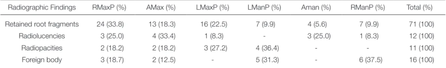
Seventy-one root fragments were detected in 60 (8%) of the 743 patients. Of the 60 patients who had root frag- ments, 50 (83%) had one root fragment, nine (15%) had two, and only one (2%) patient had three root fragments. A total of 12 radiolucencies were found in the study. One were (8%) residual cyst, 11 (92%) were idiopathic bone cav- ity. All of the radiolucent areas had well-defined borders. A total of 11 radiopacities were found in the study. Nine (82%) radiopacities had well-defined borders and two (18%) had diffuse borders. All of them were diagnosed as osteosclerosis. A total of 16 foreign bodies were found among all subjects. Three (19%) were extruded root canal materials, 13 (81%) were other foreign bodies including eleven (69%) retained amalgam fragments and two (12.5%) gunshot fragments. The distribution of the foreign bodies, root fragments and radiopaque–radiolucent conditions are shown in Table 3.
A total of 36 (5%) impacted teeth were found in 27 dif- ferent patients. Nineteen patients (70%) had only one impacted tooth, seven (26%) had two impacted teeth, and one (4%) had three impacted teeth. The distribution of the impacted teeth among regions is shown in Table 4.
DISCUSSION
One of the most important goals of panoramic imaging is to detect any underlying pathology of both maxilla and mandible before prosthetic treatment.8 Moreover, many
reports have concluded that due to the high percentage of significant radiographic findings, radiographic screening should be required in all edentulous patients.6,7 Similarly, our study showed that most of significant findings are easi- ly detected on the panoramic radiographs of edentulous jaws.
Table 2. Distribution of maxillary sinus findings
Radiographic findings Right side (%) Left side (%) Both sides (%) Total (%)
Posterior atrophic maxilla
(Maxillary sinus proximity to crest) 25 (33.8) 17 (23.0) 32 (43.2) 74 (100)
STC1 16 (69.6) 5 (21.7) 2 (8.7) 23 (100)
STC2 18 (60.0) 6 (20.0) 6 (20.0) 30 (100)
STC3 8 (72.7) 3 (27.3) - 11 (100)
Mucous retention cyst 13 (27.7) 19 (40.4) 15 (31.9) 47 (100)
STC: Soft Tissue Calcification; STC1: possible tonsilloliths, parotid calcifications; STC2: possible submandibular calcifications and lymph node calcifications; STC3:
possible carotid calcifications.
Table 3. Distribution of retained root fragments, radiolucencies, radiopacities
Radiographic Findings RMaxP (%) AMax (%) LMaxP (%) LManP (%) Aman (%) RManP (%) Total (%)
Retained root fragments 24 (33.8) 13 (18.3) 16 (22.5) 7 (9.9) 4 (5.6) 7 (9.9) 71 (100)
Radiolucencies 3 (25.0) 4 (33.4) 1 (8.3) - 3 (25.0) 1 (8.3) 12 (100)
Radiopacities 2 (18.2) 2 (18.2) 3 (27.2) 4 (36.4) - - 11 (100)
Foreign body 3 (18.7) 2 (12.5) - 5 (31.3) - 6 (37.5) 16 (100)
RMaxP: Right maxillary posterior area, AMax: Anterior maxillary area, LMaxP: Left maxillary posterior area, LManP: Left mandibular posterior area, Aman: Anterior mandibular area, RManP: Right mandibular posterior area.
Table 4. Distribution of impacted teeth Impacted teeth
Right maxillary area Left maxillary area
C: 8 C: 10
SP: 1 SP: 9
TM: 4 TM: -
T: 13 T: 19
Right mandibular area Left mandibular area
C: - C: -
SP: - SP: -
TM: 1 TM: 3
T: 1 T: 3
C: Canine, SP: Second Premolar, TM: Third Molar, T: Total number of impacted teeth
The most important limitation of this study is that the treatment planning was performed by using radiographic findings. In fact, both clinical and radiological correlation is very important to assess effective treatment planning and 3-D evaluation by using Cone-beam Computed Tomography (CBCT) can be necessary for specific conditions. Thus, it is reported that almost all of the findings on panoramic radiographs coincide with clinical findings.9 In the present study, our observations were in accordance with this hypothesis. Additionally, we did not achieve CBCT views due to comprehensive treatment planning for implant placement. Consequently, further studies can be designed by using 3D imaging methods. Moreover, several clinical conditions which require surgical treatment before applica- tion of conventional removable prosthesis in edentulous patients such as epulis fissuratum or alveolar ridge disrup- tion were excluded.
Bohay et al.10 reported 68.3% range of one or more sig- nificant radiographic findings in 375 edentulous patients. In addition, they determined 8.3% of these patients required treatment before treatment with removable dentures.
Similarly, Masood et al.4 suggested a few (3.8%) of the posi- tive radiographic findings required treatment before den- ture fabrication. Our results revealed an important part of these findings did not require surgical intervention before conventional removable prosthodontic treatment. On the other hand, a significant amount of the radiographic find- ings in edentulous patients require treatment before implant supported prosthetic treatment.
Lyman and Boucher11 reported only one impacted tooth which required extraction among 300 edentulous patients.
By this conclusion, they have not suggested routine pan- oramic examination for every edentulous patient to avoid cumulative effects of radiation exposure. Similar suggestions have been produced by Ansari12 in 1997. However, today’s implant supported prosthetic rehabilitation becomes the most preferred treatment option for edentulous patients.13 Hence, radiographic examination should be based on the concept that the edentulous patient is a candidate for implant placement.
Retained root fragments and impacted teeth are the most frequent significant radiographic findings in edentu- lous patients.4,7,12 Previous research has shown that most root fragments are localized in the molar region of the maxilla.4,7,14 In our study, retained root fragments represent- ed the second most frequent pathology. The majority of these root fragments were localized in the premolar-molar region of the maxilla. The reasons for this finding could be morphology and number of roots, as they were located posteriorly, where it is difficult to perform an operation. In addition, extraction of these roots poses several risks of complications, such as nerve injury (inferior alveolar, lin- gual, and mental nerves) and displacement of the roots into the maxillary sinus.15,16 In particular, dental surgery in older patients carries a high risk of these complications.
Impacted teeth are, of course, critically important in preoperative planning for dental prostheses and implants in
edentulous jaws, and they affect patients’ oral health and function.17 As such, patients with impacted teeth have a variety of complaints, such as carious lesions, dentigerous cysts, tooth eruption abnormalities, pain, and infections.
Stathopoulos et al.18 retrospectively investigated 7782 impacted third molars in 6182 patients and reported that the pathologic conditions related to these teeth were lower than 2.77%. Sumer et al.6 reported teeth impaction in 3.1%
of 676 edentulous patients. In our study, we found 36 impacted teeth, representing a frequency of 3.6%. This result may be related to elective procedures recommended for impacted teeth in edentulous patients by specialists.
Today, recent studies suggested an implant placement pro- tocol encroaching upon residual roots and impacted teeth.19,20 This unconventional method has been proposed to assess minimal invasive surgical procedures in implant dentistry. However, future research needs to be investigated for this procedure.
Panoramic radiographs have been used frequently for preoperative assessments of the maxillary sinus for implant placement. These assessments include the vertical dimen- sion of the alveolar crest to the maxillary sinus.21,22 In com- pletely edentulous patients, the upper alveolar ridge should be related to the floor of the maxillary sinus because of bone resorption. In these circumstances, panoramic radio- graphs simply allow an evaluation of this relation by using a lower effective dose.23 In this radiographic study, we deter- mined that 22.4% of all radiographic findings were in rela- tion of the floor of their maxillary sinuses with alveolar ridge. Therefore, open maxillary sinus augmentation was required before implant surgery for all subjects.
When maxillary sinuses are imaged, some maxillary sinus pathologies such as mucosal cysts can be detected with panoramic radiography. Sinus mucosal cysts were another frequent significant finding in our study, observed in 6.3% of all patients. As the prevalence of mucous cysts in radiographic studies has been reported as 2 - 13%, our finding was in accordance with the literature.6,24
It has been reported that a mucus retention cyst of the maxillary sinus is not a contraindication for sinus mem- brane elevation.25 Feng et al.26 retrospectively evaluated the survival rate of 21 endosseous implants placed into the ele- vated maxillary sinus area in the presence of mucus reten- tion cysts. They reported that all of the implants were func- tionally stable during the 27-months follow up period.
Nevertheless, other maxillary sinus pathologies (acute or chronic infections) accompany to positive radiological find- ings of maxillary sinuses should be evaluated carefully for implant surgery in posterior maxillary area.27
Carotid area calcifications can be detected in panoramic radiographies, and its prevalence has been reported as 3 - 5% in the general dental population. However, in a study conducted with a younger population (with a mean age of 32 - 35), the incidence was found to be very low, in a range of 0.4 - 0.8%.28 In previous studies, the frequency of radi- opaque findings which might be due to the inclusion of soft tissue calcifications was reported as 9.3 - 9.9%; thus,
our findings seem relatively low by comparison.6,7 In our study, calcifications were detected in the area considered as tonsillolith in 23 patients (3.1%), in the submandibular area and radiopaque findings considered as lymph node calcifi- cation in 30 patients (4%), and in the carotid area in 11 patients (1.5%). These additional panoramic findings did not affect the treatment planning of implant placement or prosthetic rehabilitation. Nevertheless, it seems that pan- oramic radiographs may include critical important findings in the head and neck region.
ConClusion
In conclusion, to achieve successful results in prosthetic dentistry, preprosthetic-presurgical phase of treatment planning should be made carefully. It is our opinion that due to the high frequency of significant radiographic find- ings, panoramic radiography should be analyzed, even in the absence of clinical symptoms. Within the limitation of this study, patients candidates for implant placement can more frequently require additional surgical procedures to eliminate pathological conditions of edentulous jaws.
ACknowledgements
We are grateful to Monica Malt for her efforts in the final preparation of this manuscript.
oRCid
Taha Emre Kose http://orcid.org/0000-0003-3601-0393 Nihat Demirtas http://orcid.org/0000-0001-9956-9077 RefeRenCes
1. Choi JW. Assessment of panoramic radiography as a national oral examination tool: review of the literature. Imaging Sci Dent 2011;41:1-6.
2. Awad EA, Al-Dharrab A. Panoramic radiographic examina- tion: a survey of 271 edentulous patients. Int J Prosthodont 2011;24:55-7.
3. White SC, Pharoah MJ. Oral radiology: principles and inter- pretation. 5th ed. St. Louis, MO: Mosby, 2004. p. 205-7.
4. Masood F, Robinson W, Beavers KS, Haney KL. Findings from panoramic radiographs of the edentulous population and review of the literature. Quintessence Int 2007;38:e298- 305.
5. Ezoddini Ardakani F, Navab Azam AR. Radiological findings in panoramic radiographs of Iranian edentulous patients.
Oral Radiology 2007;23:1-5.
6. Sumer AP, Sumer M, Güler AU, Biçer I. Panoramic radio- graphic examination of edentulous mouths. Quintessence Int 2007;38:e399-403.
7. Jindal SK, Sheikh S, Kulkarni S, Singla A. Significance of pre-treatment panoramic radiographic assessment of edentu- lous patients-a survey. Med Oral Patol Oral Cir Bucal 2011;
16:e600-6.
8. Nagarajan A, Perumalsamy R, Thyagarajan R, Namasivayam A. Diagnostic imaging for dental implant therapy. J Clin Imaging Sci 2014;4:4.
9. Moll MA, Seuthe M, von See C, Zapf A, Hornecker E, Mausberg RF, Ziebolz D. Comparison of clinical and dental panoramic findings: a practice-based crossover study. BMC Oral Health 2013;13:48.
10. Bohay RN, Stephens RG, Kogon SL. A study of the impact of screening or selective radiography on the treatment and postdelivery outcome for edentulous patients. Oral Surg Oral Med Oral Pathol Oral Radiol Endod 1998;86:353-9.
11. Lyman S, Boucher LJ. Radiographic examination of edentu- lous mouths. J Prosthet Dent 1990;64:180-2.
12. Ansari IH. Panoramic radiographic examination of edentu- lous jaws. Quintessence Int 1997;28:23-6.
13. Rentsch-Kollar A, Huber S, Mericske-Stern R. Mandibular implant overdentures followed for over 10 years: patient compliance and prosthetic maintenance. Int J Prosthodont 2010;23:91-8.
14. Edgerton M, Clark P. Location of abnormalities in panoram- ic radiographs of edentulous patients. Oral Surg Oral Med Oral Pathol 1991;71:106-9.
15. Arrigoni J, Lambrecht JT. Complications during and after third molar extraction. Schweiz Monatsschr Zahnmed 2004;
114:1271-86.
16. Hu YK, Yang C, Zhou Xu G, Wang Y, Abdelrehem A.
Retrieval of root fragment in maxillary sinus via anterolateral wall of the sinus to preserve alveolar bone. J Craniofac Surg 2015;26:e81-4.
17. Friedman JW. The prophylactic extraction of third molars: a public health hazard. Am J Public Health 2007;97:1554-9.
18. Stathopoulos P, Mezitis M, Kappatos C, Titsinides S, Stylogianni E. Cysts and tumors associated with impacted third molars: is prophylactic removal justified? J Oral Maxillofac Surg 2011;69:405-8.
19. Szmukler-Moncler S, Davarpanah M, Davarpanah K, Capelle- Ouadah N, Demurashvili G, Rajzbaum P. Unconventional Implant Placement Part III: Implant Placement Encroaching upon Residual Roots - A Report of Six Cases. Clin Implant Dent Relat Res 2014 Jul 17.
20. Mithridade D, Serge SM, Keyvan D, Nedjoua CO, Georgy D, Philippe R. Unconventional Implant Placement IV. Implant Placement through Impacted Teeth to Avoid Invasive Surgery. Long-term Results of 3 Cases. Open Dent J 2015;9:
15-20.
21. Kim YK, Park JY, Kim SG, Kim JS, Kim JD. Magnification rate of digital panoramic radiographs and its effectiveness for pre-operative assessment of dental implants. Dentomaxillofac Radiol 2011;40:76-83.
22. Kim YK, Park JY, Kim SG, Kim JS, Kim JD. Magnification rate of digital panoramic radiographs and its effectiveness for pre-operative assessment of dental implants. Dentomaxillofac Radiol 2011;40:76-83.
23. BouSerhal C, Jacobs R, Quirynen M, van Steenberghe D.
Imaging technique selection for the preoperative planning of oral implants: a review of the literature. Clin Implant Dent Relat Res 2002;4:156-72.
24. MacDonald-Jankowski DS. Mucosal antral cysts observed within a London inner-city population. Clin Radiol 1994;49:
195-8.
25. Mardinger O, Manor I, Mijiritsky E, Hirshberg A. Maxillary sinus augmentation in the presence of antral pseudocyst: a clinical approach. Oral Surg Oral Med Oral Pathol Oral Radiol Endod 2007;103:180-4.
26. Feng Y, Tang Y, Liu Y, Chen F, Li D. Maxillary sinus floor el- evation using the osteotome technique in the presence of an- tral pseudocysts: a retrospective study with an average fol- low-up of 27 months. Int J Oral Maxillofac Implants 2014;
29:408-13.
27. Alkan A, Celebi N, Baş B. Acute maxillary sinusitis associated with internal sinus lifting: report of a case. Eur J Dent 2008;
2:69-72.
28. Roldán-Chicano R, Oñate-Sánchez RE, López-Castaño F, Cabrerizo-Merino MC, Martínez-López F. Panoramic radio- graph as a method for detecting calcified atheroma plaques.
Review of literature. Med Oral Patol Oral Cir Bucal 2006;11:
E261-6.