Received on October 2, 2015. Revised on October 30, 2015. Accepted on November 5, 2015.
CC This is an open access article distributed under the terms of the Creative Commons Attribution Non-Commercial License (http://creativecommons.org/licenses/by-nc/4.0) which permits unrestricted non-commercial use, distribution, and reproduction in any me- dium, provided the original work is properly cited.
*Corresponding Author. YunJae Jung, Department of Microbiology, School of Medicine, Gachon University, 191 Hambakmoe-ro, Yeonsu-gu, Incheon 21936, Korea. Tel: 82-32-820-4753; Fax: 82-32-820-4744; E-mail: [email protected]
Abbreviations: DbcAMP, Dibutyryl cAMP
Comparative Analysis of Dibutyric cAMP and Butyric Acid on the Differentiation of Human Eosinophilic Leukemia EoL-1 Cells
YunJae Jung*
Department of Microbiology, School of Medicine, Gachon University, Incheon 21936, Korea
Purification of enough numbers of circulating eosinophils is difficult because eosinophils account for less than 5%
peripheral blood leukocytes. Human eosinophilic leukemia EoL-1 cells have been considered an in vitro source of eo- sinophils as they can differentiate into mature eosinophil- like cells when incubated with dibutyryl cAMP (dbcAMP) or butyric acid. In this study, the viability and phenotypic maturation of EoL-1 cells stimulated by either dbcAMP or butyric acid were comparatively analyzed. After treatment with 100 μM dbcAMP or 0.5 μM butyric acid, EoL-1 cells showed morphological signs of differentiation, al- though the number of nonviable EoL-1 cells was sig- nificantly increased following butyric acid treatment. Sti- mulation of EoL-1 cells with 0.5 μM butyric acid more ef- fectively induced the expression of mature eosinophil markers than stimulation with dbcAMP. These results sug- gest that treatment of EoL-1 cells with 0.5 μM butyric acid for limited duration could be an effective strategy for inducing their differentiation. Considering that expression of CCR3 was not sufficient in EoL-1 cells stimulated with 0.5 μM butyric acid, treatment of the chemically stimu- lated EoL-1 cells with cytokines, which primarily support eosinophil maturation, would help to obtain differentiated EoL-1 cells with greater functional maturity.
[Immune Network 2015;15(6):313-318]
Keywords: EoL-1 cells, DbcAMP, Butyric acid, In vitro differentiation, Eosinophils
INTRODUCTION
Eosinophils are multifunctional leukocytes that have been implicated in the pathogenesis of Th2-type inflammatory processes, including helminth infections and allergic disea- ses. The cytoplasm of mature eosinophils contains numer- ous secondary granules such as eosinophil peroxidase, eo- sinophil cationic protein, eosinophil-derived neurotoxin, and major basic protein, and the exocytotic release of these granule-derived cytotoxic proteins contributes to inflam- matory responses induced by eosinophil activation (1,2).
Eosinophils are produced in the bone marrow from pluri- potential stem cells, which differentiate into eosinophil progenitors marked by CD34+IL-5Rα+ expression (1).
Eosinophil lineage specification is determined by the inter- play of several transcription factors, including GATA-1 (a zinc finger family member), PU.1 (an ETS family mem- ber), and members of the CCAAT/enhancer-binding pro- tein (C/EBP) family (3-5). Following differentiation, per- missive proliferation and migration of eosinophils from the bone marrow to the circulation are regulated primarily by IL-5. However, eosinophils account for less than 5% pe-
phenotypic analysis of differentiated EoL-1 cells will con- tribute to suggesting optimal EoL-1 stimulating conditions compatible with experimental purpose. In this study, we found that stimulation with butyric acid was more effective than stimulation with dbcAMP for induction of EoL-1 cell differentiation. However, both butyric acid and dbcAMP were not sufficient for the expression of CCR3 in EoL-1 cells, and we propose subsequent cytokine treatment of chemically stimulated EoL-1 cells.
MATERIALS AND METHODS Cell culture
EoL-1 cells (DSMZ, Braunschweig, Germany) were main- tained in RPMI 1640 medium (Sigma, St. Louis, MO, USA) supplemented with 10% FBS (Gibco Laboratories, Grand Island, NY, USA) in 5% CO2 at 37oC. EoL-1 cells were induced to differentiate by the addition of dbcAMP (Sigma-Aldrich) or butyric acid (Sigma-Aldrich) for 9 days. The cell concentration was adjusted to 5×10⁵/ml ev- ery 3 days.
Morphological analysis
For morphological analysis, cultured EoL-1 cells were spun at 500 rpm for 5 min on glass slides (Cytospin 3, Shandon, Pittsburgh, PA, USA). The slides were air-dried, stained with Diff-Quik stain solution (Sysmex, Kobe, Japan) and observed using the CX41 microscope (Olym- pus, Tokyo, Japan).
Flow cytometry
Aliquots of EoL-1 cells, before and after stimulation with dbcAMP or butyric acid, were resuspended in FACS buf- fer (PBS containing 10% FBS, 10 mM EDTA, 20 mM
Real-time PCR
RNA from unstimulated and dbcAMP or butyric acid treat- ed EoL-1 cells was extracted using QIAzol lysis reagent (Qiagen, Hilden, Germany) and was column-purified with an RNeasy Mini Kit (Qiagen). The purified RNA (500 ng) was treated with DNase I (New England Biolabs, Ipswich, MA, USA), and cDNA was synthesized using the iScript cDNA synthesis kit (Bio-Rad, Hercules, CA, USA). Real- time PCR was performed using iQ SYBR Green Supermix (Bio-Rad). PCR was performed using a CFX96 Real-Time System (Bio-Rad). Sequences of primers are shown in Table S1.
Statistical analysis
The data are presented as the mean±s.e.m. All experiments were performed in triplicate. When necessary, a two-group comparison was performed using Student’s t-test. A p value
<0.05 was considered statistically significant.
RESULTS AND DISCUSSION
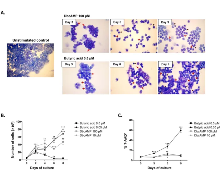
Treatment of EoL-1 cells with either 100 μM dbcAMP or 0.5 μM butyric acid for 6 to 9 days is known to induce eosinophilic maturation (9,12,13). EoL-1 cells represent undifferentiated promyelocytic eosinophils possessing a large nucleus with prominent nucleoli (Fig. 1A) (14). After treatment with dbcAMP or butyric acid, we observed mor- phological signs of EoL-1 differentiation, including nu- clear lobulation and increased proportion of cytoplasm to nucleus (Fig. 1A). However, compared to the group stimu- lated with 100 μM dbcAMP, EoL-1 cells stimulated with 0.5 μM butyric acid showed remarkable increase in apop- totic populations exhibiting cellular shrinkage and nucleus condensation (Fig. 1A). Consistent with the morphological
Figure 1. Effects of dbcAMP and butyric acid on the morphologic features and proliferation capacity of EoL-1 cells. (A) EoL-1 cells were incubated for the indicated periods in the absence or presence of 100 μM dbcAMP or 0.5 μM butyric acid. Cell number was adjusted to 5×10⁵/ml every 3 days. Diff-Quik staining of unstimulated EoL-1 cells and EoL-1 cells stimulated with dbcAMP or butyric acid. Arrows denote cells showing nuclear lobulation and arrow heads indicate cells showing shrinkage and chromatin condensation.
Original magnification, ×40. (B, C) EoL-1 cells were incubated for 8 or 9 days in medium containing indicated concentration of dbcAMP or butyric acid. The cells were then harvested and enumerated (B) and the viability of the cells was determined by flow cytometry analysis of 7-amino-actinomycin D (7-AAD) (C). All data are representative of two or more independent experiments. Data are mean±s.e.m.
values. *p<0.05, **p<0.01, and ***p<0.001 (Student’s t-test) vs. the control.
findings, stimulation with 0.5 μM butyric acid signifi- cantly inhibited the proliferation of EoL-1 cells after 2 days (Fig. 1B). The proliferation of EoL-1 cells treated with dbcAMP (100 μM and 10 μM) or 0.05 μM butyric acid increased in a time-dependent manner during 8 days of incubation (Fig. 1B). Additionally, compared to that in other groups, the number of 7-amino-actinomycin D-pos- itive nonviable cells remarkably increased in the group stimulated with 0.5 μM butyric acid (Fig. 1C). These data indicate that stimulation of EoL-1 cells with 0.5 μM buty- ric acid for prolonged period is unsuitable for EoL-1 cells
in vitro assay in terms of cell viability.
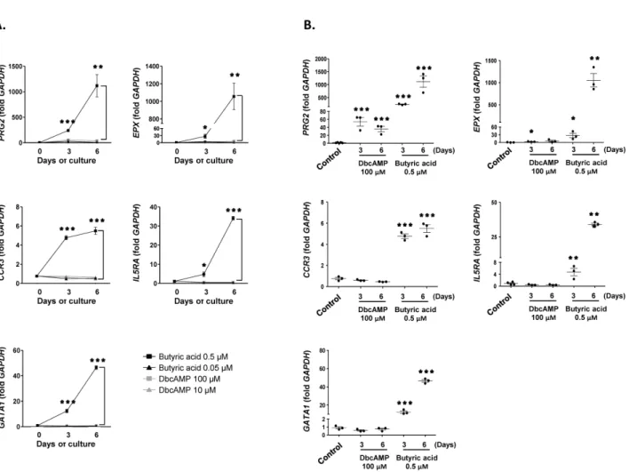
Next, we examined the expression of PRG2, EPX, CCR3, IL5RA, and GATA1, markers for mature eosino- phils, in EoL-1 cells stimulated with dbcAMP or butyric acid. As shown in Fig. 2A, 0.5 μM butyric acid effec- tively induced the expression of PRG2, EPX, CCR3, IL5RA, and GATA1 in EoL-1 cells than 100 μM dbcAMP in a time-dependent manner. Additionally, the effect of 100 μM dbcAMP stimulation was limited to the expres- sion of PRG2 and EPX, which encode cytoplasmic gran- ules of eosinophils (Fig. 2B). However, it could be plau-
Figure 2. Effects of dbcAMP and butyric acid on the differentiation of EoL-1 cells. (A) cDNA was prepared from total RNA obtained from undifferentiated EoL-1 cells (day 0) and EoL-1 cells stimulated with indicated concentrations dbcAMP or butyric acid for 3 or 6 days. mRNA expressions of PRG2, EPX, CCR3, Il5RA, and GATA1 were analyzed by real-time PCR. Data are mean±s.e.m. values. *p<
0.05, **p<0.01, and ***p<0.001 (Student’s t-test) of 0.5 μM butyric acid vs. 100 μM dbcAMP stimulation. (B) mRNA expressions of PRG2, EPX, CCR3, Il5RA, and GATA1 of undifferentiated EoL-1 cells (control) and EoL-1 cells stimulated with 100 μM dbcAMP or 0.5 μM butyric acid for 3 or 6 days. Data are mean±s.e.m. values. *p<0.05, **p<0.01, and ***p<0.001 (Student’s t-test) vs. the control.
sible that CCR3 levels could be low even in the 0.5 μM butyric acid-treated group, considering the relatively low fold increase of the transcript in these cells (Fig. 2).
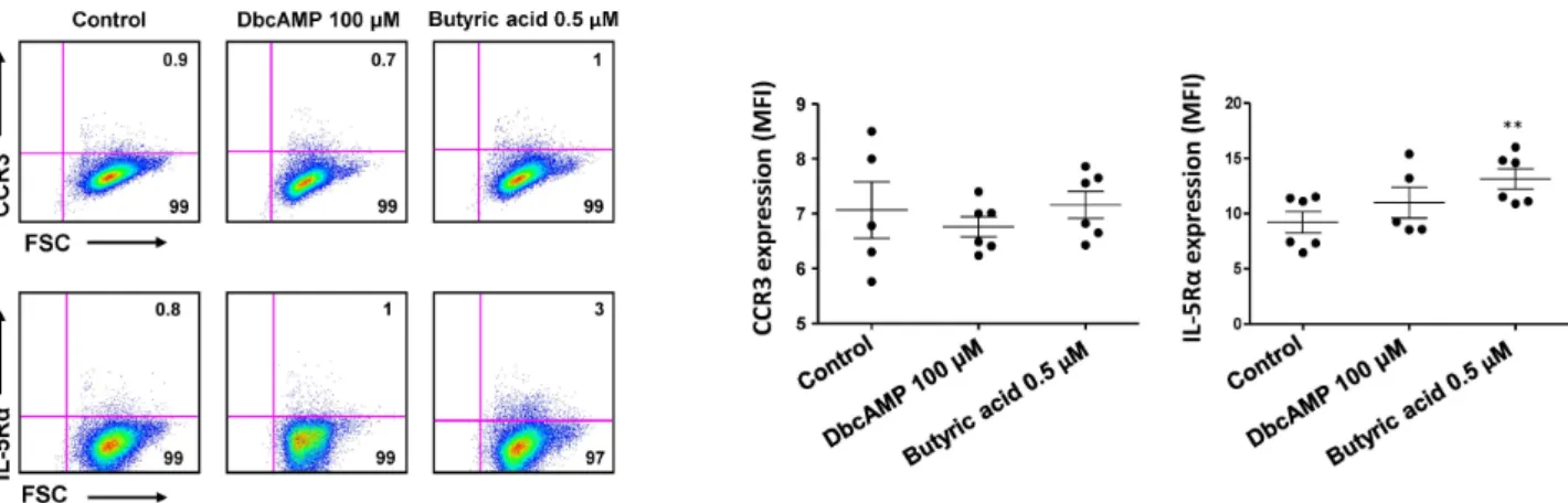
Consistent with this idea, mean fluorescence intensity (MFI) of CCR3 expression was not significantly higher in 0.5 μM butyric acid-treated EoL-1 cells than in un- stimulated control cells (Fig. 3). Technical problems re- lated to reagents used for flow cytometry analysis were ex- cluded by demonstrating relatively robust expression of CCR3 and IL-5Rα in eosinophils obtained from human peripheral blood (MFI=145± 5, MFI=309±11, respectively, Fig. S1). Collectively, these data indicate that compared
to dbcAMP treatment, treatment with 0.5 μM butyric acid was more effective in inducing phenotypic maturation of EoL-1 cells, and that the treatment duration should be less than 5 days to preserve the viability of the stimulated cells.
Eosinophil lineage-committed progenitors developed in the bone marrow are identified via surface expression of IL-5Rα and mature into eosinophil precursors containing cytoplasmic granules (15). Eosinophil precursors contain a granule-rich cytoplasm, and their permissive proliferation and differentiation into mature eosinophils is regulated by several cytokines, including IL-5, IL-3, and GM-CSF (16).
Recently it was reported that eosinophil lineage-committed
Figure 3. Effects of dbcAMP and butyric acid on the expression of CCR3 and IL-5Rα in EoL-1 cells. The expression of CCR3 or IL-5Rα in undifferentiated EoL-1 cells and EoL-1 cells stimulated with 100 μM dbcAMP or 0.5 μM butyric acid for 6 days was determined by flow cytometry. Flow cytometric expression of CCR3 or IL-5Rα in indicated cell groups was shown as mean fluorescence intensity (MFI). Data are representative of two or more independent experiments. Data are mean±s.e.m. values. **p<0.01 (Student’s t-test) vs. the control.
progenitors or eosinophil precursors in the mouse bone marrow do not express CCR3 (17). IL-5 primarily induces maturation of eosinophils and stimulates eosinophil migra- tion out of the bone marrow to the circulation mediated by eosinophils expressing CCR3. We have also reported the expression of CCR3 in EoL-1 cells treated with IL-3 and GM-CSF following stimulation with dbcAMP (6).
Therefore, we suggest that additional cytokine treatment would be needed to induce differentiation of EoL-1 cells into functionally mature phenotype following chemical stimulation.
ACKNOWLEDGEMENTS
This work was supported by the Basic Science Research Program through the National Research Foundation of Korea (NRF) funded by the Ministry of Education (2013- R1A1A2004820). The author thanks to Ye-Rang Chung (Gachon University, Korea) for technical assistance.
CONFLICTS OF INTEREST
The authors have no financial conflict of interest.
REFERENCES
1. Rothenberg, M. E., and S. P. Hogan. 2006. The eosinophil. Annu.
Rev. Immunol. 24: 147-174.
2. Rothenberg, M. E. 2004. Eosinophilic gastrointestinal disorders (EGID). J. Allergy Clin. Immunol. 113: 11-28.
3. Milanovic, M., G. Terszowski, D. Struck, O. Liesenfeld, and D.
Carstanjen. 2008. IFN consensus sequence binding protein (Icsbp) is critical for eosinophil development. J. Immunol. 181: 5045- 5053.
4. Hogan, S. P., A. Waddell, and P. C. Fulkerson. 2013. Eosinophils in infection and intestinal immunity. Curr. Opin. Gastroenterol.
29: 7-14.
5. Jung, Y., and M. E. Rothenberg. 2014. Roles and regulation of gastrointestinal eosinophils in immunity and disease. J. Immunol.
193: 999-1005.
6. Jung, Y. J., S. Y. Woo, M. H. Jang, M. Miyasaka, K. H. Ryu, H. K. Park, and J. Y. Seoh. 2008. Human eosinophils show che- motaxis to lymphoid chemokines and exhibit antigen-present- ing-cell-like properties upon stimulation with IFN-gamma, IL-3 and GM-CSF. Int. Arch. Allergy Immunol. 146: 227-234.
7. Saito, H., A. Bourinbaiar, M. Ginsburg, K. Minato, E. Ceresi, K.
Yamada, D. Machover, J. Breard, and G. Mathe. 1985. Establish- ment and characterization of a new human eosinophilic leukemia cell line. Blood 66: 1233-1240.
8. Mayumi, M. 1992. EoL-1, a human eosinophilic cell line. Leuk.
Lymphoma 7: 243-250.
9. Wong, C. K., C. Y. Ho, C. W. Lam, J. P. Zhang, and N. M.
Hjelm. 1999. Differentiation of a human eosinophilic leukemic cell line, EoL-1: characterization by the expression of cytokine re- ceptors, adhesion molecules, CD95 and eosinophilic cationic pro- tein (ECP). Immunol. Lett. 68: 317-323.
10. Yoshida, T., K. Ikuta, H. Sugaya, K. Maki, M. Takagi, H. Kanaza- wa, S. Sunaga, T. Kinashi, K. Yoshimura, J. Miyazaki, S. Takaki, and K. Takatsu. 1996. Defective B-1 cell development and im- paired immunity against Angiostrongylus cantonensis in IL-5R al- pha-deficient mice. Immunity 4: 483-494.
11. Kitaura, M., T. Nakajima, T. Imai, S. Harada, C. Combadiere, H.
M. Tanaka, K. J. Mori, M. Mayumi, and H. Mikawa. 1991. Differ-