ABSTRACT
Bone marrow-derived dendritic cells (BM-DCs) are generated from bone marrow (BM) cells cultured with granulocyte macrophage-colony stimulating factor (GM-CSF) for a week. In this study we investigated the effect of duration on the BM culture with GM-CSF. Within several months, the cells in the BM culture gradually expressed homogeneous levels of CD11c and major histocompatibility complex II on surface, and they became unable to stimulate allogeneic naïve T cells in mixed lymphocyte reaction (MLR). In addition, when the BM culture were sustained for 32 wk or longer, the BM cells acquired ability to suppress the proliferation of allogeneic T cells in MLR as well as the response of ovalbumin-specific OT-I transgenic T cells in antigen-dependent manner. We found that, except for programmed death-ligand 1, most cell surface molecules were expressed lower in the BM cells cultured with GM-CSF for the extended duration. These results indicate that BM cells in the extended culture with GM-CSF undergo 2 distinct steps of functional change; first, they lose the immunostimulatory capacity; and next, they gain the immunosuppressive ability.
Keywords: Bone marrow; Dendritic cells; Granulocyte-macrophage colony-stimulating factor;
Lymphocyte culture test, mixed; Immunosuppression
INTROCUDTION
Antigen presenting cells (APCs) such as dendritic cells (DCs) and macrophages are important players mediating innate and adaptive immunity. Particularly, DCs are the most potent professional APCs that stimulate and induce the proliferation of naïve T cells (1-3). In the bone marrow (BM), common DC progenitor (CDP) cells and monocytes are derived from monocyte and DC progenitor cells (4). CDP cells give rise to classical DCs and plasmacytoid DCs in FMS-like tyrosine kinase 3 ligand-dependent manner during the steady state (5).
On the other hand, monocytes are converted into monocyte-derived DCs in the culture with granulocyte macrophage-colony stimulating factor (GM-CSF) and IL-4, or during the inflammation caused by microbial infection (6).
Brief Communication
Received: Nov 12, 2017 Revised: Feb 2, 2018 Accepted: Feb 4, 2018
*Correspondence to Chae Gyu Park
Laboratory of Immunology, Severance Biomedical Science Institute, Brain Korea 21 PLUS Project for Medical Science, Yonsei University College of Medicine, 50-1 Yonsei-ro, Seodaemun-gu, Seoul 03722, Korea.
E-mail: [email protected]
Copyright © 2018. The Korean Association of Immunologists
This is an Open Access article distributed under the terms of the Creative Commons Attribution Non-Commercial License (https://
creativecommons.org/licenses/by-nc/4.0/) which permits unrestricted non-commercial use, distribution, and reproduction in any medium, provided the original work is properly cited.
Conflict of Interest
The authors declare no potential conflicts of interest.
Abbreviations
APC, antigen presenting cell; BM, bone marrow;
BMC, bone marrow cell; BM-DC, bone marrow- derived dendritic cell; CFSE, carboxyfluorescein succinimidyl ester; DC, dendritic cell; GM-CSF, granulocyte macrophage-colony stimulating factor; LPS, lipopolysaccharide; MHC, major histocompatibility complex; MLR, mixed lymphocyte reaction; OVA, ovalbumin; PD-L1, programmed death-ligand 1; TCR, T cell receptor
Hye Young Na1, Moah Sohn1,2, Seul Hye Ryu1,2, Wanho Choi1,2, Hyunju In1,2, Hyun Soo Shin1,2, Chae Gyu Park1,2,*
1 Laboratory of Immunology, Severance Biomedical Science Institute, Yonsei University College of Medicine, Seoul 03722, Korea
2Brain Korea 21 PLUS Project for Medical Science, Yonsei University College of Medicine, Seoul 03722, Korea
Extended Culture of Bone Marrow with Granulocyte Macrophage-Colony
Stimulating Factor Generates
Immunosuppressive Cells
Author Contributions
Conceptualization: Park CG, Na HY; Data curation: Na HY; Formal analysis: Park CG, Na HY; Funding acquisition: Park CG, Na HY;
Investigation: Na HY, Sohn M, Ryu SH, Choi W, In H, Shin HS, Park CG; Methodology: Park CG, Na HY; Supervision: Park CG; Writing - original draft: Park CG, Na HY; Writing - review &
editing: Park CG, Na HY.
DCs are efficiently generated from BM following in vitro culture with GM-CSF for a week, which has been a standard way to produce BM-derived DCs (BM-DCs) for various experiments (3,7,8). Although culturing BM-DCs is a less cumbersome method to prepare DCs in quantity than isolating in situ DCs from animal organs and tissues, it still requires freshly isolated BM cells (BMCs) and laborious cell culture procedures. To provide better convenient tools for experimenting with DCs, there have been efforts to establish immortalized cell-lines that retain the characteristics and functions of BM-DCs (9,10).
DC-like cell-lines, such as DC2.4, were generated from BM cells cultured with GM-CSF, immortalized with J2 retrovirus encoding viral myc and raf oncogenes, and selected for dendritic morphology (9). Initially, DC2.4 cell-line was described to present antigens efficiently (9) and many researchers have used DC2.4 cell-line instead of BM-DCs for various experiments in vitro and in vivo (11-15). A later study, however, revealed that DC2.4 cell-line could become poor in presenting antigens to the responding T cells (16).
In this study, we continuously propagated BM cells with GM-CSF for extended periods, i.e., up to a year or longer, and evaluated their ability to stimulate T cells. In several months of culture with GM-CSF, BM cells expressed homogeneous levels of major histocompatibility complex (MHC) class II (MHC II) and CD11c on surface but were unable to induce the proliferation of allogeneic T cells in mixed lymphocyte reaction (MLR). Then, after further extension of the culture period, we also found that those cells in the BM culture became immunosuppressive to prevent the responses of both allogeneic T cells and ovalbumin (OVA)-specific OT-I transgenic T cells.
MATERIALS AND METHODS
Animals
Mice were maintained and bred in specific pathogen-free facilities of the Department of Laboratory Animal Resources at the Yonsei University College of Medicine. OT-1 transgenic (Tg) mice carrying anti-OVA T cell receptors (TCRs) and congenic CD45.1 (B6.SJL-
PtprcaPepcb/BoyJ) C57BL/6 mouse were purchased from Jackson Laboratory (Bar Harbor, ME, USA) and bred in house. C57BL/6 and BALB/c mice were purchased from Jackson Laboratory and Orient Bio (Seongnam, Korea). Animal care and experiments were carried out according to the guidelines and protocols set by the Institutional Animal Care and Use Committee (IACUC) of the Yonsei University College of Medicine.
Cells, antibodies, and reagents
Cells were cultured in DMC7 medium (17) composed of DMEM containing L-glutamine, high glucose, and pyruvate (catalog number SH30243; HyClone, Logan, UT, USA) and 7% fetal calf serum (HyClone) supplemented with 1× solutions of non-essential amino acids (HyClone) and antibiotic-antimycotic (HyClone).
Following conjugated anti-mouse antibodies were purchased from BioLegend (San Diego, CA, USA): APC-Cy7-conjugated anti-I-A/I-E (anti-MHC II, clone: M5/114.15.2), and anti- CD45.2 (clone: 104); PE-conjugated anti-CD11c (clone: N418), anti-CD4 (clone: GK1.5), anti-CD8a (clone: 53-5.8), and anti-programmed death ligand 2 (PD-L2; clone: TY25); PerCP- Cy5.5-conjugated anti-CD11c (clone: N418), anti-CD8a (clone: 53-5.8), and anti-Vα2 (clone:
B20.1); PE-Cy7-conjugated anti-TCR β chain (clone: H57-597), and anti-CD11b (clone: M1/70);
BV421-conjugated anti-programmed death-ligand 1 (PD-L1; clone: 10F.9G2); Alexa Fluor 647-conjugated anti-IDO1 (clone: 2E2/IDO1); biotin-conjugated anti-CD19 (clone: 6D5),
2/12 https://doi.org/10.4110/in.2018.18.e16
https://immunenetwork.org
anti-CD49b (pan-NK cells, clone: DX5), and anti-I-A/I-E (clone: M5/114.15.2). Cell Trace™
carboxyfluorescein succinimidyl ester (CFSE) cell proliferation kit (Thermo Fisher Scientific, Waltham, MA, USA) and LIVE/DEAD® fixable dead cell stain kit (Thermo Fisher Scientific) were purchased and used according to the manufacturer's instructions. OVA was purchased from Sigma-Aldrich (St. Louis, MO, USA).
BM culture
Whole BM cell suspension was prepared from femurs and tibias of C57BL/6 mice as described (7,18). Then, the BM cells were counted and cultured in 24-well tissue culture plates at 1×10⁶ cells per well with DMC7 medium containing GM-CSF as described previously (17). For short-term (up to 7 days) cultures of BM cells, a half of medium in each well was carefully removed and replenished with new GM-CSF-containing DMC7 medium on every 2 or 3 days.
For long-term cultures of BM cells, the extended culture was sustained by removing non- adherent or loosely-attached BM cells periodically, i.e., every 1 or 2 wk, and maintaining only the remaining adherent BM cells in tissue culture plates. Non-adherent or loosely-attached BM cells were discarded or harvested for analysis by mild pipetting and replenished with new GM-CSF-containing DMC7 medium.
Allogeneic MLR
Cell suspensions, prepared by grinding up spleens and lymph nodes from BALB/c mice with frosted glasses and cell strainers (BD Biosciences, San Jose, CA, USA), were incubated with the mixture of biotin-conjugated monoclonal antibodies against CD19, CD49b, and MHC II.
After washing with isolation buffer (PBS containing 2% fetal bovine serum and 2 mM EDTA), antibody-reactive cells were removed using biotin-binding magnetic beads (Dynabeads;
Thermo Fisher Scientific) according to the manufacturer's instruction. The enriched T cells (1×10⁷ cells in 1 ml) were mixed with 1 μl of 0.5 mM CFSE solution and incubated for 10 min at 37°C, followed by quench with DMC7 medium and washed with PBS. Then, for MLR, 5×10⁴ of CFSE-labeled T cells were co-cultured with the graded doses of non-adherent or loosely- attached cells from the different BM cultures of C57BL/6 mice in 96-well tissue culture plates.
After 4 to 5 days, the proliferation of T cells was measured by CFSE dilution detected with FACSVerse™ flowcytometer (BD Biosciences).
Suppressor cell-mediated T cell suppression assay was performed in conjunction with allogeneic MLR. In brief, 1W-BMCs, i.e., C57BL/6 BM cells cultured with GM-CSF for 1 wk, were mixed with graded doses of control syngeneic splenocytes or suppressor cells to be tested, and further subjected to co-incubation with CFSE-labeled allogeneic T cells from BALB/c mice and analysis as described above.
Assay of suppressor cell activity on anti-OVA T cells
Cells in the BM culture were pre-treated with medium containing 100 μg/ml of soluble OVA for 1 h at 37°C. Then, the OVA-laden BMCs were washed twice with cold DMEM and co- cultured with anti-OVA OT-I TCR Tg T cells as described below. Splenic OT-I T cells were enriched by excluding CD4+, CD19+, CD49b+, and MHC II+ splenocytes using appropriate biotinylated antibodies and Dynabeads (Thermo Fisher Scientific), labeled with CFSE, and added to round bottom 96-well plates at 105/well. Then, OT-I T cells were co-cultured with OVA-laden 1W-BMCs plus graded doses of control syngeneic splenocytes or suppressor cells for 3 days. Proliferation of live OT-I T cells was evaluated by CFSE dilution and staining with the appropriate mixture of dead cell staining dye and fluorochrome-conjugated anti-CD45.2, anti-TCR β, anti-Vα2, and anti-CD8.
Stimulation of cells with lipopolysaccharide (LPS)
Cells in the BM cultured for 1 wk or longer than a year were stimulated side by side with LPS (catalog number L2630; Sigma-Aldrich) at 100 μg/ml for 18 h at 37°C. Then, cells were harvested, washed twice with PBS, stained for various surface molecules, and subjected to flow cytometric analysis.
Statistical analysis
Experiments with multiplicate samples were analyzed for statistical comparisons between different groups using unpaired Student's t-test using Prism6 (GraphPad Software, La Jolla, CA, USA). Statistical significance is denoted by the p-values equal or below 0.05, 0.01, and 0.001. Data were plotted for graphs with Prism6.
RESULTS AND DISCUSSION
In our previous study (19), we found that BM-DCs isolated from 3- or 4-wk old BM cultures were much less efficient to stimulate T cells than BM-DCs from 1-wk old BM culture.
To follow up on our previous finding that BM-DCs lose their capacity to induce the proliferation of allogeneic T cells in MLR after the extended culture with GM-CSF (19), we set out to examine the culture of BM cells from C57BL/6 mice for lengthy periods. To sustain and examine the extended culture of BM cells, non-adherent or loosely-attached cells were removed from tissue culture plates every 1 or 2 wk and only the remaining adherent cells were maintained in culture medium containing GM-CSF. Then we discovered that new non-adherent or loosely-attached cells were continually produced from the BM culture of adherent cells, and analyzed those non-adherent or loosely-attached cells periodically for longer than a year.
Every 4 wk or so, we compared the capacity to stimulate allogeneic T cells from BALB/c mice in MLR between the 1-wk cultured BM cells and the long-term cultured BM cells.
Similarly to our previous study (19), non-adherent or loosely-attached cells in the 4-wk old BM culture were found less efficient to stimulate allogeneic T cells than those in the 1-wk old BM culture (Fig. 1A). When cultured for 8 wk, BM cells in the culture (i.e., non-adherent or loosely-attached cells in the BM culture) became almost completely unable to stimulate the proliferation of allogeneic T cells in MLR (Fig. 1B). The loss of capability to stimulate allogeneic T cells in MLR was continuously observed from BM cells in the long-term culture throughout the extended periods of their culture with GM-CSF (Supplementary Fig. 1).
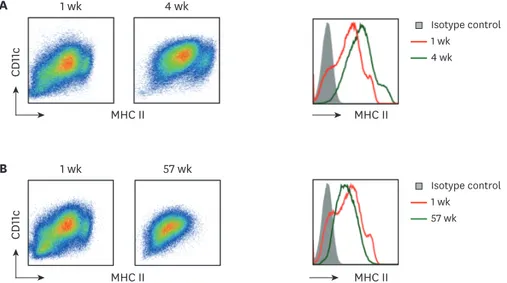
Non-adherent or loosely-attached BM cells in the 1-wk culture with GM-CSF are composed of heterogeneous populations including a large majority of CD11c+CD11b+MHC IIint/lo macrophages and a relative smaller number of CD11c+CD11b+MHC IIhi DCs (19-21). Both of the BM cells cultured with GM-CSF for 1 wk as well as for 4 wk exhibited heterogeneous populations with regard to their surface level of MHC II (Fig. 2A), whereas those cultured for 20 wk or longer became a relatively homogeneous population in terms of expressing surface MHC II (Fig. 2B).
Although cells from the long-term cultures of BM with GM-CSF were unable to stimulate allogeneic T cells in MLR (Supplementary Fig. 1), those cells expressed both MHC I and MHC II on surface throughout the extended culture periods (Figs. 2 and 3). Then, we examined whether BM cells from the long-term culture possessed the ability to suppress T cell responses.
4/12 https://doi.org/10.4110/in.2018.18.e16
https://immunenetwork.org
Allogeneic T cells mixed with BM cultured with GM-CSF
1 wk 4 wk
Allogeneic T cells
alone
CFSE CD4
CD8 A
2,000
1,000
**
0 CFSElo total allogeneic T cell number
1 wk 4 wk T cell alone
1,500
500
**
1,000
0 CFSElo CD4+ allogeneic T cell number
1 wk 4 wk T cell alone
600 400 200
**
0 CFSElo CD8+ allogeneic T cell number
1 wk 4 wk T cell alone Allogeneic T cells mixed
with BM cultured with GM-CSF
1 wk 8 wk
Allogeneic T cells
alone
CFSE CD4
CD8 B
4,000
2,000
***
3,000
1,000 0 CFSElo total allogeneic T cell number
1 wk 4 wk T cell alone
3,000
1,000
***
2,000
0 CFSElo CD4+ allogeneic T cell number
1 wk 4 wk T cell alone
1,000
500
**
0 CFSElo CD8+ allogeneic T cell number
1 wk 4 wk T cell alone Figure 1. Cells from the extended cultures of BM with GM-CSF lose their capacity to stimulate allogeneic T cells. BM cells isolated from C57BL/6 mouse are cultured with GM-CSF, and harvested in 4-wk intervals. The 5×10⁴ CFSE-labeled allogeneic T cells from BALB/c mouse are co-cultured with 1.7×10⁴ BM cells derived respectively from (A) 4-, and (B) 8-wk old cultures in comparison with the 1-wk old culture. After 4 days, CFSElow T cells are analyzed by a flow cytometer and counted. Representative data are shown from at least 2 independent experiments in quadruplicate. Error bars indicate mean ± standard error of the mean across quadruplicate samples.
**p≤0.01, ***p≤0.001.
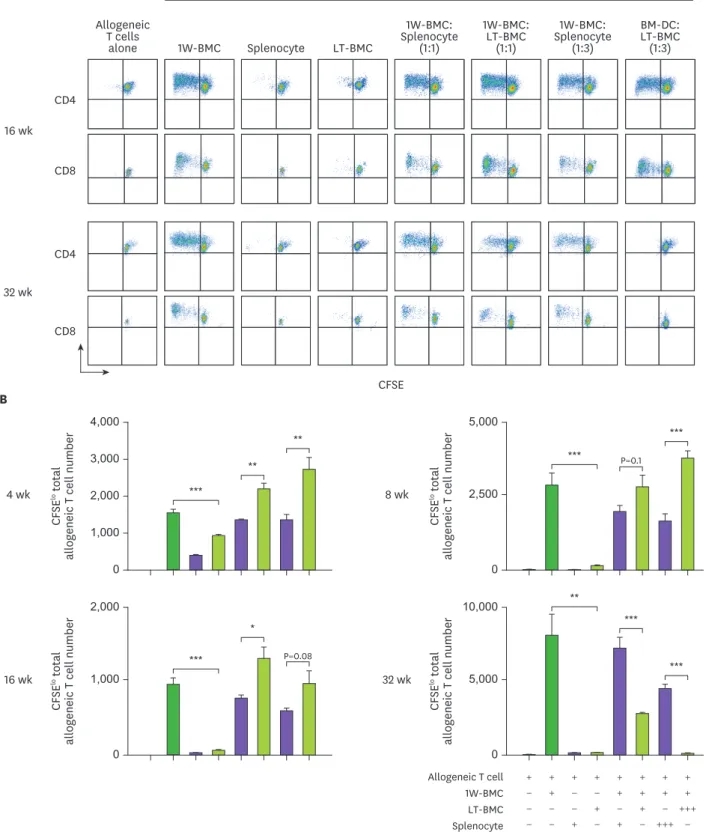
As shown in Fig. 4, cells from the BM cultured for 32 wk or longer were found to effectively suppress the proliferation of allogeneic T cells in MLR stimulated by the 1-wk cultured BM cells.
This suppressor activity of the long-term cultured BM cells was maintained throughout our examinations which lasted well over a year (Fig. 4 and Supplementary Fig. 1). Then, we further investigated whether those BM cells in the long-term culture exert their suppressor activity in antigen-dependent manner. The long-term cultured BM cells were treated with/without OVA before adding into the co-culture with the OVA-loaded 1-wk cultured BM cells and anti-OVA OT-I TCR transgenic T cells. Only the long-term cultured BM cells laden with OVA were able to suppress the response of OVA-specific OT-I T cells (Fig. 5).
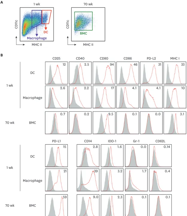
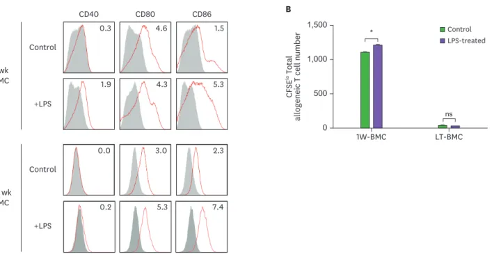
Our results indicate that BM cells cultured with GM-CSF undergo 2 distinct steps of functional change; the BM cells cultured for 8 wk or longer lose immunostimulatory capacity; and those cultured for 32 wk or longer gain immunosuppressive ability. To evaluate whether these changes are due to the altered expression of surface molecules, we compared the levels of various co- stimulatory and surface molecules between the 1-wk cultured BM cells and the 70-wk cultured BM cells. The expression of CD25, CD40, CD80, CD86, PD-L2, and MHC I were significantly diminished while the expression of PD-L1 was augmented on the surface of the 70-wk cultured BM cells (Fig. 3). We also tested whether the stimulation with LPS, a TLR4 agonist, could change the function of the long-term cultured BM cells. As shown in Fig. 6, LPS treatment increased the immunostimulatory activity of the 1-wk cultured BM cells but exhibited no such effect on the long-term cultured BM cells.
It is currently not yet clear that the expressional change of these co-stimulatory molecules during extended culture is responsible for those BM cells to lose immunostimulatory capacity or to gain immunosuppressive ability. The new features acquired by the BM cells in the extended cultures are apparently shared by the typical phenotypes of tolerogenic DCs, including the increased level of PD-L1 and the decreased levels of CD40, CD80, CD86 (22).
Further investigation is needed to define the mechanisms that cause the functional changes of BM cells in the course of the extended culture with GM-CSF.
6/12 https://doi.org/10.4110/in.2018.18.e16
https://immunenetwork.org
1 wk 4 wk
MHC II
CD11c
MHC II A
B 1 wk 57 wk
MHC II
CD11c
MHC II
Isotype control 1 wk
4 wk
Isotype control 1 wk
57 wk
Figure 2. Long-term cultured BM cells retain homogeneous levels of CD11c and MHC II. BM cells cultured with GM-CSF for 1 wk versus (A) 4 wk or (B) 57 wk are compared for their MHC II and CD11c expression. Representative data are shown from at least 2 independent experiments.
DC Macrophage
70 wk
BMC 1 wk
MHC II
CD11c
MHC II
CD11c
A
BMC
BMC
IDO-1 1.6
2.3 PD-L1
15
59
CD80 94
9.5
MHC I 33
3.1
CD14 2.8
9.0
PD-L2 31
0.0 CD40
2.5
0.2
0.0
0.1
CD62L 0.14
0.1 CD25
12
0.7
CD86 46
0.1
Gr-1
2.6 4.1
0.4 3.2
21
17 4.1 10
39 2.2
1.7 DC
Macrophage
DC
Macrophage
70 wk 70 wk 1 wk
1 wk B
Figure 3. The expression of surface molecules is evaluated on BM cells in the extended cultures with GM-CSF. BM cells in the 1-wk and 70-wk old cultures are stained side by side with antibodies against the indicated molecules. (A) In the 1-wk old BM culture, CD11c+MHC IIhi cells are gated as DCs in red tetragon and CD11c+MHC IIint cells are gated as macrophages in purple tetragon. The whole BM cells in the 70-wk old culture are gated for analysis. (B) Histograms illustrate the staining of a respective antibody (open red line) versus an isotype control (area filled with gray). The expression of each molecule is detected on cell surface but IDO-1 detected intracellularly. Mean fluorescent index is denoted in each histogram. Representative data are shown from at least 2 independent experiments.
8/12 https://doi.org/10.4110/in.2018.18.e16
https://immunenetwork.org
Allogeneic T cells mixed with
CFSE CD4
32 wk CD8 CD4
CD8 A
16 wk
Allogeneic T cells
alone 1W-BMC
1W-BMC:
Splenocyte (1:1)
1W-BMC:
LT-BMC (1:1)
1W-BMC:
Splenocyte (1:3)
BM-DC:
LT-BMC (1:3) LT-BMC
Splenocyte
2,000
1,000
0 CFSElo total allogeneic T cell number
16 wk 32 wk
+ +
− + + + + +
+ +
+ + + +
− −
− − − + − + −
− − + − + − +++
+++
− 10,000
5,000
0 CFSElo total allogeneic T cell number
Allogeneic T cell 1W-BMC LT-BMC Splenocyte
**
***
***
P=0.08
*
***
B
+ +
− + + + + +
+ +
+ + + +
− −
− − − + − + −
− − + − + − +++
+++
− 2,000
1,000
0 CFSElo total allogeneic T cell number 67 wk
Allogeneic T cell 1W-BMC LT-BMC Splenocyte
*** ***
***
4,000
2,000 3,000
1,000 0 CFSElo total allogeneic T cell number
4 wk 8 wk
5,000
2,500
0 CFSElo total allogeneic T cell number
***
***
P=0.1
**
**
***
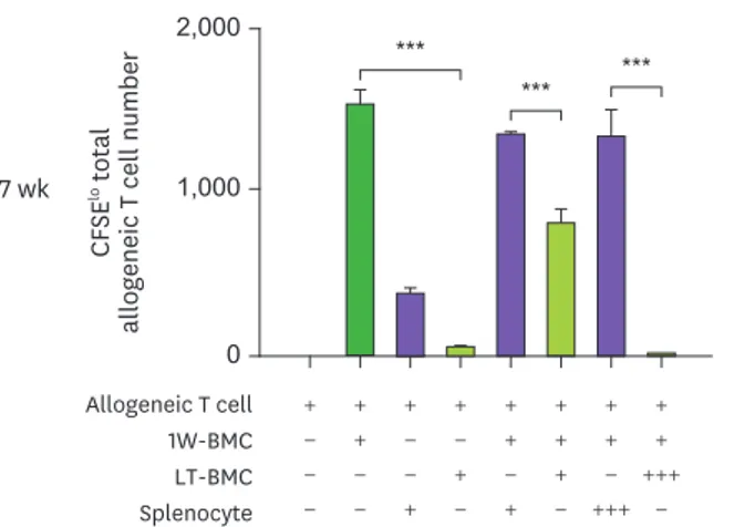
Figure 4. Cells from the extended cultures of BM with GM-CSF are suppressive to MLR. (A) BM cells cultured for 1 wk (1W-BMC) were mixed with either control syngeneic splenocytes or BM cells cultured long-term (LT-BMC) for 16 or 32 wk and co-cultured with 5×10⁴ CFSE-labeled allogeneic T cells isolated from BALB/c mice. After 4 days, CFSElow allogeneic T cells are assessed. Representative flow cytograms are shown from 2 independent experiments in quadruplicate. (B) The suppressor cell activity of BM cells in extended cultures at different time points is assessed as in (A). Assays are performed at the ratios of 1(+):1(+):3(+) and 1(+):3(+++):3(+) for 1W-BMC:LT-BMC/Splenocyte:Allogeneic T cell. The number of CFSElow T cells were counted and plotted in graphs. Data are generated from 2 independent experiments in quadruplicate. Error bars indicate mean ± standard error of the mean across quadruplicate samples.
*p≤0.05, **p≤0.01, ***p≤0.001. (continued to the next page)
Immunosuppressive Cells from Bone Marrow Culture
CFSE CD4
32 wk CD8 CD4
CD8 16 wk
Allogeneic T cells
alone 1W-BMC
1W-BMC:
Splenocyte (1:1)
1W-BMC:
LT-BMC (1:1)
1W-BMC:
Splenocyte (1:3)
BM-DC:
LT-BMC (1:3) LT-BMC
Splenocyte
2,000
1,000
0 CFSElo total allogeneic T cell number
16 wk 32 wk
+ +
− + + + + +
+ +
+ + + +
− −
− − − + − + −
− − + − + − +++
+++
− 10,000
5,000
0 CFSElo total allogeneic T cell number
Allogeneic T cell 1W-BMC LT-BMC Splenocyte
**
***
***
P=0.08
*
***
B
+ +
− + + + + +
+ +
+ + + +
− −
− − − + − + −
− − + − + − +++
+++
− 2,000
1,000
0 CFSElo total allogeneic T cell number 67 wk
Allogeneic T cell 1W-BMC LT-BMC Splenocyte
*** ***
***
4,000
2,000 3,000
1,000 0 CFSElo total allogeneic T cell number
4 wk 8 wk
5,000
2,500
0 CFSElo total allogeneic T cell number
***
***
P=0.1
**
**
***
Figure 4. (Continued) Cells from the extended cultures of BM with GM-CSF are suppressive to MLR. (A) BM cells cultured for 1 wk (1W-BMC) were mixed with either control syngeneic splenocytes or BM cells cultured long-term (LT-BMC) for 16 or 32 wk and co-cultured with 5×10⁴ CFSE-labeled allogeneic T cells isolated from BALB/c mice. After 4 days, CFSElow allogeneic T cells are assessed. Representative flow cytograms are shown from 2 independent experiments in quadruplicate. (B) The suppressor cell activity of BM cells in extended cultures at different time points is assessed as in (A). Assays are performed at the ratios of 1(+):1(+):3(+) and 1(+):3(+++):3(+) for 1W-BMC:LT-BMC/Splenocyte:Allogeneic T cell. The number of CFSElow T cells were counted and plotted in graphs. Data are generated from 2 independent experiments in quadruplicate. Error bars indicate mean ± standard error of the mean across quadruplicate samples.
*p≤0.05, **p≤0.01, ***p≤0.001.
+ +
− + + + + + + +
+ +
+ + + + + +
− −
− − − + − + − −
− − + − + − ++
++
− +++
+++
−
A
120,000
60,000
loCFSE OT-I T cell number 0
OT-I T cell OVA-laden 1W-BMC OVA-laden LT-BMC OVA-laden splenocyte
+ +
− + + + + + + +
+ +
+ + + + + +
− −
− − − + − + − −
− − + − + − ++
++
− +++
+++
−
B
5,000
2,500
loCFSE OT-I T cell number 0
OT-I T cell OVA-laden 1W-BMC LT-BMC Splenocyte
*** ***
***
ns P=0.08 **
ns ns
Figure 5. Cells from the extended culture of BM with GM-CSF are suppressive to the proliferation of OT-I T cells in antigen-dependent manner. (A) BM cells cultured for 1 wk (1W-BMC), 60-wk old BM cells cultured long-term (LT-BMC), and control syngeneic splenocytes are pre-treated with OVA (100 μg/ml), mixed as indicated, co-cultured with 105 CFSE-labeled OT-I T cells for 3 days, and analyzed for the number CFSElow OT-I T cells. (B) The suppressor cell activity is assessed as in (A) except that 62-wk old LT-BMC and control syngeneic splenocytes are not pre-treated with OVA. Assays are performed at the ratios of 1(+):1(+):100(+), 1(+):3(++):100(+), and 1(+):10(+++):100(+) for 1W-BMC:LT-BMC/Splenocyte:OT-I T cell. The number of CFSElow T cells were counted and plotted in graphs. Data are generated from 2 independent experiments in triplicate. Error bars indicate mean ± standard error of the mean across triplicate samples.
NS, not significant.
*p≤0.05, **p≤0.01, ***p≤0.001.
ACKNOWLEDGEMENTS
We were supported by grants from the National Research Foundation of Korea to Chae Gyu Park (NRF-2014R1A4A1008625, NRF-2017R1D1A1B03028385, and NRF-2017M3A9C8064887) and Hye Young Na (NRF-2017R1A6A3A11028388) and by the Brain Korea 21 PLUS Project for Medical Science, Yonsei University.
SUPPLEMENTARY MATERIAL
Supplementary Figure 1
BM cells isolated from C57BL/6 mouse are cultured with GM-CSF, and harvested in 4-week intervals.
Click here to view
REFERENCES
1. Steinman RM, Witmer MD. Lymphoid dendritic cells are potent stimulators of the primary mixed leukocyte reaction in mice. Proc Natl Acad Sci U S A 1978;75:5132-5136.
PUBMED | CROSSREF
2. Steinman RM. Decisions about dendritic cells: past, present, and future. Annu Rev Immunol 2012;30:1-22.
PUBMED | CROSSREF
10/12 https://doi.org/10.4110/in.2018.18.e16
https://immunenetwork.org
0.3 4.6 1.5
0.0 3.0 2.3
1.9 4.3 5.3
0.2 5.3 7.4
CD86 CD80
CD40 Control
+LPS BMC1 wk
Control
+LPS 70 wk
BMC
A B
1,500 1,000 500 0 CFSElo Total allogeneic T cell number
LT-BMC 1W-BMC
Control LPS-treated
ns
*
Figure 6. LPS stimulation does not affect the stimulatory function of BM cells cultured long-term (LT-BMC). BM cells in the 1-wk and 70-wk old cultures are stimulated side by side with LPS (100 μg/ml) for 18 h. After LPS stimulation, BM cells in the respective culture are stained with antibodies against the indicated molecules (A) or co-cultured with 5×10⁴ CFSE-labeled allogeneic T cells for MLR (B). Data are generated from 2 independent experiments in quadruplicate. Error bars indicate mean ± standard error of the mean across quadruplicate samples.
1W-BMC, BM cells cultured for 1 wk; NS, not significant.
*p≤0.05.
3. Park CG. Vaccine strategies utilizing C-type lectin receptors on dendritic cells in vivo. Clin Exp Vaccine Res 2014;3:149-154.
PUBMED | CROSSREF
4. Fogg DK, Sibon C, Miled C, Jung S, Aucouturier P, Littman DR, Cumano A, Geissmann F. A clonogenic bone marrow progenitor specific for macrophages and dendritic cells. Science 2006;311:83-87.
PUBMED | CROSSREF
5. Liu K, Victora GD, Schwickert TA, Guermonprez P, Meredith MM, Yao K, Chu FF, Randolph GJ, Rudensky AY, Nussenzweig M. In vivo analysis of dendritic cell development and homeostasis. Science 2009;324:392-397.
PUBMED
6. Cheong C, Matos I, Choi JH, Dandamudi DB, Shrestha E, Longhi MP, Jeffrey KL, Anthony RM, Kluger C, Nchinda G, et al. Microbial stimulation fully differentiates monocytes to DC-SIGN/CD209(+) dendritic cells for immune T cell areas. Cell 2010;143:416-429.
PUBMED | CROSSREF
7. Inaba K, Swiggard WJ, Steinman RM, Romani N, Schuler G, Brinster C. Isolation of dendritic cells. Curr Protoc Immunol 2009;Chapter 3:Unit 3.7.
PUBMED | CROSSREF
8. Lutz MB, Kukutsch N, Ogilvie AL, Rossner S, Koch F, Romani N, Schuler G. An advanced culture method for generating large quantities of highly pure dendritic cells from mouse bone marrow. J Immunol Methods 1999;223:77-92.
PUBMED | CROSSREF
9. Shen Z, Reznikoff G, Dranoff G, Rock KL. Cloned dendritic cells can present exogenous antigens on both MHC class I and class II molecules. J Immunol 1997;158:2723-2730.
PUBMED
10. Ebihara S, Endo S, Ito K, Ito Y, Akiyama K, Obinata M, Takai T. Immortalized dendritic cell line with efficient cross-priming ability established from transgenic mice harboring the temperature-sensitive SV40 large T-antigen gene. J Biochem 2004;136:321-328.
PUBMED | CROSSREF
11. Peng S, Kim TW, Lee JH, Yang M, He L, Hung CF, Wu TC. Vaccination with dendritic cells transfected with BAK and BAX siRNA enhances antigen-specific immune responses by prolonging dendritic cell life.
Hum Gene Ther 2005;16:584-593.
PUBMED | CROSSREF
12. Kang TH, Lee JH, Bae HC, Noh KH, Kim JH, Song CK, Shin BC, Hung CF, Wu TC, Park JS, et al.
Enhancement of dendritic cell-based vaccine potency by targeting antigen to endosomal/lysosomal compartments. Immunol Lett 2006;106:126-134.
PUBMED | CROSSREF
13. Ichiyanagi T, Imai T, Kajiwara C, Mizukami S, Nakai A, Nakayama T, Udono H. Essential role of endogenous heat shock protein 90 of dendritic cells in antigen cross-presentation. J Immunol 2010;185:2693-2700.
PUBMED | CROSSREF
14. Shrimpton RE, Butler M, Morel AS, Eren E, Hue SS, Ritter MA. CD205 (DEC-205): a recognition receptor for apoptotic and necrotic self. Mol Immunol 2009;46:1229-1239.
PUBMED | CROSSREF
15. Dolan BP, Li L, Takeda K, Bennink JR, Yewdell JW. Defective ribosomal products are the major source of antigenic peptides endogenously generated from influenza A virus neuraminidase. J Immunol 2010;184:1419-1424.
PUBMED | CROSSREF
16. He T, Tang C, Xu S, Moyana T, Xiang J. Interferon gamma stimulates cellular maturation of dendritic cell line DC2.4 leading to induction of efficient cytotoxic T cell responses and antitumor immunity. Cell Mol Immunol 2007;4:105-111.
PUBMED
17. Ryu SH, Na HY, Sohn M, Han SM, Choi W, In H, Hong S, Jeon H, Seo JY, Ahn J, et al. Reduced expression of granule proteins during extended survival of eosinophils in splenocyte culture with GM-CSF. Immunol Lett 2016;173:7-20.
PUBMED | CROSSREF
18. Han SM, Na HY, Ham O, Choi W, Sohn M, Ryu SH, In H, Hwang KC, Park CG. TCF4-targeting miR-124 is differentially expressed amongst dendritic cell subsets. Immune Netw 2016;16:61-74.
PUBMED | CROSSREF
19. Ryu SH, Na HY, Sohn M, Choi W, In H, Shin HS, Choi JH, Park CG. Competent antigen-presenting cells are generated from the long-term culture of splenocytes with granulocyte-macrophage colony- stimulating factor. Immunol Lett 2017;188:96-107.
PUBMED | CROSSREF
20. Helft J, Bottcher J, Chakravarty P, Zelenay S, Huotari J, Schraml BU, Goubau D, Reis e Sousa C. GM-CSF mouse bone marrow cultures comprise a heterogeneous population of CD11c(+)MHCII(+) macrophages and dendritic cells. Immunity 2015;42:1197-1211.
PUBMED | CROSSREF
21. Lutz MB, Strobl H, Schuler G, Romani N. GM-CSF monocyte-derived cells and langerhans cells as part of the dendritic cell family. Front Immunol 2017;8:1388.
PUBMED | CROSSREF
22. Yoo S, Ha SJ. Generation of tolerogenic dendritic cells and their therapeutic applications. Immune Netw 2016;16:52-60.
PUBMED | CROSSREF
12/12 https://doi.org/10.4110/in.2018.18.e16
https://immunenetwork.org