Rituximab-induced Interstitial Pneumonitis in a Young Patient:
A Case Report and Review of the Literature
Dong Mee Lee, M.D., Sung Yong Oh, M.D., Hyun Ah Yoon, M.D., Suee Lee, M.D., Sung-Hyun Kim, M.D., Hyuk-Chan Kwon, M.D.,
Soo-Keol Lee, M.D. and Hyo-Jin Kim, M.D.
Department of Internal Medicine, Dong-A University College of Medicine, Busan, Korea
Side effects of rituximab are mild in most cases, but there have been a few cases of severe pulmonary toxicity reported in elderly patients. Here we report a case of interstitial pneumonitis following rituximab treatment in a young patient. A 35-year-old woman with diffuse large B-cell lymphoma was admitted complaining of dry cough and dyspnea without fever after the 3 treatments with rituximab-CHOP (cyclophosphamide, doxorubicin, vincristine, and prednisolone) chemotherapy. Her chest CT with high- resolution CT scanning confirmed the presence of bilateral diffuse ground-glass opacities. The analysis of arterial blood gases indicated hypoxemia. The pulmonary function testing showed a restrictive pattern.
There were no other findings suggesting an infection. The findings were compatible with a rituximab-in- duced interstitial pneumonitis. After the patient was treated with prednisolone, the symptoms resolved.
Cases with rituximab-induced interstitial pneumonitis develop principally in elderly patients. However, the condition also can occur in young patients. (Korean J Hematol 2007;42:423-427.)
Key Words: Rituximab, Interstitial pneumonitis, Young patient, Diffuse large B-cell lymphoma
423 접수:2007년 9월 13일, 수정:2007년 11월 23일
승인:2007년 11월 26일
교신저자:김효진, 부산시 서구 동대신동 3가 1
602-715, 동아대학교 의과대학 내과학교실 Tel: 051-240-2951, Fax: 051-240-2088
E-mail: [email protected]
Correspondence to:Hyo-Jin Kim, M.D.
Department of Internal Medicine, Dong-A University College of Medicine
1, Dongdaeshin-dong 3-ga, Seo-gu, Busan 602-715, Korea Tel: +82-51-240-2951, Fax: +82-51-240-2088
E-mail: [email protected] INTRODUCTION
Rituximab is a monoclonal antibody against the CD20 B-cell antigen.1) It is effective in re- lapsed or refractory indolent CD 20+ B-cell non-Hodgkin’s lymphoma (NHL) and advanced stage chemoresistant or relapsed follicular lym- phoma. It is also the first-line treatment for ag- gressive NHL, including diffuse large B-cell lym- phoma in combination with CHOP.2) An evalua- tion of the efficacy of this treatment in auto- immune disorders and other malignancies has been recently conducted.3)
The most frequently observed type of non-
Hodgkin’s lymphoma is diffuse large B-cell lym- phoma, and accounts for up to 40 percent of cases. In a large, prospective randomized multi- center trial for previously untreated patients with aggressive NHL, a combination of rituximab- cy- clophosphamide, doxorubicin, vincristine, and prednisolone (R-CHOP) established a significan- tly more complete response (CR) (76% versus 63%, P=0,005), superior 2-year progression-free survival rates (PFS) (57% versus 38%, P<0.001) and overall survival (OS) rates (70% versus 57%, P=0.007), but no significant toxicity as compared to CHOP treatment alone.4)
There are no known risk factors for ritux-
Fig. 1. Rituximab-induced in- terstitial pneumonitis in chest HRCT scan and radiography.
Table 1. Analysis of arterial blood gases before and after prednisolone treatment
Parameter Before treatment After treatment
pH 7.449 7.427
pCO2 (mmHg) 38.0 33.0
pO2 (mmHg) 66.4 130.6
O2 saturation (%) 94.0 98.7
imab-induced interstitial pneumonitis, but ad- vanced age is considered to be one of its sig- nificant risk factors.5) Herein, we report an un- usual young patient with diffuse large B-cell lym- phoma who developed interstitial pneumonitis following R-CHOP chemotherapy.
CASE REPORT
A 35-year-old woman presenting with a tin- gling sensation in the lower extremities and radi- ating pain visited our hospital. The lumbar spine MRI imaging evidenced a mass around the pre- sacral and spinal canal area at the Sacral 1, 2, 3 levels. The excisional biopsy of that mass was di- agnosed as diffuse large B-cell lymphoma.
Initial chest radiography and chest computed tomography (CT) with high resolution computed tomography (HRCT) following excisional biopsy revealed nonspecific findings, with the exception of a slight pleural effusion, left lower. Pleural ef- fusion was resolved on the follow-up chest radio- graphy.
The patient was treated with R-CHOP chemo- therapy every three weeks. After the second ad- ministration of R-CHOP chemotherapy, the pa- tient developed a mild cough, but the chest ra- diography and physical examination were unre- markable.
After the third R-CHOP chemotherapy, she was admitted for dry cough and dyspnea. The physical examination showed reduced breath sounds in both lungs. Her chest radiography evi- denced bilateral diffuse haziness and a chest CT with an HRCT scan verified the presence of bi- lateral diffuse ground-glass opacity with bilateral basilar consolidation and thickening along the bronchovascular bundle (Fig. 1). The arterial blood gas analysis demonstrated a PO2 of 66 mmHg and an oxygen saturation of 94%. Pul- monary function tests revealed a restrictive pat- tern and the pulmonary diffusing lung capacity of carbon dioxide was reduced (Table 1, 2).
Other findings suggesting infection, including blood, urine, sputum culture, sputum culture, cy- tomegalovirus (CMV) IgM antibody, pneumocy- stis carinii PCR, mycoplasma antibody, antineu- trophil cytoplasmic antibody (ANCA), and anti- nuclear antibody (ANA) tests were all negative.
These results were consistent with drug (rituxi-
Table 2. Pulmonary function tests before and after prednisolone treatment
Variable Predicted value Before treatment After treatment
FVC (liters) 3.78 1.93 (51%) 3.21 (85%)
FEV1 (liters) 2.98 1.52 (51%) 2.79 (94%)
FEV1/FVC (%) 78 79 87
TLC (liters) 5.30 3.86 (73%) 4.41 (83%)
DLCO (mL/min/mmHg) 21.6 7.0 (33%) 14.1 (65%)
Abbreviations: FVC, forced vital capacity; FEV1, forced expiratory volume in one second; TLC, total lung capacity; DLCO, carbon monoxide diffusing capacity.
Fig. 2. After prednisolone treat- ment for 2 weeks, interstitial pneumonitis resolved in chest HRCT scan and radiogra- phy.
mab)-induced interstital pneumonitis with organ- izing pneumonia in both lower lobes.
She was prescribed 30mg of prednisolone daily.
Her symptoms evidenced dramatic improvement.
The follow-up chest HRCT revealed nearly to- tal resolution of drug-induced interstital pneumo- nitis in both lower lobes and bronchiolitis ob- literans (Fig. 2). Repeated pulmonary function tests recovered to normal, and the pulmonary dif- fusing lung capacity of carbon dioxide was im- proved-by 65% of the predicted value. The analy- sis of arterial blood gases revealed a PO2 of 130 mmHg and an oxygen saturation of 98.7% (Table 1, 2).
Further chemotherapy was conducted via a CHOP regimen, with the exception of rituximab.
The patient had no symptoms, including dysp- nea, dry cough.
DISCUSSION
The diagnosis of interstitial pneumonitis is possible in cases in which typical symptoms and signs such as exertional dyspnea, dry cough, ab- normal breathing sounds, and a restrictive pat- tern on pulmonary function testing are combined with radiological abnormalities including ground glass opacity and multiple infiltrations upon computed tomography (CT).6) If these findings are equivocal, a lung biopsy can help to verify the diagnosis. Smoking, occupational exposure to dusts, advanced age, diabetes mellitus, hyper- lipidemia, and obesity are known to be risk fac- tors of interstitial pneumonitis.5)
In this case, although other drugs-cyclophos- phamide and vincristine also could be an etiology of drug-induced lung injury, but interstitial
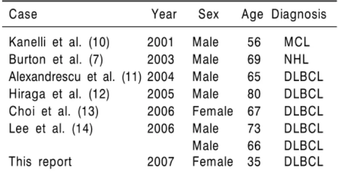
Table 3. Patients’ characteristics of reported cases
Case Year Sex Age Diagnosis
Kanelli et al. (10) 2001 Male 56 MCL Burton et al. (7) 2003 Male 69 NHL Alexandrescu et al. (11) 2004 Male 65 DLBCL Hiraga et al. (12) 2005 Male 80 DLBCL Choi et al. (13) 2006 Female 67 DLBCL
Lee et al. (14) 2006 Male 73 DLBCL
Male 66 DLBCL
This report 2007 Female 35 DLBCL
Abbreviations: MCL, mantle cell lymphoma; DLBCL, diffuse large B cell lymphoma; NHL, non-Hodgkin lymphoma.
pneumonitis did not occur when we treated with CHOP regimen. So, we described rituximab re- lated interstitial lung disease.
The adverse events of rituximab are usually mild, and include infusion-related symptoms in- cluding fever, chill, and rigors. The use of ritux- imab has not been commonly associated with pul- monary toxicity. Drug-induced lung injury asso- ciated with rituximab has been reported in less than 0.03 percent of cases, but sometimes proved fatal.7,8)
The mechanism underlying rituximab-induced interstitial pneumonitis has remained unclear un- til now. However, several immune mechanisms could be related to the acute and chronic adverse effects of rituximab treatment, which results in the activation of complement, B-lymphocyte cy- tolysis, cytotoxic T-lymphocytes, and the release of cytokines (TNF-alfa, IL-6, INF-gamma).9) Small numbers of cases of interstitial pneumo- nitis have been previously reported (Table 3).7,10-14) Rituximab-induced lung injury occurred after 2 or 3 cycles of treatment. They evidenced no his- tory of interstitial lung disease and was well con- trolled with low or high doses of steroid treat- ment. Also, they did not observe these effects un- der continued treatment, except for rituximab treatment. In this case, a similar clinical course was observed. However, all of the patient who were previously diagnosed with rituximab-in- duced interstitial lung disease were elderly (range:
56∼82 years).7,10-14) The patient described herein, however, was only 35 years old. Rituximab-in- duced interstitial lung disease usually develops only in elderly patients. However, in our case, it developed in a younger patient.
Normally, cautious monitoring for respiratory symptoms is done in cases of rituximab treatment in geriatric patients. However, we also must re- main vigilant for the occurrence of rituximab-in- duced interstitial pneumonitis, even in young patients.
요 약
대부분의 경우, 리툭시맙의 부작용은 경미하지만 고 령의 환자에서 드물게 중대한 폐합병증이 보고되었다.
본 증례는 젊은 환자에서 리툭시맙의 치료 후 생긴 간 질성 폐질환에 관해 보고하고자 한다. 미만성 대세포 B형 림프종 진단 후 리툭시맙-CHOP 치료를 3주기 시 행 받았던 35세 여자 환자로 기침과 호흡곤란을 주소 로 내원하였다. 환자는 컴퓨터 단층 촬영상 양측 폐에 미만성의 간유리 음영소견을 보였고 동맥혈 검사상 저 산소증을 나타내었으며 폐기능 검사상 제한적 형태의 폐기능 소견을 보였다. 감염의 소견은 발견되지 않았 다. 환자는 스테로이드를 사용 후 증상의 호전을 보였 고 이후 치료에서는 리툭시맙을 제외한 CHOP 요법으 로 치료하였으며 호흡곤란의 소견은 관찰되지 않았다.
이전 보고에서 리툭시맙과 관련된 폐병변의 경우 고령 에서 보고되었으나 본 증례를 통해 젊은 환자에서도 발생할 수 있음을 유념해야 하겠다.
REFERENCES
1) Maloney DG, Smith B, Rose A. Rithuximab: mecha- nism of action and resistance. Semin Oncol 2002;
29(2 Suppl):2-9.
2) Coiffier B. Rituximab therapy in malignant lym- phoma. Oncogene 2007;26:3603-13.
3) Dass S, Vital EM, Emery P. Rituximab: novel B-cell depletion therapy for the treatment of rheumatoid arthritis. Expert Opin Pharmacother 2006;7:2559- 70.
4) Coiffier B, Lepage E, Brière J, et al. CHOP chemo- therapy plus rituximab compared with CHOP alone in elderly patients with diffuse large-B-cell lym-
phoma. N Engl J Med 2002;346:235-42.
5) Enomoto T, Usuki J, Azuma A, Nakagawa T, Kudoh S. Diabetes mellitus may increase risk for idiopathic pulmonary fibrosis. Chest 2003;123:2007-11.
6) Limper AH. Drug-induced pulmonary disease. In:
Mason RJ, Murray JF, Broaddus VC, Nadel JA, eds.
4th ed. Text book of respiratory medicine. Philadel- phia: Elsevier Saunders, 2005:611-24.
7) Burton C, Kaczmarski R, Jan-Mohamed R. Intersti- tial pneumonitis related to rituximab therapy. N Engl J Med 2003;348:2690-1.
8) Herishanu Y, Polliack A, Leider-Trejo L, Grieff Y, Metser U, Naparstek E. Fatal interstitial pneumonitis related to rituximab-containing regimen. Clin Lym- phoma Myeloma 2006;6:407-9.
9) Bienvenu J, Chvetzoff R, Salles G, et al. Tumor ne- crosis factor alpha release is a major biological event associated with rituximab treatment. Hematol J
2001;2:378-84.
10) Kanelli S, Ansell SM, Habermann TM, Inwards DJ, Tuinstra N, Witzig TE. Rituximab toxicity in patients with peripheral blood mailgnant B-cell lympho- cytosis. Leuk Lymphoma 2001;42:1329-37.
11) Alexandrescu DT, Dutcher JP, O'Boyle K, Albulak M, Oiseth S, Wiernik PH. Fatal intra-alveloar hemor- rhage after rituximab in a patient with non-Hodg- kin's lymphoma. Leuk Lymphoma 2004;45:2321-5.
12) Hiraga J, Kondoh Y, Taniguchi H, Kinoshita T, Naoe T. A case of interstitial pneumonia induced by rithuximab therapy. Int J Hematol 2005;81:169-70.
13) Choi YJ, Jung WJ, Oh SI, et al. A case of interstitial lung diseasec caused by Rituximab in non-Hodgkin lymphoma. Korean J Med 2006;71:449-55.
14) Lee Y, Kyung SY, Choi SJ, et al. Two cases of inter- stitial pneumonitis caused by rituximab therapy.
Korean J Intern Med 2006;21:183-6.