36
Immune Network 서 론
항원 특이 면역기능의 활성화를 통한 항암 면역(세포) 치료법의 개발은 맞춤 치료 개념의 치료 효과의 우수성 뿐 아니라, 부작용이 현저히 감소될 수 있다는 장점을 가지고 있다. 수지상 세포는 잘 알려진 항원 특이 면역
기능 항진에 가장 강력하고 필수적인 항원 소개세포로 최근에는 자연 살해 세포 기능에도 관여하는 등 면역 기 능 전반에 큰 영향력을 행사하는 것으로 알려져 있다 (1-6). 1990년대 중반 이후 활발한 연구가 진행되어 수지 상 세포의 항암 면역 치료제로서의 가능성을 확인하고 있다. 지금까지의 연구가 주로 면역원성이 강하거나, 항 원이 알려진 흑색종, 신장암, 전립선암을 대상으로 진행 되어 기대되는 임상 실험 결과(7-21)를 보고하고 있으나 폐암을 대상으로 하는 수지상 세포 치료에 관한 보고는 제한적이다(22-24).
흡연 또는 서구화되어 가는 식생활 등에 의해 발암 물
이용한 Adjuvant Therapy 가능성 연구
1을지대학교 의과대학 흉부외과, 성균관대학교 의과대학 삼성서울병원 암센터
이석재
1․김명주․인소희․백소영․이현아
Immunocell Therapy for Lung Cancer: Dendritic Cell Based Adjuvant Therapy in Mouse Lung Cancer Model
Seog Jae Lee
1, Myung Joo Kim, So Hee In, Soyoung Baek and Hyunah Lee
1Department of Thoracic & Cardiovascular Surgery, Eulji University School of Medicine, The Cancer Center, Samsung Medical Center, Sungkyunkwan University School of Medicine, Seoul, Korea
ABSTRACT
Background: The anti-tumor therapeutic effect of autologous tumor cell lysate pulsed- dendritic cells (DCs) was studied for non-immunogenic and immune suppressive lung cancer model. To test the possibility as an adjuvant therapy, minimal residual disease model was considered in mouse in vivo experiments. Methods: Syngeneic 3LL lung cancer cells were inoculated intravenously into the C57BL/6 mouse. Autologous tumor cell (3LL) or allogeneic leukemia cell (WEHI-3) lysate pulsed-DCs were injected twice in two weeks. Intraperitoneal DC injection was started one day (MRD model) after tu- mor cell inoculation. Two weeks after the final DC injection, tumor formation in the lung and the tumor-specific systemic immunity were observed. Tumor-specific lym- phocyte proliferation and the IFN-r secretion were analyzed for the immune monitoring.
Therapeutic DCs were cultured from the bone marrow myeloid lineage cells with GM-CSF and IL-4 for 7 days and pulsed with tumor cell lysate for 18 hrs. Results:
Compared to the saline treated group, tumor formation was suppressed in 3LL tumor cell lysate pulsed-DC treated group, while 3LL-specific immune stimulation was mini- mum. WEHI-3-specific immune stimulation occurred in WEHI-3 lysate-pulsed DC treated group, which had no correlation with tumor regression. Conclusion: The data suggest the possible anti-tumor effect of cultured DCs as an adjuvant therapy for mini- mal residual disease state of lung cancer. The significance of immune modulation in DC therapy including the possible involvement of NK cell as well as antigen-specific cytotoxic T cell activity induction was discussed. (Immune Network 2005;5(1):36-44) Key Words: Dendritic cells, anti-cancer immunotherapy, lung cancer
책임저자:이현아, 성균관의대 삼성서울병원 암센터 ꂕ 135-710, 서울시 강남구 일원동 50번지 Tel: 02-3410-3455, Fax: 02-3410-6808 E-mail: [email protected]
본 연구의 일부 실험은 식품의약품안전청(04092세치안081) 연구 비 지원으로 수행되었음.
질에 노출정도가 심해지면서 현대 사회에서 암은 지속 적인 증가를 보이는 난치성 질환으로 두려움의 대상이 되고 있다. 특히 폐암은 전 세계적으로뿐 아니라 우리나 라에서도 발생순위 1, 2위를 다투며, 사망률 수위를 차지 하고 있다. 폐암 사망률은 지속적으로 증가하는 추세이 며 우리나라에서도 다른 암에 비해서 낮은 생존율을 보 이고 있다. 현재 치료법으로는 외과적 수술, 약물치료 및 방사선치료 등이 사용되고 있고 필요에 따라 상호보완 적으로 사용되고 있다. 새로운 항암제의 개발이 꾸준히 진행되어왔음에도 불구하고 일반적으로 조기에 발견되 지 못하는 폐암의 경우 치료 효과는 상당히 낮은 상황이 다. 또한 정상세포에 대한 부작용이 해결되지 못한 현실 에서 새로운 치료법 개발 필요성이 계속 대두되고있다.
이에 맞추어 최근의 표적 치료제 개발에 많은 연구가 집 중되고 있다. 대표적으로 상피세포 성장인자 수용체 (EGFR)에 작용하는 “이레사(Iressa)”가 개발되어 임상 응 용이 시작되었다. 또한 폐암 세포를 이용한 vaccine (BCG, GM-CSF transfection), Muc-1, Mage-3 등의 종양항원 peptide vaccine, 유전자 치료 등이 연구되고 있으나 만족 할 만한 임상 실험 결과는 도출되지 않고 있다(25-32).
수지상 세포 항암 면역(세포) 치료의 가능성과 활발한 연구에도 불구하고 폐암 치료법으로의 개발에는 적극적 이지 못한 이유는, 폐암의 낮은 면역원성과 알려지지 않 은 종양 항원 뿐 아니라 폐암 세포 자체가 만들어내는 IL-10 등에 의한 면역억제 환경(33-35)때문에 좋은 효과 를 기대하기 힘들기 때문이다. 소세포암 세포에서 분비 되는 면역 억제 물질 IL-10은 수지상 세포의 분화 성숙 을 방해하고, apoptosis를 일으키는 것으로 알려져 있다.
그러나 제한된 연구 결과들에서 수지상 세포를 이용한 폐암 치료의 가능성을 확인할 수 있다. 비소세포암을 대상으로 mutant p53 peptide를 항원소재로 하는 임상 시 험이 미국 NCI에서 시행되고 있다. 폐암 환자에서 수지 상 세포가 종양 부위로 많이 유입된 경우 예후(36) 및 생존율(7)이 좋은 것으로 보고되어 있다. In vitro실험에 서는, 수지상 세포가 폐암 세포를 항원으로 섭취하여 항 암 면역 반응을 성공적으로 유도하는 것이 확인되었다 (38). 인터루킨-7과 herpes simplex thymidine kinase 유전 자(31), beta-galactosidase 유전자(39), cytokine 유전자(30) 또는 mutant p53 peptid 유전자(32)를 주입한 수지상 세포 의 면역 기능 유도와 항암 효과가 관찰되었다. 그러나 수지상 세포가 최종 분화 세포임을 고려할 때 유전자 주 입의 효율이 낮고 유전자 전달 virus에 의한 부작용들이 보고되고 있어 보다 간편하고 안전하며 효과적인 방법 이 필요할 것으로 생각된다.
본 실험에서는 마우스 폐암 모델에서 수지상 세포를 이용한 항암 면역 치료법의 가능성을 확인하고자 하였 다 수술 등의 치료로 암 조직을 제거한 후 잔류암세포에
의한 재발을 방지하기 위한 adjuvant therapy로서 수지상 세포 치료의 가능성을 확인하기 위해 미세 잔류암 모델 을 만들었다. 또한 폐암 세포 용해액을 항원의 소재로 사용하여(40), 알려지지 않은 폐암 특이 항원의 문제점 을 해결하면서 특이 항원 peptide만을 사용했을 때 나타 나는 약한 면역 기능 유도의 문제를 용해액 내에 포함되 어 있는 면역 adjuvant인 heat-shock protein 등의 도움으로 해결하고자 하였다. 면역 치료 개념에서는 면역억제 물 질의 분비, 낮은 면역원성 등으로 인해 단점을 가진 폐암 세포가 수지상 세포에 탑재되어 종양 특이 면역 기능을 유도하는지 관찰하였고, 실제 in vivo 항암 효과와 연관 시켜 폐암의 수지상 세포 치료 가능성을 확인하였다.
재료 및 방법
동물. 암컷 순계인 C57BL/6 mice (5∼6 주령)를 대한 Bio-Link (충청북도 음성)로부터 구입하였다. Mice는 특 정 병원균이 없는(specific pathogen -free, SPF) 동물로 실 험동물 연구실에서 ILAR (Institute of Laboratory Animal Resources) guideline에 따라 사육되었다. 실험 기간 중 사 료와 물은 자유롭게 섭취시켰고, 12시간 명: 암 조건을 유지하였다. 모든 동물은 실험을 시작하기 전 일주일 동 안 적응 기간을 거쳤다.
시약. RPMI-1640 medium, fetal bovine serum 및 penicil- lin-streptomycin은 미국 GIBCO laboratories (Grand Island, NY)로부터 구입하였다. 아래의 시약들은 미국 SIGMA Chemical Co. (St.Louis, MO)로부터 구입하였다; lipopoly- saccharide (LPS, from E. Coli 055:B5), concanavalin A (ConA), mitomycin-C. Carboxyfluorescein diacetate succini- midyl ester (CFSE)는 Molecular Probes Inc., Eugene, OR) 로부터 구입하였다. (Flow cytometric phenotyping을 위하 여 BD-Pharmingen (Sandiego, CA, USA)으로부터 다음과 같은 항체들을 구입하였다. : fluorescein isothiocyanate (FITC)-or phycoerythrin (PE)-labeled monoclonal Abs for MHC class I (H-2Kb), MHC class II (IAb), CD4, CD8, CD11c, CD80 and CD86. Low-Tox-M 보체 및Lympho- lyte-M은 CEDARLANE (Ontario, Canada)으로부터 구입 하였다.
세포주. C57BL/6 syngeneic 폐암 세포주인 Lewis Lung Carcinoma cell line (3LL) 및 Balb/C syngeneic 혈액암 세 포주인 WEHI-3 cell line을 미국 세포주 은행(ATCC, Rockville, MD)으로부터 구입하였다. 세포 주의 배양은 RPMI-1640 medium (10% heat inactivated FBS, 2 mM glutamine, 100 U/ml penicillin, 그리고 100 g/ml strep- tomycin을 첨가한 complete medium)으로 유지되었다. 생 쥐 골수 세포로부터 myeloid계 세포를 분리하기 위하여 사용한 항체는 hybridoma cell lines에서 얻었다(GK1.5 for anti-L3T4, 53.672 for anti-Lyt-2, RA3 for anti-B220, J11d
for anti-B cells/neutrophills). 각 세포주는 미국 세포주 은 행 (ATCC, Rockville, MD)으로부터 구입하였다.
생쥐 골수 세포 분리. 경추 탈골 후 얻은 대퇴골을 주사 기에 담긴 RPMI-1640으로 씻어 골수를 분리한 후 배양 액 중에서 screen mesh를 통해 단일 세포로 만들었다. 적 혈구는 Tris-buffered 0.15 M ammonium chloride solution (pH 7.2)으로 씻어 용해시키고 남아있는 유핵 세포는 hemocytometer에서 계수하였다. 세포 생존율은 Trypan Blue exclusion (routinely >90%)으로 확인하였다.
골수성 수지상 세포(myeloid-DC)의 Ex vivo 배양. 생쥐 의 골수 단세포로부터 시작하는 골수성 수지상 세포의 배양은 본 실험실에서 변형시킨 Inaba et al(41). 법으로 시행하였다. 분리된 골수 단세포를 L3T4, Lyt-2, B220 및 B 세포 항체와 보체로 panning하여 골수성 세포(mye- loid-lineage cells)만 분리하고 1×10⁶/ml의 세포에 GM- CSF 와 IL-4 (1×10³ units/ml)를 넣고 6일간 배양하였다.
배양 6일째 tumor cell lysate 50μg/ml을 넣고 18시간 더 배양하였다. Tumor cell lysate는 3LL (폐암 세포주) 및 WEHI-3 (혈액암 세포주)을 liquid nitrogen (-180oC)과 incubator (37oC)에서 freezing-thawing과정을 거친 후 1,500 rpm에서 15분간 원심 분리하여 얻어진 상등액의 단백질 함유를 Bradford 법으로 확인하여 정량하였다. 배 양 7일째 수지상 세포를 거두어 표현형(Table I) 및 syn- geneic splenocyte를 responder로 하는 autologous mixed lymphocyte response (MLR) assay로 기능을 검색하였다.
또한 배양액 중의 IL-10 및 IL-12의 양을 측정하여 수지 상 세포를 정성하였다. 치료용으로 주사하기 위한 수지 상 세포는 종양 세포 용해액과 함께 배양 한 후 수확하 여 주사용 생리 식염수에 부유시킨 후(1×10⁶/200μl/
mouse) 2시간 이내에 생쥐 복강으로 투여하였다.
종양 세포 이식 및 치료용 수지상 세포 주입. 배양된 3LL cells을 거두어 정맥내(1×10⁵ cells/100μl/mouse)로 주입하였다. 치료용 수지상 세포의 투여는(1×10⁶ DCs/
mouse), 종양 세포 주입 24시간 후부터 시작하였다. 일주 일 간격으로 2회 치료용 수지상 세포를 복강내로 투입하 였다(LDC). 대조군은 saline을 투여한 군과, 종양세포 용 해액으로 pulsing하지 않은 수지상 세포 투여군(UDC) 및 혈액암 세포 용해액으로 pulsing한 수지상 세포 투여군 (WDC)을 설정하였다. 종양 형성은 마지막 치료 2주 후 생 쥐를 희생시켜 확인하였다. 즉 3LL 폐암 세포 주사 후 21 일째까지 생존여부와 독성반응을 관찰하였으며, 21일째 에 살아있는 모든 실험동물들을 희생시켜 폐암 형성 및 전신전이 양상을 육안으로 관찰하였다. 형성된 폐암은 수 지상 세포 치료에 반응을 보인 일부의 마우스를 제외하고 폐에 생성된 종양의 결절들이 상당히 크고 서로 뭉쳐 있어 서 각개 결절의 수를 세는 것이 불가능하여 종양의 형성 정도를 평가하기 위해 몇 개의 등급으로 구분하여 반 정
량적으로 평가하는 방법(42)을 활용하였다(Table 2).
세포 표현형 검색(Flow cytometric analysis). 수지상 세 포 또는 비장 세포의 표현형 검색은 기 발표된 Yoon 등(43)의 방법을 따랐다. 비장 단핵세포는(1×10⁵ cells/
100μl) 형광 물질로 표식된 항체들을 함유한PBS with 0.1% sodium azide and 1% FBS (PBS-CS)에 부유시킨 후 4oC 에서 40분간 배양하였다. 사용된 항체들은 다음과 같다: total T cell을 위한 hamster anti-mouse CD3-FITC, 각각 CD4및 CD8 T cell subset을 위한 rat anti-mouse L3T4-PE, rat anti-mouse Lyt-2-PE, 활성 대식세포 확인을 위한 rat anti-mouse Mac3-PE. Negative control은 rat IgG2b-PE (Pharmingen, San Diego, CA)와 hamster IgG- FITC (Jackson ImmunoResearch Laboratories, Inc., West Grove, PA). 수지상 세포의 확인은 같은 방법으로 Table 1에 표시된 항체를 사용하였다. 배양이 끝난 후 세포를 세척하고, 500μl의 PBS-CS에 풀어 Flow cytometer (FACSVantage, Becton-Dickson, Mountain View, CA)로 분 석하였다. 분석은 세포 염색 1시간 이내에 시행하였다.
수지상 세포의 cytokine 분비 기능 평가. 배양 수지상 세포의 성숙도 및 기능을 예측하는 방법으로 배양액 중 에 분비된 IL-12 및 IL-10의 양을 ELISA assay kit (Becton Dickinson, Sunnyvale, CA, USA)를 이용하여 측정하였다.
즉 배양 7일째 수지상 세포 수확과 함께 배양액을 취해 cytokine의 양을 측정한 후 배양액 중에 부유되어 있던 1×10⁶개의 수지상 세포가 분비한 양으로 환산한다.
비장 림프구 증식 반응. 배양 수지상 세포의 기능을 확 인(auto-MLR)하기 위하여 또는 치료 후 종양 특이 면역 기능 증가를 확인(immune monitoring)하기 위하여 비장 단핵세포의 증식을 확인하는 실험이다. 형광 물질인 car- boxyfluorescein diacetate succinimidyl ester (CFSE)를 세포 에 결합시킨 후 수지상 세포와 같이 배양하거나(auto- MLR), 수지상 세포 pulsing에 사용한 종양 세포 용해액 첨가로 세포증식을 유도하여(immune monitoring), 분열 한 세포내의 형광 발현도가 증식유도 이전에 비해 감소 하는 정도를 flow cytometer로 측정하는 방법을 사용하였 다. 분석할 비장세포들을 HBSS (Gibco BRL, Life Tech- nologies, NY, USA)에 5×10⁶ cells/ml의 농도로 맞추어 CFSE 0.5μM을 첨가하여 37oC 암소에서 10분간 반응시 킨 후, 5% FBS가 첨가된 차가운 HBSS로 2회 세척한 후 2×10⁶ cells/mL의 농도로 24 well plate의 각 well에 넣고 37oC, 5% CO2 및 가습 조건에서 96시간 배양한다. 배양 후 수확한 비장세포의 형광 발생 정도를 flow cytometer 로 측정한다.
종양 항원 특이 effector cytokine (IFN-γ)의 분비 측정.
수지상 세포 치료 후 생체 내에 종양 특이 항암 면역 반 응에서 주 역할을 담당하는 것으로 알려진 IFN-γ (44)가 비장 면역 세포에서 분비되는지 확인하였다. 비장 림프
구 1×10⁷을 종양세포 용해액 20μg/ml 과 함께 24 well plate에서 37oC, 5% CO2 및 가습 조건으로 24시간 배양한 상등액 중에서 분비된 IFN-γ를 ELISA assay kit (Becton Dickinson, Sunnyvale, CA, USA)로 측정하였다.
통계 처리. In vivo 실험은 그룹당 6마리씩으로, 2번 반 복하였다. 통계적 유의성의 검색은 analyses of variance (ANOVA) using the Fisher protected least significant dif- ference test를 이용하였다. P value가 0.05 이하일 때 통계 적 유의성이 있는 것으로 인정하였다.
결 과
치료용 수지상 세포의 배양.
수지상 세포 면역 표현형 발현: 마우스의 골수성 세포
로부터 배양한 수지상 세포의 표현형(Table I) 발현을 flow cytometry로 측정하여 확인하였다. 항원 소개 기전 을 담당하는 MHC class I/II, CD80 & 86이 고농도로 발현 되고, DC의 marker로 알려진 CD11c도 잘 발현되는 전형 적인 골수성 수지상 세포로 항원 특이 세포 면역 기능 증가에 관여할 수 있음을 확인하였다. CD80 (B7.1)의 발 현이 약간 증가되는 경향 외에 종양 세포 용해액 pul- sing에 의한 영향은 관찰되지 않았다(Fig. 1).
수지상 세포의 Cytokine 분비: 배양된 치료용 수지상 세포는 IL-12를 분비하는 성숙된 수지상 세포로 확인되 었다(Fig. 2). 폐암 세포 주 용해액 pulsing에 의해 IL-12의 분비가 증가하는 경향을 관찰하였고, 혈액암 세포주 용 해액에 의해서는 약간 감소하는 경향을 나타내었다. IL- 12가 종양특이 세포 면역(Th1/Tc1)활성화에 관여하는 것 으로 알려져 있어, 배양된 수지상세포에 의한 항암 치료 효과를 기대할 수 있을 것으로 생각된다.
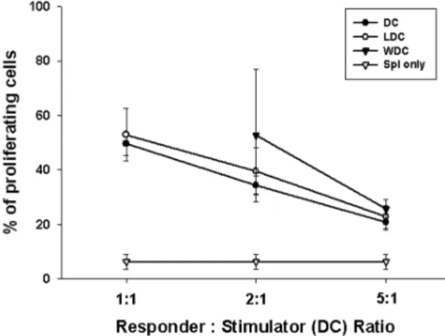
수지상 세포 자극에 의한 비장 임파구의 증식 반응 (Auto-MLR): C57BL/6 마우스에서 채취한 비장 임파구 를 같은 C57BL/6 마우스의 골수로부터 분화시킨 수지상 세포와 같이 배양하여 유도되는 비장 세포의 증식 반응 을 CFSE assay로 관찰하였다. 배양된 수지상 세포는 비 장 세포의 증식을 유도하였고 폐암(LDC) 및 혈액암 세 포(WDC) 용해액 탑재에 의해 통계적으로 유의한 변화 는 나타나지 않았다(Fig. 3).
폐암의 미세 잔류암 모델에서 수지상 세포의 항암 효 과. Lewis lung carcinoma (3LL) 세포주를 주사한 24시간 후부터 일주일 간격으로 2회 폐암(3LL) 또는 혈액암 (WEHI-3) 세포 용해액으로 탑재한 수지상 세포로 치료 한 후 2주 후에 모든 실험 동물을 안락사시켜 종양의 생 성, 전이양상을 육안으로 관찰하였다(Fig. 4). 육안 및 Table I. List of surface marker antibodies for DC char-
acterization
ꠚꠚꠚꠚꠚꠚꠚꠚꠚꠚꠚꠚꠚꠚꠚꠚꠚꠚꠚꠚꠚꠚꠚꠚꠚꠚꠚꠚꠚꠚꠚꠚꠚꠚꠚꠚꠚꠚꠚꠚꠚꠚꠚꠚꠚꠚꠚꠚꠚꠚꠚ Antibodies Fluorescence Specificity
ꠏꠏꠏꠏꠏꠏꠏꠏꠏꠏꠏꠏꠏꠏꠏꠏꠏꠏꠏꠏꠏꠏꠏꠏꠏꠏꠏꠏꠏꠏꠏꠏꠏꠏꠏꠏꠏꠏꠏꠏꠏꠏꠏꠏꠏꠏꠏꠏꠏꠏꠏ
CD4 PE Primed or unprimed T cells
CD8 PE Activated macrophages,
some dendritic cells CD11c FITC Dendritic cells, CD4-CD8+
intraepithelial lymphocytes CD80 FITC B7-1 co-stimulatory molecule CD86 FITC B7-2 co-stimulatory molecule
H2kb PE MHC class I molecule
IAb PE MHC class II molecule
ꠏꠏꠏꠏꠏꠏꠏꠏꠏꠏꠏꠏꠏꠏꠏꠏꠏꠏꠏꠏꠏꠏꠏꠏꠏꠏꠏꠏꠏꠏꠏꠏꠏꠏꠏꠏꠏꠏꠏꠏꠏꠏꠏꠏꠏꠏꠏꠏꠏꠏꠏ
Figure 1. Flow cytometric phenotyping of cultured-DCs. DCs were cultured from the bone marrow of C57BL/6 mice with GM-CSF and IL-4 for 7 days (UDC) and pulsed with either 3LL (LDC) or WEHI-3 (WDC) cell lysate. Cells were stained with fluorescent labeled surface marker antibodies (Table I) and an- alyzed with FACSVantageTM (Becton-Dickson, Mountain View, CA, USA). Antigen presentation related markers like MHC class I/II (H2kb/IAb), co-stimulatory molecules B7.1 (CD80) and B7.2
(CD86), as well as CD11c were also expressed in high level. Data supported that the cultured cells were DCs.
Figure 2. IL-12 secretion from the cultured-DCs. Cytokines se- creted into the culture media of DCs were determined by ELISA (Becton Dickinson, Sunnyvale, CA, USA). Amount of cytokines (pg/ml) was converted to pg/1×106 cells using # of cells in 1ml of culture media.
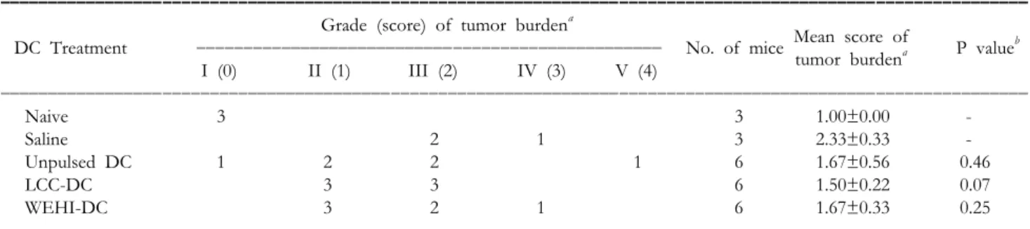
현미경으로 관찰한 폐암의 생성 정도를 De Matos (42)를 참고하여 마련한 기준에 따라 정량화하였다(Table II). 생 리식염수만으로 치료한 양성 대조군(saline)에 비해 수지
상 세포 치료 군에서 종양 생성이 적은 것을 확인 하였 다. 그러나 정량적으로 통계적 유의성은 관찰되지 않았 다(Table III). 실험 종료 전 사망한 마우스는 종양 세포 용해액을 탑재하지 않은 unpulsed-DC (UDC) 투여 군에 서만 1마리 있었다. 수지상 세포의 폐암 치료 효과는 폐 암 세포주 용해액을 탑재한 수지상 세포 투여군(LDC)의 종양 생성 억제 효과가 가장 뚜렷하였고(tumor scale 2.33 vs. 1.50 for saline vs. LDC, respectively) 관련이 없는 혈액 암 세포주 탑재 수지상 세포(WDC)의 투여 효과는 UDC 투여군과 동일하였다(tumor scale 1.67 for both UDC and WDC). 생성된 종양 세포와 같은 세포의 용해액을 탑재 한 수지상 세포의 항암 효과가 가장 큰 것은 종양 특이 면역 반응에 의한 작용 기전의 중요성을 확인한 것이다.
그러나 치료 군에서도 종양 생성이 완전히 억제되지 않 고 약간의 종양 생성이 관찰되었다. 이것은 폐암의 낮은 면역원성과 폐암에서 분비되는 면역 억제 물질에 의한 항암 작용 억제의 가능성을 시사하는 것으로 생각된다.
종양 항원 특이 면역 반응 검색.
종양 항원 특이 임파구 증식 반응: 종양을 발생시키고 수지상 세포로 치료한 마우스의 비장세포에서 종양 세 포 용해액 자극에 반응하여 증식하는 종양 항원 특이 면 역 기능을 검색하였다. T 세포 mitogen인 ConA 자극에 따른 증식 반응이 수지상 세포 치료 군에서 증가하였다.
그러나 이 반응은 종양 항원에 대한 특이 반응이 아닌
Table II. Seimiquantitiative rating system of pulmonary Lewis lung carcinoma in C57BL/6 mice
ꠚꠚꠚꠚꠚꠚꠚꠚꠚꠚꠚꠚꠚꠚꠚꠚꠚꠚꠚꠚꠚꠚꠚꠚꠚꠚꠚꠚꠚꠚꠚꠚꠚꠚꠚꠚꠚꠚꠚꠚꠚꠚꠚꠚꠚꠚꠚꠚꠚꠚꠚꠚꠚꠚꠚꠚꠚꠚꠚꠚꠚꠚꠚꠚꠚꠚꠚꠚꠚꠚꠚꠚꠚꠚꠚꠚꠚꠚꠚꠚꠚꠚꠚꠚꠚꠚꠚꠚꠚꠚꠚꠚꠚꠚꠚꠚꠚꠚꠚꠚꠚꠚꠚꠚꠚꠚꠚꠚ
Grade Score Description of pulmonary tumor burden
ꠏꠏꠏꠏꠏꠏꠏꠏꠏꠏꠏꠏꠏꠏꠏꠏꠏꠏꠏꠏꠏꠏꠏꠏꠏꠏꠏꠏꠏꠏꠏꠏꠏꠏꠏꠏꠏꠏꠏꠏꠏꠏꠏꠏꠏꠏꠏꠏꠏꠏꠏꠏꠏꠏꠏꠏꠏꠏꠏꠏꠏꠏꠏꠏꠏꠏꠏꠏꠏꠏꠏꠏꠏꠏꠏꠏꠏꠏꠏꠏꠏꠏꠏꠏꠏꠏꠏꠏꠏꠏꠏꠏꠏꠏꠏꠏꠏꠏꠏꠏꠏꠏꠏꠏꠏꠏꠏꠏ I 0 No tumor mass or colonies are observed grossly or microscopically. No enlargement of lung was observed.
II 1 Tumor mass or colonies account for less than 10% of the pulmonary mass.
III 2 Tumor mass or colonies account for approximately 10∼50% of the pulmonary mass.
IV 3 Tumor mass or colonies covered over 50% of pulmonary mass.
V 4 Death by heavy tumor burden.
ꠏꠏꠏꠏꠏꠏꠏꠏꠏꠏꠏꠏꠏꠏꠏꠏꠏꠏꠏꠏꠏꠏꠏꠏꠏꠏꠏꠏꠏꠏꠏꠏꠏꠏꠏꠏꠏꠏꠏꠏꠏꠏꠏꠏꠏꠏꠏꠏꠏꠏꠏꠏꠏꠏꠏꠏꠏꠏꠏꠏꠏꠏꠏꠏꠏꠏꠏꠏꠏꠏꠏꠏꠏꠏꠏꠏꠏꠏꠏꠏꠏꠏꠏꠏꠏꠏꠏꠏꠏꠏꠏꠏꠏꠏꠏꠏꠏꠏꠏꠏꠏꠏꠏꠏꠏꠏꠏꠏ Figure 4. Gross findings of lewis lung carcinoma formed in the C57BL/6 mouse lung. Representative lung tissues were obtained from naive mice (Naive) control mice treated with saline (Sa- line), and mice treated with unpulsed- DC (DC), 3LL lysate pulsed-DC (LLC- DC), and WEHI-3 lysate pulsed-DC (WEHI-DC), respectively.
Figure 3. In vitro induction of autologous -lymphocyte prolifera- tion (Auto-MLR) by cultured myeloid-DCs. As a responder, CFSE labeled splenocytes (2×10⁵ cells/well) from the syngeneic C57BL/6 mice were co-cultured with mitomycin-C treated DCs (stimulator) pulsed with 3LL (LDC) or WEHI-3 (WDC) cell lysates for 96 hrs. Decreased fluorescence intensity of prolif- erating splenocytes were detected by flow cytometer. The con- centrations of DCs were 2×10⁵, 1×10⁵ and 4×10⁴ for the ratio of responder: stimulator 1:1, 2:1 and 5:1 respectively. The proliferative response was proportional to the DC concentrations.
비 특이적 T 세포 증식 반응으로 수지상 세포 투여에 의 한 비 특이적 면역 반응 증가를 유추할 수 있었다. 반면 에 자가 폐암 세포 용해액으로 자극한 비장 세포의 증식 은 media만 넣은 대조군과 유사한 정도로 폐암 항원 특 이 면역 반응이 충분히 유도되지 못했음을 확인하였다.
폐암과 무관하며 동종인 혈액암 세포 용해액에 대한 반 응은 자가 폐암 용해액에 의한 반응보다 우수하여, allo- geneic 면역 반응이 일어났음을 시사하였다. 이 결과는 종양 생성 억제 효과와 상관성을 보이지 않았다(Fig. 5).
종양 항원 특이 IFN-γ분비 반응: 임파구에서 분비하는 IFN-γ는 수지상 세포의 항암 작용 기전에서 중심 역할 을 담당하는 것으로 알려져 있다(44). 종양을 발생시키 고 수지상 세포로 치료한 마우스의 비장세포를 종양 세
포 용해액으로 자극하여 배양액 중으로 분비된 IFN-γ를 ELISA로 측정하였다. ConA에 의해 비 특이적 자극을 받 았을 때 수지상 세포를 치료한 군에서 IFN-γ의 분비가 증가하였다. 그러나 종양 항원 특이 비장 세포 증식 반응 에서와 같이 자가 폐종양 항원에 대한 반응은 미미하였 고, 특히 폐암 세포 용해액을 탑재한 수지상 세포로 치료 한 군에서는 폐종양 항원 특이 IFN-γ의 분비가 오히려 저하되는 것을 관찰하였다. Alloantigen인 혈액암 세포 용해액에 대한 반응은 수지상 세포 투여 군에서 현저히 증가하였다. 종양항원 특이 임파구 증식 반응에서와 마 찬가지로 IFN-γ의 분비 반응도 종양 생성 억제 효과와 상관성을 보이지 않았다(Fig. 6).
Table III. Quantitation of anti-tumor effect of DCs for lewis lung carcinoma formed in lung
ꠚꠚꠚꠚꠚꠚꠚꠚꠚꠚꠚꠚꠚꠚꠚꠚꠚꠚꠚꠚꠚꠚꠚꠚꠚꠚꠚꠚꠚꠚꠚꠚꠚꠚꠚꠚꠚꠚꠚꠚꠚꠚꠚꠚꠚꠚꠚꠚꠚꠚꠚꠚꠚꠚꠚꠚꠚꠚꠚꠚꠚꠚꠚꠚꠚꠚꠚꠚꠚꠚꠚꠚꠚꠚꠚꠚꠚꠚꠚꠚꠚꠚꠚꠚꠚꠚꠚꠚꠚꠚꠚꠚꠚꠚꠚꠚꠚꠚꠚꠚꠚꠚꠚꠚꠚꠚꠚꠚ
Grade (score) of tumor burdena Mean score of
DC Treatment I (0)ꠏꠏꠏꠏꠏꠏꠏꠏꠏꠏꠏꠏꠏꠏꠏꠏꠏꠏꠏꠏꠏꠏꠏꠏꠏꠏꠏꠏꠏꠏꠏꠏꠏꠏꠏꠏꠏꠏꠏꠏꠏꠏꠏꠏꠏꠏꠏꠏꠏII (1) III (2) IV (3) V (4) No. of mice tumor burdena P valueb ꠏꠏꠏꠏꠏꠏꠏꠏꠏꠏꠏꠏꠏꠏꠏꠏꠏꠏꠏꠏꠏꠏꠏꠏꠏꠏꠏꠏꠏꠏꠏꠏꠏꠏꠏꠏꠏꠏꠏꠏꠏꠏꠏꠏꠏꠏꠏꠏꠏꠏꠏꠏꠏꠏꠏꠏꠏꠏꠏꠏꠏꠏꠏꠏꠏꠏꠏꠏꠏꠏꠏꠏꠏꠏꠏꠏꠏꠏꠏꠏꠏꠏꠏꠏꠏꠏꠏꠏꠏꠏꠏꠏꠏꠏꠏꠏꠏꠏꠏꠏꠏꠏꠏꠏꠏꠏꠏꠏ
Naive 3 3 1.00±0.00 -
Saline 2 1 3 2.33±0.33 -
Unpulsed DC 1 2 2 1 6 1.67±0.56 0.46
LCC-DC 3 3 6 1.50±0.22 0.07
WEHI-DC 3 2 1 6 1.67±0.33 0.25
ꠏꠏꠏꠏꠏꠏꠏꠏꠏꠏꠏꠏꠏꠏꠏꠏꠏꠏꠏꠏꠏꠏꠏꠏꠏꠏꠏꠏꠏꠏꠏꠏꠏꠏꠏꠏꠏꠏꠏꠏꠏꠏꠏꠏꠏꠏꠏꠏꠏꠏꠏꠏꠏꠏꠏꠏꠏꠏꠏꠏꠏꠏꠏꠏꠏꠏꠏꠏꠏꠏꠏꠏꠏꠏꠏꠏꠏꠏꠏꠏꠏꠏꠏꠏꠏꠏꠏꠏꠏꠏꠏꠏꠏꠏꠏꠏꠏꠏꠏꠏꠏꠏꠏꠏꠏꠏꠏꠏ
a Pulmonary metastatic tumor burden is classified according to the subjective grading system (Table 2), b P values of <0.05 are considered significant in comparison to control (saline-treated) mice group.
Figure 5. Splenocyte proliferations induced by tumor cell lysates.
CFSE labeled splenocytes from the Lewis lung carcinoma bearing and tumor cell lysate pulsed-DC treated mice were cultured with 3LL and WEHI-3 cell lysates or ConA for 96 hrs to observe the proliferations. Splenocytes were prepared from the naive mice (Naive), control mice treated with saline (Saline), and mice treated with unpulsed-DC (UDC), 3LL lysate pulsed-DC (LDC), and WEHI-3 lysate pulsed-DC (WDC), respectively.
Figure 6. IFN-r secretions induced by tumor cell lysates from the splenocytes. Splenocytes from the Lewis lung carcinoma bearing and tumor cell lysate pulsed-DC treated mice were stimulated with 3LL and WEHI-3 cell lysates or ConA for 24 hrs to observe the IFN-r secretion into the culture media which is quantitated by ELISA. Asterisks (*) indicate that the concen- trations of IFN-γ secreted from each tumor cell lysate pulsed- DC treated group were higher than that of saline treated group with statistical significance (P<0.05).
고 찰
우리나라를 비롯한 전 세계의 많은 나라에서 발병률 또 는 암으로 인한 사망률의 수위(45)를 차지하고 있는 폐 암은 새로운 치료제 개발에 많은 노력을 기울이게 하고 있다. 새로운 기전의 항암제 또는 EGFR을 대상으로 하 는 표적 치료제 등(46)의 연구가 활발히 진행되고 있으나 여전히 부작용의 문제는 남아 있다. 정상세포에 대한 부 작용을 해결할 수 있을 것으로 고려되는 종양 특이 면역 기능의 증진을 치료에 응용하려는 항암 면역(세포) 치료 법의 연구가 활발함에도 불구하고 폐암 치료법으로의 개발에는 적극적이지 못한 이유는, 폐암의 낮은 면역원 성과 알려지지 않은 종양 항원 뿐 아니라 폐암 세포 자 체가 만들어내는 면역억제 환경(33-35)때문에 좋은 효과 를 기대하기 힘들기 때문이다. 수지상 세포는 잘 알려진 항원 특이 면역 기능 항진에 가장 강력하고 필수적인 항 원 소개세포로 최근에는 자연 살해 세포 기능에도 관여 하는 등 면역 기능 전반에 큰 영향력을 행사하는 것으로 알려져 있다(1-6). 1990년대 중반 이후 활발한 연구가 진 행되어 수지상 세포의 항암 면역 치료제로서의 가능성 을 확인하고 있다. 지금까지의 연구가 주로 면역원성이 강하거나, 항원이 알려진 흑색종, 신장암, 전립선암 등을 대상으로 진행되어 기대되는 임상 실험 결과(7-21)를 보 고하고 있으나 폐암 등 낮은 면역원성 종양을 대상으로 하는 수지상 세포 치료에 관한 연구는 제한되어 있다.
본 실험에서는 암 조직의 외과적 적출 후에 잔류 할 것 으로 생각되는 암세포를 제거하기 위한 adjuvant therapy 로서 수지상 세포를 이용한 면역 세포 치료법의 가능성 을 확인하고자 하였다. 또한 폐암 세포 용해액을 항원의 소재로 사용하여(40), 알려지지 않은 폐암 특이 항원의 문제점을 해결하면서 특이 항원 peptide만을 사용했을 때 나타나는 약한 면역 기능 유도의 문제를 용해액 내에 포함되어 있는 면역 adjuvant인 heat-shock protein 등의 도 움으로 해결하고자 하였다. 임상에서 시도 가능한 자가 수지상 세포에 자가 암 조직을 사용하는 개념의 마우스 모델을 만들기 위해, C57BL/6 마우스에서 수지상 세포 를 배양하고, syngeneic 세포인 Lewis lung carcinoma 세 포주(3LL)로 암을 유발하고, 또 그 용해액으로 항원 소 재를 삼아 수지상 세포에 탑재하였다. 잔류 암세포 치료 모델로, 3LL세포 주입 후 24시간 후에 수지상 세포 치료 를 시작하였다. 종양을 유발하고 saline만을 투여한 대조 군에서보다 수지상 세포 치료를 시도한 군에서 종양 생 성이 억제되었다. 특히 폐암 세포 용해액으로 탑재한 수 지상 세포 투여군의 종양 생성 억제 효과가 가장 월등하 였다. 그러나 통계적인 유의성을 나타내지 못하였고, 종 양 생성을 완전히 차단하지 못하였다. 수지상 세포 치료 군의 비장 임파구를 통해 분석한 폐암 항원 특이 면역
반응이 미약한 것이 약한 항암 효과와 상관성을 가질 것 으로 생각된다. 비록 배양된 치료용 수지상 세포는 in vitro 확인 실험을 통해 성숙도가 높고 수지상 세포의 면 역 증진 기능을 담당하는 IL-12를 잘 분비하는 등 세포 면역 기능 활성화에 적합한 기능을 가지고 있는 것으로 확인되었으나, 실제 생체 내 폐암 환경에서는 수지상 세 포의 apoptosis가 일어날 뿐 아니라 T 세포 등도 기능이 억제되므로 원하는 종양 항원 특이 면역 기능 유도가 제 대로 일어나지 못했음을 유추 할 수 있었다. 특히 종양 항원 특이 CD8+ 세포가 분비하고 항암 효과의 주역으 로 알려져 있는 IFN-γ의 분비가 유도되지 못한 것을 관 찰하였다.
이 같은 종양 항원 특이 면역기능 유도가 불완전한 것 은 in vitro에서 stimulator로 이용한 폐암 세포 용해액 중 에 포함된 IL-10, TGF-β 등의 면역 억제 물질(47)도 한가 지 이유가 될 수 있을 것으로 생각된다. 폐암과 연관이 없고 allogeneic인 Balb/c syngeneic 혈액암 세포의 용해액 을 탑재한 수지상 세포 치료군의 혈액암 항원 특이 면역 반응은 폐암을 가지고 있는 마우스에서도 증가한 것을 관찰하였다. 그러나 이 같은 반응은 실제 폐암의 생성과 성장을 막는데 큰 역할을 하지 못한 것으로 확인되었다.
즉 in vitro면역 반응에서와 다르게, 실제 in vivo 항암 효 과와 관련된 반응은 종양 특이 반응일 것으로 유추할 수 있고 이는 폐암 세포 용해액 탑재 수지상 세포 치료 군 에서 관찰된 미미한 면역 반응이 부분적으로 in vitro stimulator로 첨가된 폐암 용해액의 영향 때문일 가능성 을 추측하게 한다. 또한 ConA로 자극한 실험의 결과 비 특이적 T 세포 기능이 현저히 증가하여 있는 것을 관찰 하였고, 이 같은 수지상 세포 투여로 인한 반응이 일부 종양 생성 억제에 관여하였을 가능성도 배제할 수 없다.
실제 수지상 세포의 항암 면역 기전은 종양 항원 특이 살해세포의 활성화 유도에 그치지 않고, 자연 살해 세포 등 innate와 adaptive 면역계 전반에 영향을 미치는 것과 연관되어 있기 때문이다. 이 같은 가능성을 확인하기 위 하여, 종양 항원 비특이적 세포 면역 반응 즉, 자연 살해 세포 등의 활성을 검토할 예정이다.
지금까지 발표된 수지상 세포를 이용한 폐암 치료는 유전자 삽입을 통한 치료법 연구와 최근의 자가 수지상 세포의 비소세포 암에 대한 임상 결과로 제한되어 있다 (22). 폐암을 대상으로 면역 기능 유도와 항암 효과가 관 찰된 유전자 주입 수지상 세포 연구들은(25-32) 폐암이 만들어내는 면역 억제 환경을 피해 가거나 적극적으로 대처하려는 시도이다. 즉 폐암 세포의 apoptosis를 유도 하거나, 면역원성을 증강시키거나, 폐암 세포에서 분비 되는 면역억제 물질의 작용을 억제하려는 시도와 함께 수지상 세포의 종양 항원 소개 기능을 이용하여 항암 반 응을 강화하려는 시도이다. 자가 수지상 세포에 자가 종
양 세포 용해액을 항원으로 탑재한 수지상 세포는 폐암 과 같은 낮은 면역원성과 면역억제 환경을 만들어 내는 종양의 치료에는 좋은 결과를 기대하기 힘들 가능성 때 문이다. 그러나 수지상 세포와 같은 분화 말기 세포는 유전자의 삽입이 매우 어렵고, 또한 유전자 치료에 수반 되는 부작용이 대두되고 있는 현 상황에서는 임상 응용 에는 많은 문제점을 포함하고 있다. 따라서, 폐암의 환경 을 조절하면서, 자가 종양 세포 용해액을 항원으로 하는 수지상 세포 자체를 이용한 치료법의 가능성을 시도할 필요가 있을 것이다. 수술 등으로 종양의 주 조직을 제거 한 후 잔류하는 부분의 치료를 위한 adjuvant therapy로서 의 가능성을 고려할 수 있는 것은 제거해야 할 tumor burden이 적어, 낮은 면역원성 과 면역억제 환경을 극복 하고 수지상 세포의 항암 면역 반응을 이용할 수 있을 것으로 예상되기 때문이다. 면역원성이 강한 흑색종 (48)이나 신장암(49)의 동물 실험에서 보고한 수지상 세 포의 강한 항암 효과 및 종양 항원 특이 면역 기능 유도 에는 미치지 못하나, 본 실험은 자가 종양 세포 용해액을 탑재한 자가 수지상 세포의 폐암 치료 가능성을 확인하 였다. 보다 효과적인 치료용 수지상 세포 배양법 확립 등 치료법의 개선을 위한 연구와 함께 폐암과 관련된 면 역억제 환경 조절을 위한 IL-2, IL-12 등과의 병용 투여 등의 방법을 통해 보다 확실한 폐암의 치료제로서 수지 상 세포를 이용할 수 있을 것으로 생각된다.
감사의 글
폐암의 반 정량적 평가 지표 설정에 도움주신 이영준 박사님께 감사드립니다.
참 고 문 헌
1. Steinman RM: The dendritic cell system and its role in im- munogenicity. Annu Rev Immunol 9;271-296, 1991 2. Paglia P, Chiodoni C, Rodolfo M, Colombo MP: Murine
dendritic cells loaded in vitro with soluble protein prime cytotoxic T lymphocytes against tumor antigen in vivo. J Exp Med 183;317-322, 1996
3. Banchereau J, Steinman RM: Dendritic cells and the control of immunity. Nature 392;245-252, 1998
4. Palucka K, Banchereau J: Dendritic cells: a link between in- nate and adaptive immunity. J Clin Immunol 19;12-25, 1999 5. Palucka K, Banchereau J: Linking innate and adaptive immu-
nity. Nat Med 5;868-870, 1999
6. Fernandez N, Lozier A, Flament C, Ricciardi-Castagnoli P, Bellet D, Suter M, Perricandet M, Tursz T, Maraskovsky E, Zitvogel L: Dendritic cells directly trigger NK cell functions:
Cross-talk relevant in innate anti-tumor immune responses in vivo. Nature Medicine 4;405-410, 1999
7. Azuma T, Horie S, Tomita K, Takahashi T, Tanaka Y, Kashiwase K, Nieda M, Takeuchi T, Ohta N, Shibata Y, Hirai H, Kitamura T: Dendritic cell immunotherapy for patients with metastatic renal cell carcinoma: University of Tokyo experience. Int J Urol 9;340-6, 2002
8. Oosterwijk-Wakka J, Tiemessen D, Bleumer I, de Varies I,
Jongmans W, Adema G, Debruyne F, de Mulder PH, Oosterwijk E, Mulders PF: Vaccination of patients with me- tastatic renal cell carcinoma with autologous dendritic cells pulsed with autologous tumor antigen in combination with interleukin-2: A phase I study. J Immunother 25;500-508, 2002
9. Holtl L, Zelle-Rieser C, Gander H, Papesh C, Ramoner R, Bartsch G, Rogatsch H, Barsoum AL, Coggin JH Jr, Thurn- her M: Immunotherapy of metastatic renal cell carcinoma with tumor lysate-pulsed autologous dendritic cells. Clin Can- cer Res 8;3369-3376, 2002
10. Marten A, Renoth S, Heinicke T, Albers P, Pauli A, Mey U, Caspari R, Flieger D, Hanfland P, Von Ruecker A, Eis- Hubinger A, Muller S, Schwaner I, Lohmann U, Heylmann G, Sauerbruch T, Schmidt-Wolf I: Allogeneic dendritic cells fused with tumor cells: preclinical results and outcome of a clinical phase i/ii trial in patients with metastatic renal cell carcinoma. Hum Gene Ther 14;483-494, 2003
11. Gitlitz B, Belldegrun A, Zisman A, Chao D, Pantuck A, Hinkel A, Mulders P, Moldawer N, Tso C, Figlin R: A pilot trial of tumor lysate-loaded dendritic cells for the treatment of metastatic renal cell carcinoma. J Immunother 26;412- 419, 2003
12. Nestle F, Alijagic S, Gilliet M, Sun Y, Grabbe S, Dummer R, Burg G, Schadendorf D: Vaccination of melanoma patients with peptide- or tumor lysate-pulsed dendritic cells.
Nat Med 3;328-332, 1998
13. Banchereau J, Palucka A, Dhodapkar M, Burkeholder S, Ta- quet N, Rolland A, Taquet S, Coquery S, Wittkowski K, Bhardwaj N, Pineiro L, Steinman R, Fay J: Immune and clin- ical responses in patients with metastatic melanoma to CD34 (+) progenitor-derived dendritic cell vaccine. Cancer Res 61;6451-6458, 2001
14. Krause S, Neumann C, Soruri A, Mayer S, Peters J, An- dreesen R: The treatment of patients with disseminated ma- lignant melanoma by vaccination with autologous cell hybrids of tumor cells and dendritic cells. J Immunother 25;421-428, 2002
15. Smithers M, O'Connell K, MacFadyen S, Chambers M, Greenwood K, Boyce A, Abdul-Jabbar I, Barker K, Grimmett K, Walpole E, Thomas R: Clinical response after intradermal immature dendritic cell vaccination in metastatic melanoma is associated with immune response to particulate antigen. Cancer Immunol Immunother 52;41-52, 2003 16. Hersey P, Menzies S, Halliday G, Nguyen T, Farrelly M,
DeSilva C, Lett M: Phase I/II study of treatment with den- dritic cell vaccines in patients with disseminated melanoma.
Cancer Immunol Immunother 53;125-134, 2004
17. Haenssle H, Krause S, Emmert S, Zutt M, Kretschmer L, Schmidberger H, Andreesen R, Soruri A: Hybrid cell vac- cination in metastatic melanoma: clinical and immunologic results of phase I/II study. J Immunother 27;147-155, 2004 18. Tjoa B, Erickson S, Bowes V, Ragde H, Kenny G, Cobb
O, Ireton R, Troychak M, Boynton A, Murphy G: Follow-up evaluation of prostate cancer patients infused with auto- logous dendritic cells pulsed with PSMA peptides. Prostate 32;272-278, 1997
19. Tjoa B, Simmons S, Bowes V, Ragde H, Rogers M, Elgamal A, Kenny G, Cobb O, Ireton R, Troychak M, Salgaller M, Boynton A, Murphy G: Evaluation of phase I/II clinical trials in prostate cancer with dendritic cells and PSMA pep- tides. Prostate 36;39-44, 1998
20. Murphy G, Tjoa B, Simmons S, Jarisch J, Bowes V, Ragde H, Rogers M, Elgamal A, Kenny G, Cobb O, Ireton R, Troychak M, Salgaller M, Boynton A: Infusion of dendritic cells pulsed with HLA-A2-specific prostate-specific mem-
brane antigen peptides: a phase II prostate cancer vaccine tri- al involving patients with hormone-refractory metastatic dis- ease. Prostate 38;73-78, 1999
21. Barrou B, Benit G, Ouldkaci M, Cussenot O, Salcedo M, Agrawal S, Massicard S, Bercovici N, Ericson M, Thiounn N: Vaccination of prostatectomized prostate cancer patients in biochemical relapse with autologous dendritic cells pulsed with recombinant human PSA. Cancer Immunol Immuno- ther 53;453-460, 2004
22. Hirschowitz E, Foody T, Kryscio R, Dickson L, Sturgill J, Yannelli J: Autologous dendritic cell vaccine for non-small- cell lung cancer. J Clin Oncol 22;2808-2815, 2004 23. Hege K, Carbone D: Lung cancer vaccines and gene therapy.
Lung Cancer 41;S103-113, 2003
24. Fong L, Hou Y, Rivas A: Altered peptide ligand vaccination with Flt3 ligand expanded dendritic cells for tumor immu- notherapy. Proc Natl Acad Sci USA 98;8809-8814, 2001 25. 서광욱, 이봉호, Choti M: 쥐의 전이성 간암 모델에서 자가
종양 백신을 이용한 면역 치료. J Korean Cancer Assoc 31;360-366, 1999
26. 권희충, 정재민, 김정현, 함용호, 서지숙, 이기호, 김창민, 이 한수, 이춘택: Lewis 폐암 마우스 모델에서 Retroviral Vec- tor나 Adenoviral Vector로 이입된 Herpes Simplex Virus Thymidine Kinase 유전자 치료. 결핵 및 호흡기 질환 49;
298-309, 2000
27. Park K, Kim G, Jang S, Kim C, Kwon S, Yoo C, Kim Y, Kwon H, Kim C, Han S, Shim Y, Lee C: Genetherapy with GM-CSF, interleukin-4 and herpes simplex virus thymidine kinase shows strong antitumor effect on lung cancer. Anti- cancer Res 23;1559-1564, 2003
28. Kojima T, Yamazaki K, Tamura Y, Ogura S, Tani K, Konishi J, Shinagawa N, Kinoshita I, Hizawa N, Yamaguchi E, Do- saka-Akita H, Nishimura M: Granulocyte-macrophage colony- stimulating fctor gene-transduced tumor cells combined with tumor-derived gp96 inhibit tumor growth in mice. Hum Gene The 14;715-728, 2003
29. Salgia R, Lynch T, Skarin A, Lucca J, Lynch C, Jung K, Dranoff G: Vaccination with irradiated autologous tumor cells engineered to secrete granulocyte-macrophage colony- stimulating factor augments antitumor immunity in some patients with metastatic non-small-cell lung carcinoma. J Clin Oncol 21;624-630, 2003
30. Sharma S, Yang S, Batra R, Dubinett S: Intratumoral therapy with cytokine gene-modified dendritic cells in murine lung cancer models. Methods Mol Med 75;711-22, 2003 31. Sharma S, Miller P, Stolina M, Zhu L, Huang M, Paul R,
Dubinett S: Multicomponent gene therapy vaccines for lung cancer : effective eradication of established murine tumors in vivo with interleukin-7/herpes simplex thymidine kinase- transduced autologous tumor and ex vivo activated dendritic cells. Gene Ther 4;1361-1370, 1997
32. Hege K, Carbone D: Lung cancer vaccines and gene therapy.
Lung Cancer 41;S103-113, 2003
33. Young M, Wright M, Vellody K, Lathers D: Skewed differ- entiation of bone marrow CD34+ cells of tumor bearers from dendritic toward monocytic cells, and the redirection of differentiation toward dendritic cells by 1 alpha, 25-dihy- droxyvitamin D3. Int J Immunopharmaol 21;675-688, 1999 34. Katsenelson NS, Shurin GV, Bykovskaia SN, Shogan J, Shurin MR: Human small cell lung carcinoma and carcinoid tumor
regulate dendritic cell maturation and function. Mol Pathol 14;
40-45, 2001
35. Neuner A, Schindel M, Wildenberg U, Muley T, Lahm H, Fischer J: Cytokine secretion: clinical relevance of immuno- suppression in non-small cell lung cancer. Lung Cancer 34;
S79-S82, 2001
36. Inoshima N, Nakanishi Y, Minami T, Izumi M, Takayama K, Yoshino I, Hara N: The influence of dendritic cell in- filtration and vascular endithelial growth factor expression on the prognosis of non-small-cell lung cancer. Clin Cancer Res 8;3480-3486, 2002
37. Zeid N, Muller H: S100 positive dendritic cells in human lung tumors associated with cell differentiation and enhanced survival. Pathology 25;338-343, 1993
38. Zhou Y, McEarchern J, Howard E, Prestano G, Salgaller M, Bosch M: Dendritic cells efficiently acquire and present an- tigen derived from lung cancer cells and induce antigen- specific T-cell responses. Cancer Immunol Immunother 52, 413-422, 2003
39. Specht J, Wang G, Do M, Lam J, Royal R, Reeves M, Ro- senberg S, Hwu P: Dendritic cells retrovirally transduced with a model antigen gene are therapeutically effective against es- tablished pulmonary metastasis. J Exp Med 186;1213-1221, 1997
40. Fields R, Shimizu K, Mule JJ: Murine dendritic cells pulsed with whole tumor lysates mediate potent antitumor immune responses in vitro and in vivo. Proc Natl Acad Sci USA 95;
9482-9487, 1998
41. Inaba K, Inaba M, Romani N, Aya H, Dequchi M, Ikehara S, Steinman RM: Generation of large numbers of dendritic cells from mouse bone marrow cultures supplemented with granulocyte/macrophage colony-stimulating factor. J Exp Med 176;1693-1702, 1992
42. Dematos P, Abdel-Wahab Z, Vervaert C, Hester D, Seigler H: Pulsing of dendritic cells with cell lysates from either B16 melanoma or MCA-106 fibrosarcoma yields equally dffective vaccines against B16 tumors in mice. J Surg Oncol 68;79-91, 1998
43. Yoon HL, Singh K, Ratner S, Reiners JJ: Phorbol ester ef- fects on splenic lymphocyte composition and cytotoxic T cell activities of SSIN mice: a strain deficient in CD8+ T cells.
Carcinogenesis 17;2617-2624, 1996
44. Kemp R, Ronchese F: Tumor-specific Tc1, but not Tc2, cells deliver protective antitumor immunity. J Immunol 167; 6497- 6502, 2001
45. 한국중앙암등록사업 연례 보고서. 보건복지부 한국중앙 암 등록본부, 2002
46. 이진수: 폐암: Overview. 대한의사협회지 1;l7-11, 2003 47. Lee H, In S, Kim M, Baek S, and Lee S: Antitumor immunity
modulated by IL-10 producing dendritic cells pulsed with Lewis lung carcinoma cell lysates: Proceedings of 95th An- nual meeting of American Association for Cancer Research 45, 2004
48. Lee H: Dendritic cell based cancer immunotherapy: The 2nd International Symposium on Stem Cells and Cell Therapy May 1, 2004,
49. 이현아, 최광민, 백소영, 이홍기, 정철원: 수지상 세포를 이 용한 항암 면역 치료: 생쥐 신장암 모델을 이용한 연구.
Immune Network 4;44-52, 2004