661 서 론
전립선암을 일으키는 인자들에 대한 역학조사 결과 유전 적 소인, 내인성 요인 및 환경인자들이 거론되고 있으나 실 질적인 위험인자로 확인된 것은 없다. 전립선암은 주로 말 초부위에서 시작되며 초기에는 안드로겐 의존적인 세포가 대부분이나 많은 경우 진행된 상태에서 발견되므로 궁극적 으로 호르몬에 반응하지 않는 세포의 성장이 전립선암 환 자의 예후를 결정하는 중요한 인자가 된다.1,2 현재 호르몬
치료에 반응하지 않는 암세포를 죽이고자 고안된 호르몬 이외의 치료방법에 대한 연구가 이루어지고 있으며 특히 세포의 사멸기전 결함이나 적절한 세포사멸 신호의 부족에 의해서 전립선암 세포의 증식이 유도될 수 있으므로 암세 포 분화를 촉진하거나 세포사멸 기전을 복원할 수 있는 새 로운 약물요법이나 식이요법의 효과에 대한 연구가 활발히 진행되고 있다.2
생선에 고농도로 함유된 ω-3 고도불포화 지방산 (pol- yunsaturated fatty acid; PUFA)이 전립선암에 미치는 영향에 대해서는 억제적으로 작용할 가능성을 제시한 선행연구들3-6
Receptor (PPAR)α, PPARγ 및 Phosphoglycerate Mutase 2의 발현 감소
Downregulation of Peroxisome Proliferator-Activated Receptor (PPAR)α, PPARγ, and Phosphoglycerate Mutase 2 in Prostate Cancer
Hyuk Jin Cho, Duk Yoon Kim, Jung Wook Kim1, Tag Keun Yoo2, Eun Kyoung Yang1
From the Department of Urology, Catholic University of Daegu School of Medi- cine, Daegu, 1Department of Physiology, Kyungpook National University School of Medicine, Daegu, 2Department of Urology, Eulji University School of Medicine, Seoul, Korea
Purpose: To evaluate whether factors related to lipid and glucose meta- bolism have a potential role in the progression of prostate cancer, we measured the mRNA levels of the peroxisome proliferator-activated recep- tor (PPAR), fatty acid elongase (ELOVL), and two glycolytic enzymes in prostate cancer (CaP) tissues.
Materials and Methods: Prostate tissues, obtained from radical prosta- tectomy (n=10) and transurethral resection of prostate (n=18), were quickly frozen in liquid nitrogen for RNA measurements. Transcript signals of PPARα, PPARγ, ELOVL2, ELOVL5, phosphoglycerate kinase 1 (PgK1) and phosphoglycerate mutase 2 (PgM2) were measured using a reverse- transcription polymerase chain reaction.
Results: The transcript signals of PPARα and PPARγ were down-regu- lated in CaP tissues. In addition, the mRNA level of PgM2 in CaP tissues was lower than that in benign prostatic hyperplasia (BPH) tissues. How- ever, the messages for ELOVL2, ELOVL5, and PgK1 were not significantly changed.
Conclusions: These results suggest that lowering of the PPARα, PPARγ and PgM2 messages may be involved in aberrant and uncontrolled prostate cell growth and differentiation. (Korean J Urol 2006;47:661-666) ꠏꠏꠏꠏꠏꠏꠏꠏꠏꠏꠏꠏꠏꠏꠏꠏꠏꠏꠏꠏ
Key Words: Prostate cancer, Peroxisome proliferator-activated receptors, Phosphoglycerate mutase
대한비뇨기과학회지 제 47 권 제 6 호 2006
대구가톨릭대학교 의과대학 비뇨기과학교실,
1경북대학교 의과대학 생리학교실,
2을지의과대학교 비뇨기과학교실
조혁진․김덕윤․김정욱1
유탁근2․양은경1
접수일자:2006년 1월 4일 채택일자:2006년 4월 25일
교신저자: 김덕윤
대구가톨릭대학교 의과대학 비뇨기과학교실
대구광역시 남구 대명 4동 3056-6
ꂕ 705-034 TEL: 053-650-4663 FAX: 053-623-4660 E-mail: dykim@mail.
cu.ac.kr
본 연구는 보건복지부 보건의료기술진흥사 업의 지원 (과제고유번호:A050584)에 의 하여 이루어진 것임.
과 ω-3 PUFA 섭취와 호르몬에 반응하는 암들 간에 상관관계 가 거의 없다는 상반된 보고7도 있어 지방 식이가 전립선암에 미치는 영향은 아직 확실하지 않다. 유전자 전사 조절을 통해 세포의 증식과 분화를 조절8하는 것으로 알려져 있는 peroxi- some proliferator-activated receptor (PPAR)는 α, β/δ 및 γ의 세 가지 아형으로 구분되며9,10, PPARα와 PPARγ는 지방산 과 eicosanoid에 의해 활성화된다.11 특히 탄소 20-22개를 지 닌 PUFA인 eicosapentaenoic acid와 docosahexaenoic acid에 의해 PPARα의 활성이 민감하게 조절되므로,12 지방산 elongase (ELOVL) 중 탄소 18-20개를 지닌 PUFA의 연장에
관여13,14하는 것으로 알려져 있는 ELOVL2와 ELOVL5도 전
립선조직의 증식이나 분화 및 세포사멸에 영향을 미칠 가 능성이 있다.
또한 Narayanan 등15에 의해 정상 전립선 세포에서의 증 식이나 세포사멸에는 관여하지 않는 당대사효소인 phospho- glycerate mutase 2 (PgM2)가 전립선암 세포의 사멸에 관여 할 가능성도 제시되었다. 그리고 안드로겐은 전립선 조직 의 정상적인 분화와 증식에 밀접하게 관련되어 있으므로 안드로겐 의존적인 유전자들은 전립선암의 발생과 진행에 관여할 가능성이 크며, 암 조직에서의 유전자 발현 양상은 상피세포인 전립선암 세포주를 사용한 실험결과와 다를 수
있다.16,17 본 연구에서는 전립선세포주를 사용한 선행연구
결과나 안드로겐 의존적인 특성으로 미루어 볼 때 전립선 조직에서 세포의 증식과 분화 조절에 관여할 가능성이 클 것으로 기대되는 PPAR과 지방 및 당대사 관련 효소들의 mRNA 발현 양상을 전립선암 환자와 전립선비대증 환자의 전립선조직에서 비교하여, 전립선암의 성장과 분화에 미치 는 이들 인자들의 역할을 알아보고자 하였다.
재료 및 방법
근치전립선절제술을 시행받은 환자 10명과 경요도전립 선절제술을 시행받은 환자 18명으로부터 조직을 얻었으며, 전립선암 환자는 연령분포가 64-71세이며, Gleason score는 7-9점, 병기는 T2N0M0-T3N0M0였다. 술 전 혈청 전립선특이 항원 수치는 5.8-17.6ng/ml였다. 전립선 조직은 절제 즉시 액 체 질소에 담가 급속 냉각한 다음 역전사 중합효소 연쇄반 응과 면역탁본법을 사용하여 RNA와 단백발현을 측정할 때 까지 -70oC에 보관하였다. 전립선암 조직은 육안적으로 전립선암이 의심되는 부위를 분리한 다음, 중심부 조직은 상기 방법으로 저장하여 실험에 사용하였고, 남은 조직은 병리조직검사로 전립선암을 확인하였다.
Trizol reagent (Invitrogen, Carlsbad, USA)를 사용하여 전립 선 조직의 총 RNA를 분리하였고, UV/visible spectrophoto-
meter (Pharmacia Biotech. Milwaukee, USA)로 260nm 및 280nm에서의 흡광도를 측정하여 총 RNA의 순도와 농도를 계산하였다. 전립선 조직 내 다양한 유전자들의 mRNA 발 현을 측정하고자 DNA thermal cycler (MJ research, Water- town, USA)를 사용하여 다음의 조건에서 중합효소 연쇄반 응을 시행하였다. 총 RNA 20μg을 oligo (dT) primer와 M- MLV reverse transcriptase (Promega, Madison, USA)로 역전 사시켜 얻은 cDNA 주형을 primer (Table 1)와 함께 변성 (94oC, 2분), 붙임 (primer에 따라 56-60oC, 1분), 중합 (72oC, 2분)하는 단계를 25-35회 수행하고 최종적으로 72oC에서 5 분간 두어 증폭하였다. 증폭된 산물을 1% agarose 겔 상에 서 전기영동한 다음 ethidium bromide로 염색시켜 gel docu- mentation system (Biorad, Hercules, USA)으로 영상 분석하였 다. Primer 제작에는 Primer3 software18를 사용하였다.
적출한 전립선조직을 150mM NaCl, 25mM Tris-HCl (pH 7.4), 1% Triton X-100, 1% sodium deoxycholate, 0.1% SDS, 1%
Nonidet P-40, protease inhibitor (Roche, Basel, Switzerland)를 포함한 완충액에 담가 homogenizer로 마쇄한 다음, 12,000 rpm, 4oC에서 20분간 원심분리 한 후 상층액을 취하여 Bradford 방법으로 단백 농도를 측정하였다. 추출한 총 단백 40μg을 10% polyacrylamide gel에서 전기영동한 후 poly- Table 1. Primer sequences for the polymerase chain reaction (PCR)
ꠚꠚꠚꠚꠚꠚꠚꠚꠚꠚꠚꠚꠚꠚꠚꠚꠚꠚꠚꠚꠚꠚꠚꠚꠚꠚꠚꠚꠚꠚꠚꠚꠚꠚꠚꠚꠚꠚꠚꠚꠚꠚꠚꠚꠚꠚꠚꠚꠚꠚꠚꠚꠚꠚꠚ Gene bank Gene Primer sequence (5'→3')
accession no.
ꠏꠏꠏꠏꠏꠏꠏꠏꠏꠏꠏꠏꠏꠏꠏꠏꠏꠏꠏꠏꠏꠏꠏꠏꠏꠏꠏꠏꠏꠏꠏꠏꠏꠏꠏꠏꠏꠏꠏꠏꠏꠏꠏꠏꠏꠏꠏꠏꠏꠏꠏꠏꠏꠏꠏ PPARα 5'-CTCGTCCACAGGAGGTTTGT-3' BC071932
5'-GACTCGGAAGCAGGAAGGT-3' PPARγ 5'-TGAATGTCGTGTCTGTGGAG-3' L40904
5'-CCTCGCCTTTGCTTTGGT-3'
ELOVL2 5'-GCTGGGAGGAGAAAGATGG-3' BC060809 5'-AGGCACTCTGGACGCTAATG-3'
ELOVL5 5'-CCAACCATCCCTGAAGAAGA-3' BC067123 5'-GCTGTGATGACACCAGGATG-3'
PgK1 5'-CTGTGGGGGTATTTGAATGG-3' NM_000291
5'-CTTCCAGGAGCTCCAAACTG-3'
PgM2 5'-GGAGATTGTTCCCCAGATCA-3' BC001904
5'-GTTTCCTCATCACCCAGGAA-3'
β-actin 5'-GGACTTCGAGCAAGAGATGG-3' NM_001101 5'-AGCACTGTGTTGGCGTACAG-3'
18S rRNA 5'-GTGGAGCGATTTGTCTGGTT-3' M10098 5'-ACGCTGAGCCAGTCAGTGTA-3'
ꠏꠏꠏꠏꠏꠏꠏꠏꠏꠏꠏꠏꠏꠏꠏꠏꠏꠏꠏꠏꠏꠏꠏꠏꠏꠏꠏꠏꠏꠏꠏꠏꠏꠏꠏꠏꠏꠏꠏꠏꠏꠏꠏꠏꠏꠏꠏꠏꠏꠏꠏꠏꠏꠏꠏ PPAR: peroxisome proliferator-activated receptor, ELOVL: fatty acid elongase, PgK1: phosphoglycerate kinase 1, PgM2: phospho- glycerate mutase 2
vinylidene fluoride membrane에 옮겼다. Membrane은 3%
skim milk를 함유한 tris buffered saline (50mM Tris (pH 7.4), 10mM NaCl)으로 1시간 동안 실온 (15-30oC)에서 반응시킨 후, 1:500 또는 1:1,000으로 희석한 peroxisome proliferator- activated receptor (PPAR)α와 PPARγ, phosphoglycerate ki- nase (PgK) 1/2에 대한 일차항체 (Santa Cruz Biotechnolog, Santa Cruz, USA)와 4oC에서 14-16시간 동안 반응시켰다. 이 후 membrane을 과산화효소와 접합된 단클론 이차 항체와 반응시킨 후 ECL system (Amersham Biosciences, Bucking- hamshire, England)으로 단백질을 확인하였다.
실험결과는 평균과 표준오차로 나타냈다. 두 군 간의 비교 에는 Mann-Whitney test (SPSS 12.0)를 사용하였으며, p값이 0.05 미만일 경우 유의한 차이가 있는 것으로 판정하였다.
결 과
Peroxisome proliferator-activated receptor (PPAR)α와 PPARγ 의 mRNA 발현이 전립선암 조직 (PPARα, 0.498±0.175, n=6, p=0.022; PPARγ, 0.335±0.058, n=6, p=0.011)에서 전립 선비대증 조직 (PPARα, 1.065±0.104, n=10; PPARγ, 0.803
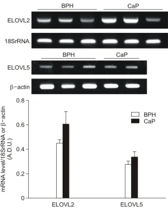
±0.140, n=10)에 비해 모두 유의하게 낮았다 (Fig. 1). 고도 불포화 지방산 (polyunsaturated fatty acid)을 기질로 하는 지 방산 elongase (ELOVL)인 ELOVL2와 ELOVL5의 mRNA 발 현을 측정한 결과, ELOVL2 mRNA는 전립선암 조직에서 증가하는 경향을 보였으나 통계적인 유의성은 없었고, ELOVL5 mRNA 발현도 두 군 간에 차이가 없었다 (Fig. 2).
당대사효소 중 선행연구들에서 안드로겐에 의해 발현이 조절되는 것으로 알려져 있는 phosphoglycerate kinase 1 (PgK1)의 mRNA 발현은 두 군 간에 유의한 차이를 보이지 않았으나, phosphoglycerate mutase 2의 mRNA 발현은 전립 선암 조직 (0.152±0.040, n=6, p=0.001)에서 전립선비대증 조직 (0.703±0.073, n=10)에 비해 현저히 낮았다 (Fig. 3).
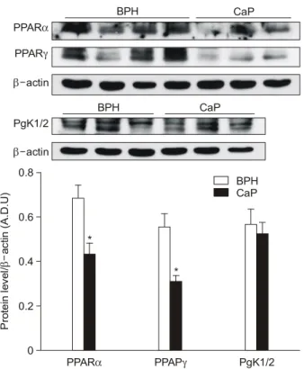
면역탁본법으로 PPARα, PPARγ, PgK1/2의 단백발현 정 도를 측정한 결과, 전립선암조직에서 PPARα (0.435±0.050, n=6, p=0.035)와 PPARγ (0.314±0.025, n=6, p=0.035)의 단 백발현이 전립선비대증 조직 (PPARα, 0.684±0.061, n=7;
PPARγ, 0.556±0.062, n=7)에 비해 낮았으나 PgK1/2의 단 백발현은 두 군 간에 차이가 없어 (Fig. 4), 두 조직에서의 Fig. 1. Representative examples (upper panel) and densitometric
analyses (lower panel) of the polymerase chain reaction (PCR) products for peroxisome proliferator-activated receptor (PPAR)α and PPARγ, corrected for the 18S rRNA value, in each sample.
The values are expressed in arbitrary densitometric units (A.D.U.) as the mean±SE of human prostate tissue; either benign pro- static hyperplasia (BPH) or prostate cancer (CaP). *p<0.05, vs.
BPH (Mann-Whitney test).
PPARα PPARγ
mRNA level/18SrRNA (A.D.U)
0 0.2 0.4 0.6 0.8 1 1.2
CaP BPH
*
BPH CaP
PPARα
PPARγ
18SrRNA
*
Fig. 2. Representative examples (upper panel) and densitometric analyses (lower panel) of the polymerase chain reaction (PCR) products for fatty acid elongase (ELOVL) 2 and ELOVL5, cor- rected for 18S rRNA or β-actin, respectively, in each sample.
The values are expressed in arbitrary densitometric units (A.D.U.) as the mean±SE of human prostate tissue; either benign prostatic hyperplasia (BPH) or prostate cancer (CaP).
BPH CaP
ELOVL2
18SrRNA
ELOVL2 ELOVL5
mRNA level/18SrRNA or actin (A.D.U.)β- 0 0.2 0.4 0.6 0.8
CaP BPH
BPH CaP
ELOVL5
β-actin
PPARα, PPARγ, PgK1/2의 단백발현과 유전자 발현 양상 이 일치하였다.
고 찰
본 연구 결과 PPARα와 PPARγ 및 phosphoglycerate mutase 2 (PgM2)의 mRNA 발현은 전립선암 조직에서 전립 선비대증 조직에 비해 현저히 낮았으며, 지방산 elongase (ELOVL)2, ELOVL5, 그리고 phosphoglycerate kinase 1 (PgK1)의 mRNA 발현은 두 군 간에 유의한 차이를 보이지 않았다. 또한 전립선암 조직의 PPARα와 PPARγ의 단백발 현이 전립선비대증 조직에 비해 낮았고 PgK1/2의 단백발현 은 두 군 간에 차이가 없어, 두 조직에서의 유전자 발현 양 상이 일치하였다.
전립선암 세포주에서 PPARγ mRNA 발현이 정상 전립 선세포보다 높으며19 전립선암 조직에서 PPARγ 단백의 면 역 반응력이 정상 조직에서보다 높다는 보고20도 있으나, Mueller 등21에 의하면 정상 전립선조직에 비해 전립선암 조 직에서 PPARγ mRNA 발현이 낮았으며, LNCaP 세포나
DU145 세포보다 높은 PPARγ 단백 발현을 보인 PC3 세포 에서 인산화되어 불활성인 PPARγ 단백이 상당한 수준으 로 측정되었다고 하였다. 또한 Jiang 등17은 linoleic acid로부 터 13-hydroxyoctadecadienoic acid (13-HODE)의 생성에 관여 하는 15-lipoxigenase-1의 발현은 전립선암에서 오히려 증가 되어 있으며, 그 결과 13-HODE가 mitogen activated protein kinase의 활성을 증가시키므로 PPARγ의 인산화가 촉진되 어 전립선 조직 내 PPARγ 함량은 증가되나 PPARγ 배위 자에 대한 민감도는 오히려 저하된다고 하였다. PPARγ 배 위자인 troglitazone21과 rosiglitazone22은 당뇨병에서 흔한 고 지질증의 치료제로 개발되었으며 암세포 분화 촉진에 의한 항암효과에 대한 2상 임상연구 결과 단독투여만으로는 전 립선암 치료효과가 낮으며, 전립선암에서의 PPARγ의 작 용에 대한 연구가 더 수행되면 향후 다른 약제와의 병행요 법이 가능할 것으로 보고되었다.23 그리고 PPARγ 배위자 는 부작용이 적으므로 보다 효과적인 새로운 PPARγ 배위 자를 개발하거나 현재 사용 중인 PPARγ 배위자에 반응하 는 환자들의 특성을 파악하는 것도 좋은 시도가 될 수 있다.
PPARα는 간세포에서 암 유발에 관여하고, 안드로겐에 Fig. 3. Representative examples (upper panel) and densitometric
analyses (lower panel) of the polymerase chain reaction (PCR) products of phosphoglycerate kinase 1 (PgK1) and phosphogly- cerate mutase 2 (PgM2), corrected for the 18S rRNA value, in each sample. The values are expressed in arbitrary densitometric units (A.D.U.) as the mean±SE of human prostate tissue; either benign prostatic hyperplasia (BPH) or prostate cancer (CaP). *p<
0.01, vs. BPH (Mann-Whitney test).
PgM2 PgK1
mRNA level/18SrRNA (A.D.U)
0 0.2 0.4 0.6 0.8
CaP BPH
BPH CaP
PgM2
PgK1
18SrRNA
*
Fig. 4. Expressions of peroxisome proliferator-activated receptor (PPAR)α, PPARγ and phosphoglycerate kinase 1 (PgK1), cor- rected for β-actin, in each sample. Values are shown in arbitrary densitometric units (A.D.U.) as the mean±SE of human prostate tissue; either benign prostatic hyperplasia (BPH) or prostate can- cer (CaP). *p<0.05, vs. BPH (Mann-Whitney test).
BPH CaP
PPARα PPARγ
β-actin
BPH CaP
PgK1/2 β-actin
Protein level/actin (A.D.U)β-
0 0.2 0.4 0.6 0.8
CaP BPH
PPARα PPAPγ PgK1/2
*
*
의존적으로 조절되며, 지방산과 스테로이드 호르몬의 신호 전달계간의 통합에 관여할 것으로 보고되었다.24 전립선암 세포주를 사용한 실험에서 Mueller 등21은 PPARγ 배위자에 의해서는 암세포의 성장이 용량 의존적으로 억제되었으나 PPARα 배위자나 PPARδ 배위자는 암세포의 성장에 영향 을 미치지 않음을 보고하였다. Collett 등24 은 전립선비대증 조직 기저상피세포에서는 PPARα mRNA가 거의 발현되지 않거나 매우 낮게 발현되었고, Gleason score가 8-10인 전립 선암 조직에서는 높게 발현되었다고 한다. 본 연구에서는 전립선암의 경우 전립선비대증에 비해 PPARα mRNA와 단 백발현이 낮았으며, 전립선암의 분화도나 병기에 따른 PPARα의 발현양상에 대해서는 좀 더 연구해야 할 것이다.
암세포는 Krebs cycle에 결함이 있으므로 거의 대부분의 에너지를 해당과정에 의존하고 있으며,15 해당과정에 관여 하는 당대사 효소 중 하나인 PgK1은 serine proteinase인 plasmin을 환원시켜 angiostatin 유리를 유도하여 혈관신생 과 종양 성장을 억제하는 것으로 보고되었으나,25 본 연구에 서는 PgK1 mRNA와 단백발현에서 전립선암 조직과 전립선 비대증 조직 간에 차이가 없었다. 그러나 resveratrol의 항암 작용에 대한 선행연구15에서 전립선암세포의 성장을 억제 하고 세포사멸에 관여할 것으로 보고된 PgM2의 mRNA 발 현은 전립선비대증 조직에 비해 전립선암 조직에서 현저히 낮았다.
상피세포인 전립선암 세포주를 사용한 실험과 전립선암 조직에서의 결과가 다른 것은 선행연구26에서도 보고되었 으며, 암 조직에서의 유전자 발현 양상의 변화는 대부분의 경우 유전자 염기서열의 변화보다는 DNA 메틸화 (methyla- tion)를 포함하는 후성유전적 변화 (epigenetic modification) 로 환경요인들의 영향을 받게 된다.16 또한 일부 유전자들의 변화는 상피세포와 주변의 다른 세포들과의 상호작용에 의 해 영향을 받는 경우16,17도 있어, 전립선암 세포와 암 조직 에서 다른 결과를 보일 가능성도 고려해 볼 수 있으나, 전립 선에서의 PPARα와 PgM2의 생물학적 활성에 대해서는 좀 더 연구해야 하겠다.
결 론
전립선암 조직에서의 PPARα와 PPARγ, 및 phosphogly- cerate mutase 2의 mRNA 발현은 전립선비대증 조직에 비해 현저히 낮았으며, 이들 인자들의 발현 변화가 전립선 암세 포의 비정상적인 증식과 분화 유발에 관여할 가능성을 보 여주고 있다.
REFERENCES
1. Gleave M, Miyake H, Chi K. Beyond simple castration:
targeting the molecular basis of treatment resistance in advanced prostate cancer. Cancer Chemother Pharmacol 2005;
56(Suppl 1):47-57
2. Pienta KJ, Smith DC. Advances in prostate cancer chemo- therapy: a new era begins. CA Cancer J Clin 2005;55:300-18 3. Freeman VL, Meydani M, Hur K, Flanigan RC. Inverse
association between prostatic polyunsaturated fatty acid and risk of locally advanced prostate carcinoma. Cancer 2004;
101:2744-54
4. Augustsson K, Michaud DS, Rimm EB, Leitzmann MF, Stampfer MJ, Willett WC, et al. A prospective study of intake of fish and marine fatty acids and prostate cancer. Cancer Epidemiol Biomarkers Prev 2003;12:64-7
5. Mamalakis G, Kafatos A, Kalogeropoulos N, Andrikopoulos N, Daskalopoulos G, Kranidis A. Prostate cancer vs hyperpla- sia: relationships with prostatic and adipose tissue fatty acid composition. Prostaglandins Leukot Essent Fatty Acids 2002;
66:467-77
6. Yang YJ, Lee SH, Hong SJ, Chung BC. Comparison of fatty acid profiles in the serum of patients with prostate cancer and benign prostatic hyperplasia. Clin Biochem 1999;32:405-9 7. Terry PD, Rohan TE, Wolk A. Intakes of fish and marine fatty
acids and the risks of cancers of the breast and prostate and of other hormone-related cancers: a review of the epidemio- logic evidence. Am J Clin Nutr 2003;77:532-43
8. Schoonjans K, Staels B, Auwerx J. Role of the peroxisome proliferator-activated receptor (PPAR) in mediating the effects of fibrates and fatty acids on gene expression. J Lipid Res 1996;37:907-25
9. Green S, Wahli W. Peroxisome proliferator-activated recep- tors: finding the orphan a home. Mol Cell Endocrinol 1994;
100:149-53
10. Dreyer C, Krey G, Keller H, Givel F, Helftenbein G, Wahli W. Control of the peroxisomal beta-oxidation pathway by a novel family of nuclear hormone receptors. Cell 1992;68:
879-87
11. Kliewer SA, Sundseth SS, Jones SA, Brown PJ, Wisely GB, Koble CS, et al. Fatty acids and eicosanoids regulate gene expression through direct interactions with peroxisome pro- liferator-activated receptors alpha and gamma. Proc Natl Acad Sci USA 1997;94:4318-23
12. Pawar A, Xu J, Jerks E, Mangelsdorf DJ, Jump DB. Fatty acid regulation of liver X receptors (LXR) and peroxisome proli- ferator-activated receptor alpha (PPARalpha) in HEK293 cells.
J Biol Chem 2002;277:39243-50
13. Wang Y, Botolin D, Christian B, Busik J, Xu J, Jump DB.
Tissue-specific, nutritional, and developmental regulation of rat
fatty acid elongases. J Lipid Res 2005;46:706-15
14. Horton JD, Shah NA, Warrington JA, Anderson NN, Park SW, Brown MS, et al. Combined analysis of oligonucleotide microarray data from transgenic and knockout mice identifies direct SREBP target genes. Proc Natl Acad Sci USA 2003;
100:12027-32
15. Narayanan NK, Narayanan BA, Nixon DW. Resveratrol- induced cell growth inhibition and apoptosis is associated with modulation of phosphoglycerate mutase B in human prostate cancer cells: two-dimensional sodium dodecyl sulfate-poly- acrylamide gel electrophoresis and mass spectrometry evalua- tion. Cancer Detect Prev 2004;28:443-52
16. Rose A, Xu Y, Chen Z, Fan Z, Stamey TA, McNeal JE, et al. Comparative gene and protein expression in primary cul- tures of epithelial cells from benign prostatic hyperplasia and prostate cancer. Cancer Lett 2005;227:213-32
17. Jiang M, Shappell SB, Hayward SW. Approaches to under- standing the importance and clinical implications of peroxi- some proliferator-activated receptor gamma (PPARgamma) signaling in prostate cancer. J Cell Biochem 2004;91:513-27 18. van Baren MJ, Heutink P. The PCR suite. Bioinformatics
2004;20:591-3
19. Nwankwo JO, Robbins ME. Peroxisome proliferator-activated receptor-gamma expression in human malignant and normal brain, breast and prostate-derived cells. Prostaglandins Leukot Essent Fatty Acids 2001;64:241-5
20. Segawa Y, Yoshimura R, Hase T, Nakatani T, Wada S,
Kawahito Y, et al. Expression of peroxisome proliferator- acti- vated receptor (PPAR) in human prostate cancer. Prostate 2002;51:108-16
21. Mueller E, Smith M, Sarraf P, Kroll T, Aiyer A, Kaufman DS, et al. Effects of ligand activation of peroxisome proliferator- activated receptor gamma in human prostate cancer. Proc Natl Acad Sci USA 2000;97:10990-5
22. Smith MR, Manola J, Kaufman DS, George D, Oh WK, Mueller E, et al. Rosiglitazone versus placebo for men with prostate carcinoma and a rising serum prostate-specific antigen level after radical prostatectomy and/or radiation therapy.
Cancer 2004;101:1569-74
23. Strother JM, Beer TM, Dreicer R. Novel cytotoxic and biological agents for prostate cancer: where will the money be in 2005? Eur J Cancer 2005;41:954-64
24. Collett GP, Betts AM, Johnson MI, Pulimood AB, Cook S, Neal DE, et al. Peroxisome proliferator-activated receptor alpha is an androgen-responsive gene in human prostate and is highly expressed in prostatic adenocarcinoma. Clin Cancer Res 2000;6:3241-8
25. Lay AJ, Jiang XM, Kisker O, Flynn E, Underwood A, Cond- ron R, et al. Phosphoglycerate kinase acts in tumour angio- genesis as a disulphide reductase. Nature 2000;408:869-73 26. Stamey TA, Warrington JA, Caldwell MC, Chen Z, Fan Z,
Mahadevappa M, et al. Molecular genetic profiling of Gleason grade 4/5 prostate cancers compared to benign prostatic hyper- plasia. J Urol 2001;166:2171-7