- 281 -
Imaging Science in Dentistry 2017; 47: 281-4 https://doi.org/10.5624/isd.2017.47.4.281
Dermal filling injection is a technique extensively used in modern therapeutic approaches for cosmetic tissue aug- mentation and the correction of skin depressions1 in the maxillofacial area. The increased demand has led to the development of a variety of commercial cosmetic fillers,2 with polymethyl methacrylate microspheres suspended in a solution of bovine collagen2,3 being widely used. Al- though cosmetic fillers are non-toxic, non-immunogenic,4 and minimally invasive,1 complications are associated with their use, such as foreign body granuloma forma- tion,3,5 which is rare and has a delayed onset.
The clinical presentation of foreign body granuloma varies, ranging from a painful firm swelling to a painful nodule,6 and patients usually seek treatment from dental care practitioners and oral surgeons. Therefore, clinicians in both of those categories should be prepared to evaluate
these patients accurately. This report presents the unusu- al case of a 52-year-old woman with a facial granuloma mimicking a benign neoplasm, underscoring the impor- tance of an appropriate diagnosis in order to avoid con- founding it with a true pathological entity.
Case Report
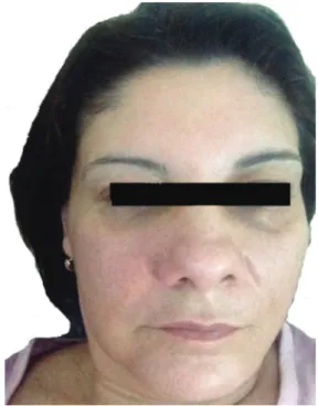
A 52-year-old woman visited a private dentist com- plaining of a painless swelling in the face characterised by unilateral volume of the nasolabial area. The swelling had been present for nearly 3 months and the patient re- ported that it had not increased in size since developing (Fig. 1). The patient’s medical history and habits were non-informative. Upon an oral examination, the right and left central incisors, left maxillary lateral incisor, and ca- nine responded to thermal and electric pulp testing within normal limits. Periodontal probing showed normal and healthy gingiva. On palpation, swelling was non-tender, firm in consistency, and not fixed. There was no detect- able lesion on periapical radiographs. Clinically, a benign
Magnetic resonance imaging appearance of foreign-body granulomatous reactions to dermal cosmetic fillers
Andre Luiz Ferreira Costa
1, Rubens Caliento
2, Glauber Bareia Liberato da Rocha
2, Joao Pedro Perez Gomes
3, Alison Jhisel Calle Mansmith
1, Claudio Froes de Freitas
1,4, Paulo Henrique Braz-Silva
3,5,*
1Department of Radiology and Orthodontics, School of Dentistry, University City of Sao Paulo, Sao Paulo, Brazil
2Division of Oral and Maxillofacial Pathology, Department of Stomatology, School of Dentistry, University of Sao Paulo, Sao Paulo, Brazil
3Division of General Pathology, Department of Stomatology, School of Dentistry, University of Sao Paulo, Sao Paulo, Brazil
4Division of Radiology, Department of Stomatology, School of Dentistry, University of Sao Paulo, Sao Paulo, Brazil
5Laboratory of Virology, Institute of Tropical Medicine of Sao Paulo, University of Sao Paulo, Sao Paulo, Brazil
AbstRACt
Foreign body granulomas can develop after the injection of various cosmetic filling materials into the facial area to flatten wrinkles. Clinically, reactive lesions are easily mistaken for soft-tissue neoplasms or cysts. This report presents a case of foreign body granuloma in a 52-year-old female patient complaining of a painless swelling in the nasolabial region. Both clinical and histological features are described, underscoring the diagnostic role of magnetic resonance imaging findings.(Imaging Sci Dent 2017; 47: 281-4)
Key woRds: Magnetic Resonance Imaging; Granuloma, Foreign-Body; Dermal Filllers
Copyright ⓒ 2017 by Korean Academy of Oral and Maxillofacial Radiology
This is an Open Access article distributed under the terms of the Creative Commons Attribution Non-Commercial License(http://creativecommons.org/licenses/by-nc/3.0) which permits unrestricted non-commercial use, distribution, and reproduction in any medium, provided the original work is properly cited.
Imaging Science in Dentistry·pISSN 2233-7822 eISSN 2233-7830 Received September 8, 2017; Revised October 21, 2017; Accepted October 31, 2017
*Correspondence to : Prof. Paulo Henrique Braz-Silva
Division of General Pathology, Department of Stomatology, School of Dentistry, University of São Paulo, Av. Prof. Lineu Prestes, 2227 Cidade Universitária, São Paulo - SP 05508-000, Brazil
Tel) 55-11-2648-8144, Fax) 55-11-30917902, E-mail) pbraz@usp.br
Magnetic resonance imaging appearance of foreign-body granulomatous reactions to dermal cosmetic fillers
- 282 -
neoplasm of the soft tissue was hypothesised.
Magnetic resonance imaging(MRI) of the head was performed as an additional assessment, revealing a low-intensity rounded lesion on T2-weighted images (axial view)(Fig. 2A) and anterior wall involvement of the ipsilateral maxillary sinus with slight bulging. On the T1-weighted post-gadolinium images with spectral pre-saturation with inversion recovery(SPIR), the coronal scan showed a region with a low and homogeneous signal and well-circumscribed limits(Fig. 2B). The T1-weighted sagittal image with an iso-hypointense signal revealed an
augmented mass in the medial canthal area(Fig. 2C). The signal characteristics and morphology conflicted with those of a tumour.
After MRI, the patient was further questioned about her medical history regarding the nasolabial folds, and she re- ported undergoing an aesthetic procedure with filling ma- terial performed by an aesthetic plastic surgeon 15 years ago, but she was unable to say which material was used.
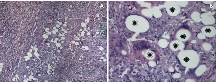
An incisional biopsy was performed and histopatholog- ical analysis showed a well-circumscribed granulomatous reaction without central necrosis, characterized by an ep- ithelioid histiocytic organization, numerous multinucle- ated giant cells with peripheral disposition of the nuclei, and optically clear vacuoles in the cytoplasm, suggesting that polymethyl methacrylate was the foreign body. The peripheral areas of the granulomas were surrounded by a collagenous capsule with mononuclear inflammatory cell infiltration(Fig. 3).
The patient was scheduled for an excisional biopsy. A nodule, measuring 2.7cm×1.6cm×1.1cm, of fibrous consistency, whitish colouration, lobular surface, and irregular growth was easily removed under local anaes- thesia, including a small amount of healthy surrounding tissue. The post-operative period was uneventful, and her recovery was uncomplicated.
discussion
Injectable cosmetic fillers are widely used in cosmetic surgery for their lasting effects and few complications.5,7,8 However, some complications such as inflammatory gran- uloma may occur at the injection site or other sites, even several years after the operation.7 Granulomas are caused
Fig. 1. Swelling and mild redness is seen of the right nasolabial fold.
Fig. 2. A. An axial T2-weighted MR image shows a cluster of low-intensity fluid collection. B. A coronal T1-weighted spectral pre-satura- tion with inversion recovery(SPIR) image with contrast surrounded by a thin hypo-intense capsule in the nasolabial fold. C. A T1-weighted sagittal image reveals an iso-hypointense collection.
A B C
- 283 -
Andre Luiz Ferreira Costa et al
by granulomatous inflammation after the aggregation of macrophages in response to large foreign bodies that can- not be phagocytosed by macrophages.9
Foreign body granulomas can arise following the injec- tion of dermal fillers, manifesting with various clinical and histological features depending on the type of inject- ed filler,7 frequently several years after the original cos- metic treatment.1 Therefore, because of the period elapsed between the surgical procedure and the complications, it is common that these patients do not remember the filler they received, or the origin of the lesion.1
A strong female tendency is evident among all previ- ously published reports, possibly reflecting the tendency of women to seek cosmetic care more often than men,5,10 as in the present case. In addition, the patient’s age and the location of the lesion reflect the fact that physiologi- cal lengthening and loss of volume are expected to occur with aging.10
The differential diagnosis may encompass a wide range of conditions. Labial cases presenting well-defined nod- ules suggest salivary gland cysts and tumours, in addition to soft-tissue neoplasms and cysts.10,11 The MRI appear- ance of facial fillers varies according to the type of filler used.3 In this case, the T1- and T2-weighted MRI scans showed that the lesion was well-circumscribed and typ- ically iso-intense or hypo-intense to the superficial and deep layers of the facial fat.
The characteristics of soft-tissue facial tumours on MRI depend on the histological grade of the tumour,12 but in general, these lesions present an intermediate signal on T1-weighted images and hyper-intensity on T2-weight-
ed images with enhancement after contrast administra- tion.12,13
The advent of long-standing foreign body granulomas due to cosmetic fillers can cause confusion, as patients may not remember the previous facial filling treatment or when it occurred. The clinical features include erythema- tous and indurated painless nodules or painful swelling,2,6 but such features are non-specific. This means that they are often difficult to distinguish from other pathological conditions.
Imaging is important, not only to confirm the diagnosis of foreign body granuloma lesions, but also in the differ- ential diagnosis of other lesions.5,14 Ultrasound has been reported to be useful for identifying granuloma lesions,14 but this procedure has weaknesses, such as the absence of certain anatomical landmarks, the lack of consolidated criteria to diagnose inflammatory reactions, and depen- dence on the operator’s skill.1
MRI seems to be the best diagnostic tool, allowing a correct assessment of filler dislocation due to multipla- nar acquisitions and determination of anatomical land- marks.14 Various studies have investigated MRI1,5,14,15 as a diagnostic modality for accurately identifying the presence of foreign material. Grippaudo et al.14 showed that contrast-enhanced MRI enabled the identification of sub-cutaneous abscesses or granulomas characterized, re- spectively, by circular or diffuse enhancement.
In conclusion, clinicians should keep in mind that there are several clinical similarities between granulomatous reactions due to dermal fillers and salivary gland cysts or tumours. The integration of clinical examinations and
Fig. 3. Histopathological view(hematoxylin and eosin stain; original magnification A. 100×. B. 400×): A well-circumscribed granulo- matous reaction without necrosis, showing a large number of multinucleated giant cells with peripheral disposition of the nuclei associated with areas without substance, indicating foreign body particles(*).
A B
Magnetic resonance imaging appearance of foreign-body granulomatous reactions to dermal cosmetic fillers
- 284 -
imaging techniques, particularly MRI, enables a correct diagnosis to be made.
References
1. Di Girolamo M, Mattei M, Signore A, Grippaudo FR. MRI in the evaluation of facial dermal fillers in normal and compli- cated cases. Eur Radiol 2015; 25: 1431-42.
2. Vargas-Machuca I, González-Guerra E, Angulo J, del Carmen Fariña M, Martin L, Requena L. Facial granulomas secondary to Dermalive microimplants: report of a case with histopatho- logic differential diagnosis among the granulomas secondary to different injectable permanent filler materials. Am J Derma- topathol 2006; 28: 173-7.
3. Ginat DT, Schatz CJ. Imaging features of midface injectable fillers and associated complications. AJNR Am J Neuroradiol 2013; 34: 1488-95.
4. Shahrabi Farahani S, Sexton J, Stone JD, Quinn K, Woo SB.
Lip nodules caused by hyaluronic acid filler injection: report of three cases. Head Neck Pathol 2012; 6: 16-20.
5. Tal S, Maresky HS, Bryan T, Ziv E, Klein D, Persitz A, et al.
MRI in detecting facial cosmetic injectable fillers. Head Face Med 2016; 12: 27.
6. Shahrabi-Farahani S, Lerman MA, Noonan V, Kabani S, Woo SB. Granulomatous foreign body reaction to dermal cosmetic fillers with intraoral migration. Oral Surg Oral Med Oral Pathol Oral Radiol 2014; 117: 105-10.
7. Lee JM, Kim YJ. Foreign body granulomas after the use of
dermal fillers: pathophysiology, clinical appearance, histolog- ic features, and treatment. Arch Plast Surg 2015; 42: 232-9.
8. Kawamura JY, Domaneschi C, Migliari DA, Sousa SO. For- eign body reaction due to skin filler: a case report. Oral Surg Oral Med Oral Pathol Oral Radiol Endod 2006; 101: 469-71.
9. Bentkover SH. The biology of facial fillers. Facial Plast Surg 2009; 25: 73-85.
10. Jham BC, Nikitakis NG, Scheper MA, Papadimitriou JC, Levy BA, Rivera H. Granulomatous foreign-body reaction involving oral and perioral tissues after injection of bioma- terials: a series of 7 cases and review of the literature. J Oral Maxillofac Surg 2009; 67: 280-5.
11. Ficarra G, Mosqueda-Taylor A, Carlos R. Silicone granuloma of the facial tissues: a report of seven cases. Oral Surg Oral Med Oral Pathol Oral Radiol Endod 2002; 94: 65-73.
12. Razek AA, Huang BY. Soft tissue tumors of the head and neck: imaging-based review of the WHO classification. Ra- diographics 2011; 31: 1923-54.
13. Abdel Razek AA. Computed tomography and magnetic res- onance imaging of lesions at masticator space. Jpn J Radiol 2014; 32: 123-37.
14. Grippaudo FR, Di Girolamo M, Mattei M, Pucci E, Grippaudo C. Diagnosis and management of dermal filler complications in the perioral region. J Cosmet Laser Ther 2014; 16: 246-52.
15. Josse G, Haftek M, Gensanne D, Turlier V, Mas A, Lagarde JM, et al. Follow up study of dermal hyaluronic acid injection by high frequency ultrasound and magnetic resonance imag- ing. J Dermatol Sci 2010; 57: 214-6.