https://doi.org/10.5624/isd.2020.50.1.45
Introduction
Diagnostic imaging is an essential component of treat
ment planning in oral rehabilitation through implant pla
cement.1 Clinicians can use conventional radiography or conebeam computed tomography(CBCT) for diagnostic imaging. CBCT enables clinicians to obtain 3dimension
al images.2 However, since the radiation doses of dental CBCT are usually higher than those of conventional(2di
mensional) radiography, it is very important to consider the risks of radiation exposure when using CBCT for di
agnostic purposes in dentistry.3
Clinical guidelines are systematically developed state
ments that assist clinicians and patients in making deci
sions about the most appropriate care for specific clinical circumstances. In other words, guidelines can be helpful in situations that require clinicians to choose an appropriate imaging modality. Guidelines are often referred to as “se
lection criteria” or “referral criteria.”
In the field of radiology, developed countries utilize evi
dencebased clinical imaging guidelines(CIG) to augment
Development of an evidence-based clinical imaging diagnostic guideline for implant planning: Joint recommendations of the Korean Academy of Oral and Maxillofacial Radiology and National Evidence-based Healthcare Collaborating Agency
MinJi Kim 1, SamSun Lee 1,*, Miyoung Choi 2, Eun Ju Ha 3, Chena Lee 4, JoEun Kim 1, MinSuk Heo 1
1Department of Oral and Maxillofacial Radiology and Dental Research Institute, School of Dentistry, Seoul National University, Seoul, Korea
2Division for Healthcare Technology Assessment Research, National Evidence-based Healthcare Collaborating Agency, Seoul, Korea
3Department of Radiology, Ajou University School of Medicine, Suwon, Korea
4Department of Oral and Maxillofacial Radiology, Yonsei University College of Dentistry, Seoul, Korea
ABSTRACT
Purpose: This study was conducted to develop an evidencebased clinical imaging diagnostic guideline for implant planning, taking into account efficacy, benefits, and risks.
Materials and Methods: The guideline development process employed the adaptation methodology used for Korean clinical imaging guidelines(KCIG). Core databases(OvidMedline, OvidEmbase, National Guideline Clearinghouse, Guideline International Network) and domestic databases(KoreaMed, KMbase, and KoMGI) were searched for guidelines. The retrieved articles were analyzed by 2 reviewers, and articles were selected using wellestablished inclusion criteria.
Results: The search identified 294 articles, of which 3 were selected as relevant guidelines. Based on those 3 guide
lines, 3 recommendations for implant planning were derived.
Conclusion: We recommend radiography or conebeam computed tomography(CBCT) scanning for individual pat
ients judged to require a crosssectional image after reading of a panoramic Xray image and a conventional intraoral radiological image. Various steps should be taken to raise awareness of these recommendations among clinicians and the public, and KCIG should be regularly reviewed and revised.(Imaging Sci Dent 2020; 50: 45-52)
KEY WORDS: Implant; Imaging; Radiography; ConeBeam Computed Tomography
Copyright ⓒ 2020 by Korean Academy of Oral and Maxillofacial Radiology
This is an Open Access article distributed under the terms of the Creative Commons Attribution NonCommercial License(http://creativecommons.org/licenses/bync/3.0) which permits unrestricted noncommercial use, distribution, and reproduction in any medium, provided the original work is properly cited.
Imaging Science in Dentistry·pISSN 22337822 eISSN 22337830
*This work received financial support from the Research Program funded by the Korea Centers for Disease Control and Prevention(fund code: 2017E3600100). This study was a coinvestigation of the National Evidencebased Collaborating Agency and the Korean Society of Radiology (research number: NECAS17005).
Received September 19, 2019; Revised November 20, 2019; Accepted December 3, 2019
*Correspondence to : Prof. SamSun Lee
Department of Oral and Maxillofacial Radiology, School of Dentistry, Seoul National University, 101 Daehakro, Jongnogu, Seoul 03080, Korea
Tel) 82220723978, Email) [email protected]
the clinical decisionmaking of physicians when requesting or prescribing a radiological examination. In Korea, the methodology of developing guidelines is to adapt CIG by modifying previously developed guidelines to make them suitable for the local healthcare environment.4 Through this process, this study aimed to develop an evidencebased Korean clinical imaging guidelines(KCIG) for implant planning, taking into account efficacy, benefits, and risks.
Materials and Methods
Development of Korean CIG for implant planning The guideline development process involved a collab
oration between the Korean Academy of Oral and Maxil
lofacial Radiology(KAOMFR) and the Korean Society of Radiology, and the National Evidencebased Healthcare Collaborating Agency(NECA) organized a development committee and working group to develop this guideline.
Three experts in oral and maxillofacial radiology experts comprised the working group. The working group, research methodology specialists, and clinical guideline specialists who supported the overall planning and research method
ology comprised the development committee.4 They pub
lished a description of the methodology of the guideline adaptation process that was applied in this study. A consen
sus group consisted of 5 nominated members from the final 5 related academic societies who participated in the sym
posium conducted to establish a consensus.
Defining the key question
Questions were generated in the form of population/
patient, intervention/index test, comparator/control, and outcome(PICO) questions by the working group and were reviewed by the development committee and the consen
sus group. The following key question was identified: for a patient scheduled for implantation, what is the appropriate imaging modality?
Guideline search
Core databases such as OvidMedline, OvidEmbase, National Guideline Clearinghouse, and Guideline Inter
national Network were searched for guidelines. Addition
ally, 3 domestic research databases(KoreaMed, KMbase, and KoMGI) were searched from 2000 to the first week of March 2017. The presearch yielded 51 article abstracts.
The extensive searches of databases used the terms “dental implant,” “radiograph,” “guideline,” “recommendation,”
and “practice guideline.” The working group reviewed
the search strategy and results and performed additional searches to ensure the inclusion of any important omitted guidelines.
Selection of the searched guidelines
According to predefined selection criteria, 2 members of the working group independently reviewed the literature during the primary screening process and secondary selec
tion process to ensure objectivity. The primary screening process involved reviewing the title and abstract of the identified studies and guidelines. In the secondary selection process, the full text of the identified studies was reviewed, and the reasons for excluding studies were noted.
The inclusion criteria for guidelines were as follows:
1) the study population included patients scheduled for implantation, 2) the study intervention was CBCT, 3) the study comparators were panoramic and periapical radio
graphs, 4) the study assessed the effectiveness of CBCT for evaluating alveolar bone morphology in edentulous re
gions and its surrounding structures, 5) the study presented a practice guideline, 6) the study presented recommenda
tions, 7) the study utilized an evidencebased method, and 8) the study was published in Korean or English.
The exclusion criteria were as follows: 1) patients of interest for the key question were not included, 2) a key questionrelated imaging examination was not included, 3) appropriate results(diagnostic accuracy, efficacy, safety, prognosis, and patients’ preferences) were not reported, 4) the study presented nonclinical practical guidelines, 5) recommendations were not suggested, 6) the guidelines were not produced via an evidencebased method, 7) the guidelines were reported in neither English nor Korean, 8) the study was an overlapping publication, and 9) the full text was not obtainable.
Disagreements between reviewers were resolved either by consultation between the reviewers or by obtaining in
put from a third reviewer.
Search for recent literature
Randomized controlled trials(RCTs) and observational studies were searched, and the recent literature(since 2011) was reviewed.
Quality assessment
The finally selected guidelines underwent quality ap
praisal using the Korean Appraisal of Guidelines for Re
search and Evaluation II(AGREE II) tool.5 Two appraisers from the development committee independently assessed
the selected literature. Each evaluation category was scored on a scale ranging from 1 to 7 points, and the reasoning behind the scores was noted to ensure clarity and reproduc
ibility of the assessment results. If there was a difference
>4 in scores for any of the categories among the apprais
ers, the study was reexamined. In essence, guidelines that scored 50 or above in the “rigor of development” domain were considered candidates for inclusion in the develop
ment process of Korean CIG(KCIG).4
Grading the level of evidence and drafting the recommendation document
This step assessed whether an identified guideline was uptodate, acceptable, and applicable. The level of evi
dence of the KCIG was merged with the evidence level of individual studies, and was categorized as high(I), moder
ate(II), low(III), or very low(IV).
A draft of the recommendation document consisted of recommendations for the key question, summary, and ev
idence; considerations for the recommendation and refer
ences; and each recommendation with its overall evidence level. The recommendations in the KCIG were graded as A, B, C, or I, indicating the strength of the recommenda
tion.
External review and approval of the clinical guideline
The finalized recommendation document was reviewed both internally by clinical imaging experts who did not participate in the development of the guideline and exter
nally by related society members(endusers of the guide
line). Appropriate modifications were made after collect
ing opinions.
Results
PICO
The guideline was developed based on the key ques
tion, which was generated from the PICO questions by the working group. In this study, the population comprised patients scheduled for implantation. The intervention was CBCT. The comparators were panoramic and periapical radiographs. The outcome was the effectiveness of CBCT for evaluating alveolar bone morphology in edentulous pa
tients and its surrounding structures.
Search for guidelines
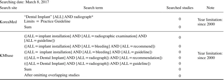
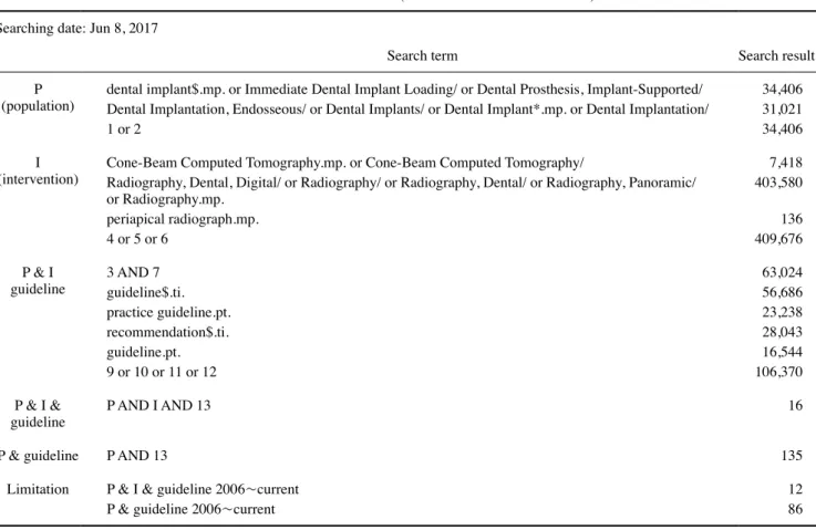
The search results from domestic databases are shown in Table 1. No results were obtained from KoMGI. The search results from international databases are shown in Tables 2 and 3. By searching for “dental implant,” 5 search results were obtained from the Guideline International Network and National Guideline Clearinghouse databases.
Selection of searched guidelines
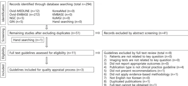
A total of 294 guidelines were retrieved from the data
bases. After the exclusion of duplicates, 51 guidelines re
mained. Finally, 3 guidelines were selected in accordance with the inclusion and exclusion criteria(Fig. 1).
Search for recent studies
The recent literature(since 2011) was reviewed. This starting point was 3 years earlier than the most recent guideline. RCTs and observational studies that were identi
fied by applying condition number 4 in Table 4 were read;
as a result, 6 studies were selected.
Table 1. Search results from domestic literature databases Searching date: March 8, 2017
Search site Search term Searched studies Note
KoreaMed “Dental Implant” [ALL] AND radiograph*
Limits = Practice Guideline 0 Year limitation:
since 2000
Sum 0
KMbase
([ALL=implant installation] AND [ALL=radiographic examination] AND
[ALL=guideline]) 0
Year limitation:
since 2000 ([ALL=implant installation] AND [ALL=bleeding] AND [ALL=recommend]) 0
([ALL= implant installation] AND [ALL=bleeding] AND [ALL=guideline]) 0 (([ALL=Dental Implant] AND [ALL=radiograph]) AND [ALL=recommendation]) 0 (([ALL=Dental Implant] AND [ALL=radiograph]) AND [ALL=guideline]) 0
Sum 0
After omitting overlapping studies 0
Table 3. Search results from international databases: OvidEmbase (1974 to week 23 of 2017) Searching date: Jun 8, 2017
Search title Search result
(population)P edentulousness/ or tooth prosthesis/ or denture/ or dentistry/ or tooth implant/ or tooth implantation/
or dental implant*.mp. or dental surgery/ 155,281
(intervention)I ConeBeam Computed Tomography.mp. or computer assisted tomography/ or conebeam computed tomography/ or single photon emission computer tomography/
tooth radiography/ or Radiography.mp. or radiography/
periapical radiograph.mp.
2 or 3 or 4
683,365 548,309 1,099,180155 P & I
guideline 1 AND 5 guideline$.ti.
recommendation$.ti.
7 or 8
10,569 56,686 28,043 118,152 P & I &
guideline P AND I AND 9 33
P & guideline P AND 9 657
Limitation P & I & guideline 2006~current
P & guideline 2006~current 21
272 Table 2. Search results from international databases: OvidMedline (1946 to first week of June 2017)
Searching date: Jun 8, 2017
Search term Search result
(population)P dental implant$.mp. or Immediate Dental Implant Loading/ or Dental Prosthesis, ImplantSupported/
Dental Implantation, Endosseous/ or Dental Implants/ or Dental Implant*.mp. or Dental Implantation/
1 or 2
34,406 31,021 34,406 (intervention)I ConeBeam Computed Tomography.mp. or ConeBeam Computed Tomography/
Radiography, Dental, Digital/ or Radiography/ or Radiography, Dental/ or Radiography, Panoramic/
or Radiography.mp.
periapical radiograph.mp.
4 or 5 or 6
7,418 403,580 409,676136 P & I
guideline 3 AND 7 guideline$.ti.
practice guideline.pt.
recommendation$.ti.
guideline.pt.
9 or 10 or 11 or 12
63,024 56,686 23,238 28,043 16,544 106,370 P & I &
guideline P AND I AND 13 16
P & guideline P AND 13 135
Limitation P & I & guideline 2006~current
P & guideline 2006~current 12
86
Quality assessment
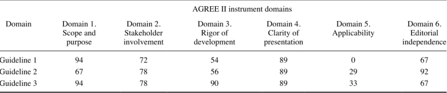
Table 5 presents the results of the quality assessment of the 3 guidelines using the AGREE II instrument.5 All 3
guidelines received scores >50 in the “rigor of develop
ment” domain, and the committee recommended consult
ing them. The titles of the 3 guidelines are presented below.
Table 4. Search results of recent literature through OvidMedline InProcess and Other NonIndexed Citations and OvidMedline from 1946 to present
Searching date: Oct 12, 2017
Search term Search result
(population)P dental implant$.tw. or Immediate Dental Implant Loading/ or Dental Prosthesis, ImplantSupported/
Dental Implantation, Endosseous/ or Dental Implants/ or Dental Implantation/
1 or 2
17,818 28,672 33,862 (intervention)I ConeBeam Computed Tomography.tw. or ConeBeam Computed Tomography/
Radiography, Dental, Digital/ or Radiography/ or Radiography, Dental/ or Radiography, Panoramic/ or Radiography.tw.
periapical radiograph$.tw.
4 or 5 or 6
9,540 375,388 1,542 384,195
P & I 3 AND 7 3,742
Year
limitation limit 8 to yr=“2011Current” 1,727
RCTfilter exp Randomized Controlled Trials/ or Random*.mp. or RCT.mp. or exp Controlled Clinical Trial/ or
exp Placebo Effect/ or exp Placebos/ or Placebo.mp. or trial.mp. 1,759,548 Observational
study filter exp Epidemiologic Studies/ or exp CaseControl Studies/ or exp Cohort Studies/ or exp
Seroepidemiologic Studies/ or Case control.mp. or cohort stud*.mp. or cohort analys*.mp. or Follow up stud*.mp. or observational stud*.mp. or Longitudinal.mp. or Retrospective.mp.
9 and 10RCT
9 and 11Observational study
After omitting overlapping studies(#12) After omitting overlapping studies(#13)
2,563,395
310733 296697 RCT: randomized controlled trial
Fig. 1. The guidelines identified by searching were selected by stepbystep screening. This is the flow diagram of guideline selection.
Guideline 1: CBCT in implant dentistry: a systematic review focusing on guidelines, indications, and radiation dose risks6
Guideline 2: Consensus statements and recommended clinical procedures regarding contemporary surgical and radiographic techniques in implant dentistry7
Table 6. Recommendation matrix of the existing guidelines
Source guidelines Recommendation Grade of recommendation
Guideline 1: CBCT in implant dentistry: a systematic review focusing on guidelines, indications, and radiation dose risks
Practitioners who prescribe or use CBCT units should design specific CBCT equipment protocols that are task specific and incorporate the imaging goal for patient’s specific presenting circumstances.
The protocol should include considerations of exposure (mA and kVp), minimum imagequality parameters (e.g., number of basis images, resolution), and restriction of the FOV to visualize adequately the region of interest.
Not available
Guideline 2: Consensus statements and recommended clinical procedures regarding contemporary surgical and radiographic techniques in implant dentistry
The clinician performing or interpreting CBCT scans for implant dentistry should take into consideration current radiologic guidelines.
The decision to perform CBCT imaging for treatment planning in implant dentistry should be based on individual patient needs following thorough clinical examination.
• When crosssectional imaging is indicated, CBCT is preferable over CT.
CBCT imaging is indicated when information supplemental to the clinical examination and conventional radiographic imaging is considered necessary.
CBCT may be an appropriate primary imaging modality in specific circumstances (e.g., when multiple treatment needs are anticipated or when jawbone or sinus pathology is suspected).
• The use of a radiographic template in CBCT imaging is advisable to maximize surgical and prosthetic information.
• The FOV of the CBCT examination should be restricted to the ROI whenever possible.
• Patient and equipmentspecific dose reduction measures should be used at all times.
• To improve image data transfer, clinicians should request radiographic devices and thirdparty dental implant software applications that offer fully compliant DICOM data export.
Not available
Guideline 3: Radiation No. 172 CBCT for dental and maxillofacial radiology(evidencebased guidelines)
CBCT is indicated for crosssectional imaging prior to implant placement as an alternative to existing crosssectional techniques where the radiation dose of CBCT is shown to be lower.
DFor crosssectional imaging prior to implant placement, the advantage of CBCT with adjustable fields of view, compared with MSCT, becomes greater where the region of interest is a localized part of the jaws, as a similarsized field of view can be used
GP
D,GP
CBCT: conebeam computed tomography, FOV: field of view, ROI: region of interest, DICOM: Digital Imaging and Communications in Medicine, MSCT:
multislice computed tomograph
Table 5. Results of the quality assessment of the guidelines using the Korean version of the Appraisal of Guidelines for Research and Evaluation II(AGREE II) instrument(%)
AGREE II instrument domains
Domain Domain 1.
Scope and purpose
Domain 2.
Stakeholder involvement
Domain 3.
Rigor of development
Domain 4.
Clarity of presentation
Domain 5.
Applicability Domain 6.
Editorial independence
Guideline 1 94 72 54 89 0 67
Guideline 2 67 78 56 89 29 92
Guideline 3 94 78 90 89 33 67
Guideline 3: Radiation No. 172 CBCT for dental and maxillofacial radiology(evidencebased guidelines)8
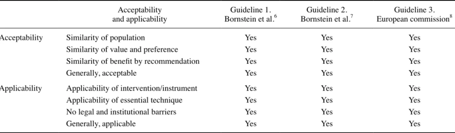
Conventional imaging was recommended in all 3 guide
lines as an appropriate examination modality for patients scheduled for implant placement. If insufficient informa
tion is obtained via conventional imaging, CBCT can be a next step. The domestic acceptability and applicability of the 3 guidelines were acceptable(Table 7).
Grading the level of evidence and drafting the recom
mendation document
Based on the 3 guidelines, 3 recommendations were proposed, for which the recommendation grade and evi
dence level are as follows:
Recommendation 1. In the absence of a clinical abnor
mality in the oral cavity, a panoramic Xray examination and periapical radiological examination of the relevant part of the alveolar bone are necessary to determine the status of the bone and the shape of adjacent anatomical structures(recommendation grade A, evidence level II).
Recommendation 2. A CBCT scan should be performed for each patient judged to require a crosssectional image after reading of the panoramic Xray image and intraoral radiological image(recommendation grade B, evidence level II).
Recommendation 3. A CBCT scan can be used as the primary test for patients clinically suspected to have pathological abnormalities of the jaw or the maxillary si
nus(recommendation grade B, evidence level II).
Finalizing the recommendation document
Reviews can be performed using different methods, such as conducting a seminar to hear directly from the users and holding a public meeting with the head of the Consumer Protection Committee, newspaper reporters, and healthcare officials. To make this guideline useful for clinicians who
request imaging examinations, the recommendations will be disseminated widely through diverse methods, such as academic presentations and public communication. The developed Korean clinical imaging diagnostic guideline (KCIG) will be reassessed annually, and may be revised if new key evidence is presented.
Discussion
This study aimed to develop a guideline for the appropri
ate use of various radiographical modalities for Korean pa
tients scheduled for implantation. In the future, we will use this method to create a Korean guideline for more than 50 PICO questions that clinicians would like to be clarified.
Joint recommendations were made by the KAOMFR and NECA, following the adaptation process of evidencebased CIGs. One of the 3 selected guidelines was identified through a manual search, because the guideline developed by the SEDENTEXCT research project is only provided on their website. Therefore, the compiled guideline has been posted for easy access worldwide. Furthermore, we will create a mobile application for this guideline, which will make it easy for endusers to see the guideline on their mo
bile phones.
All 3 guidelines for the method of examination of pa
tients scheduled for implantation, which received scores
>50 scores in the “rigor of development” domain, uni
formly recommended conventional imaging, such as pan
oramic radiography. It was recommended to use a pan
oramic radiograph to decide whether a CBCT scan is nec
essary.
We recommended CBCT scanning in individual patients judged to require a crosssectional image after reading of a panoramic Xray image and a conventional intraoral radiological image.6,9 More specifically, a crosssectional
Table 7. Results of the assessment of acceptability and applicability Acceptability
and applicability Guideline 1.
Bornstein et al.6 Guideline 2.
Bornstein et al.7 Guideline 3.
European commission8
Acceptability Similarity of population Yes Yes Yes
Similarity of value and preference Yes Yes Yes
Similarity of benefit by recommendation Yes Yes Yes
Generally, acceptable Yes Yes Yes
Applicability Applicability of intervention/instrument Yes Yes Yes
Applicability of essential technique Yes Yes Yes
No legal and institutional barriers Yes Yes Yes
Generally, applicable Yes Yes Yes
image after panoramic radiography is needed in the fol
lowing conditions in the maxilla:8 (a) an incisive canal, (b) descent of the maxillary sinus, (c) doubt regarding the si
nus septum in sinus grafting, (d) doubt about the shape of the alveolar ridge, and (e) pathosis. In the mandible, the conditions requiring a crosssectional image are: (a) doubt about the position of the mandibular canal or mental fo
ramen,(b) doubt about the shape of the alveolar ridge, (c) severe resorption, and(d) pathosis.
A benefit of CBCT is that it provides a crosssectional view of the residual alveolar bone with a lower radiation dose than multislice computed tomography(MSCT). Ad
ditionally, when acquiring images using a radiological marker, an appropriate plan can be made considering the implant direction. However, CBCT is inadequate for eval
uating bone quality. Unlike MSCT, the grayscale values in CBCT images are not reliable; thus, evaluating density objectively is challenging. However, the grayscale values of CBCT images have been reported to be correlated with implant retention.10 Evaluating the residual alveolar bone using CBCT has the advantages of less radiation exposure than conventional MSCT and an adjustable field of view so that clinicians can observe only the necessary part.8
The radiation dose for each examination was 7.2μSv for panoramic radiography,11 18.3μSv for periapical ra
diography,12 and 11674μSv for CBCT of the alveolar bone.8 Since CBCT has a large effective dose difference depending on the region of interest, it is recommended to adjust the exposed site based on the ROI.13
The working group gathered guidelines and determined their domestic acceptability and applicability through con
sensus.
We have prepared a plan for effective dissemination of this guideline by consensus of the committee to strength
en its application. To improve the applicability of the gen
erated guideline, we will publish articles in leading jour
nals and create and use a clinical decision support system as a domestic mobile application.
In conclusion, this study was the first to develop an evidencebased CIG for implant planning in Korea. As subsequent activities, applicability and monitoring are rec
ommended to ensure that the application of the guideline in clinical settings is fully justified. Additionally, KCIG should be regularly reviewed and revised.
References
1. Harris D, Horner K, Gröndahl K, Jacobs R, Helmrot E, Benic GI, et al. E.A.O. guidelines for the use of diagnostic imaging in implant dentistry 2011. A consensus workshop organized by the European Association for Osseointegration at the Medical Uni
versity of Warsaw. Clin Oral Implants Res 2012; 23: 124353.
2. Fienitz T, Schwarz F, Ritter L, Dreiseidler T, Becker J, Roth
amel D. Accuracy of cone beam computed tomography in as
sessing periimplant bone defect regeneration: a histologically controlled study in dogs. Clin Oral Implants Res 2012; 23: 882
3. Bornstein MM, Horner K, Jacobs R. Use of cone beam com7.
puted tomography in implant dentistry: current concepts, indications and limitations for clinical practice and research.
Periodontol 2000 2017; 73: 5172.
4. Choi SJ, Jeong WK, Jo AJ, Choi JA, Kim MJ, Lee M, et al.
Methodology for developing evidencebased clinical imaging guidelines: joint recommendations by Korean Society of Ra
diology and National EvidenceBased Healthcare Collaborat
ing Agency. Korean J Radiol 2017; 18: 20816.
5. Brouwers MC, Kho ME, Browman GP, Burgers JS, Cluzeau F, Feder G, et al. AGREE II: advancing guideline development, reporting and evaluation in health care. CMAJ 2010; 182:
E83942.
6. Bornstein MM, Scarfe WC, Vaughn VM, Jacobs R. Cone beam computed tomography in implant dentistry: a systematic review focusing on guidelines, indications, and radiation dose risks. Int J Oral Maxillofac Implants 2014; 29 Suppl: 5577.
7. Bornstein MM, Al Nawas B, Kuchler U, Tahmaseb A. Consen
sus statements and recommended clinical procedures regarding contemporary surgical and radiographic techniques in implant dentistry. Int J Oral Maxillofac Implants 2014; 29 Suppl: 7882.
8. SEDENTEXCT Guideline Development Panel. Radiation pro
tection No 172. Cone beam CT for dental and maxillofacial radiology. Evidence based guidelines. Luxembourg: European Comminssion DirectorateGeneral for Energy; 2012.
9. Jacobs R, Salmon B, Codari M, Hassan B, Bornstein MM.
Cone beam computed tomography in implant dentistry: recom
mendations for clinical use. BMC Oral Health 2018; 18: 88.
10. Arisan V, Karabuda ZC, Avsever H, Özdemir T. Conventional multislice computed tomography(CT) and conebeam CT (CBCT) for computerassisted implant placement. Part I: rela
tionship of radiographic gray density and implant stability. Clin Implant Dent Relat Res 2013; 15: 893906.
11. Lee C, Lee SS, Kim JE, Symkhampha K, Lee WJ, Huh KH, et al. A dose monitoring system for dental radiography. Imaging Sci Dent 2016; 46: 1038.
12. Gijbels F, Jacobs R, Sanderink G, De Smet E, Nowak B, Van Dam J, et al. A comparison of the effective dose from scanogra
phy with periapical radiography. Dentomaxillofac Radiol 2002;
31: 15963.
13. Jacobs R, Quirynen M. Dental cone beam computed tomogra
phy: justification for use in planning oral implant placement.
Periodontol 2000 2014; 66: 20313.