수직 불안정 골반환 손상에서 S1, S2 나사못을 이용한 후방 고정술의 수술 결과
여광희ㆍ문남훈*ㆍ안재민
†ㆍ정재윤*ㆍ장재훈*
부산대학교병원 외상외과, 정형외과*, 부산고려병원 정형외과†
Surgical Outcome of Posterior Pelvic Fixation Using S1, S2 Screws in Vertically Unstable Pelvic Ring Injury
Kwang Hee Yeo, M.D., Nam Hoon Moon, M.D.*, Jae Min Ahn, M.D.†, Jae Yoon Jeong, M.D.*, Jae Hoon Jang, M.D.*
Departments of Trauma Surgery and Orthopaedic Surgery*, Pusan National University Hospital, Department of Orthopaedic Surgery, Busan Korea Hospital†, Busan, Korea
Received July 18, 2017 Revised August 11, 2017 Accepted November 10, 2017 Correspondence to:
Jae Hoon Jang, M.D.
Department of Orthopaedic Surgery, Pusan National University Hospital, 179 Gudeok-ro, Seo-gu, Busan 49241, Korea
Tel: +82-51-240-8749 Fax: +82-51-247-8395 E-mail: [email protected] Financial support: This work was supported by clinical research grant in 2016 from Pusan National University Hospital.
Conflict of interests: None.
Purpose: Iliosacral screw fixation is an effective and less invasive method that is used widely for the definitive treatment of unstable pelvic ring injuries. On the other hand, fixation failures after iliosacral screw fixation have been reported in vertically unstable pelvic ring injuries. This study examined the surgical outcomes of posterior pelvic fixation using S1 and S2 screws in vertically unstable pelvic ring injuries.
Materials and Methods: Between January 2011 and April 2016, 17 patients with vertically unstable pelvic ring injuries who met the minimum 1 year follow-up criteria were treated with internal fixation using posterior pelvic S1 and S2 screws. Their mean age was 43.9 years. According to the AO/OTA clas- sification, 10 patients had C1, 6 had C2, and 1 had C3 injuries. Surgical treatments of single or multiple steps, where necessary, were performed by two surgeons. The clinical and radiologic outcomes were assessed retrospectively using radiographs and medical records.
Results: Overall, 16 patients had bone healing without screw loosening; however, one patient could not maintain anterior pelvic fixation because of an open fracture and deep infection in the anterior pelvic ring. Of five patients who complained of neurological symptoms after injury, three had partially recovered from their neurological deficit. At the last follow-up, the clinical outcomes according to the Majeed score were excellent in 5, good in 6, fair in 4, and poor in 2 patients. The postoperative radio- logic outcomes by Matta and Tornetta’s method were excellent in 5, good in 8, and fair in 4 patients.
Malposition of the S2 screw was identified in one case. The mean time to union was 14.6 weeks after surgery.
Conclusion: S1 and S2 screw fixation can be an effective treatment option for posterior pelvic stabiliza- tion in vertically unstable pelvic ring injuries when considering the surgical outcomes, such as screw loosening and loss of reduction.
Key Words: Vertically unstable pelvic ring injury, Posterior pelvic fixation, S1 and S2 screw
Copyright © 2018 The Korean Fracture Society. All rights reserved.
This is an Open Access article distributed under the terms of the Creative Commons Attribution Non-Commercial License (http://creativecommons.org/licenses/by-nc/4.0) which permits unrestricted non-commercial use, distribution, and reproduction in any medium, provided the original work is properly cited.
서 론
수직 불안정(AO/OTA classification C type, C형) 골반환 손상은 대부분 고 에너지 손상으로 발생하게 되는 외상으로 골반의 후방 골-인대 복합체의 손상으로 인해 회전 및 수직 불안정성을 보인다. 또한 회전 불안정(AO/OTA classification B type, B형) 골반환 손상에 비해 많은 동반 손상과 함께 높 은 사망률과 이환율을 보이고 수술적 치료가 어려우며 불량 한 방사선적, 임상적 결과를 보인다고 보고되고 있다.1,2) 치료 에 있어 비수술적 치료나 외고정술만을 시행했을 경우 그 결 과가 좋지 않아3,4) 정확한 정복과 견고한 내고정술이 필수적
이다.2,5-7) 이러한 골반환 손상의 후방 고정을 위해 접근 방법
과 고정물의 선택에 따라 여러 방법들이 소개되고 있으나,8-11) 이들 중 천장 나사못을 이용한 내고정술은 효과적이고 비침 습적인 술식으로12) 널리 사용되고 있다. 그러나 일부 연구에 서 C형 골반환 손상에서 천장 나사못 고정술은 나사 이완, 고 정 실패 및 정복 소실 등의 위험이 있다고 보고하였다.13,14) 이 에 대해 본 연구에서는 C형 골반환 손상의 후방 고정을 위해 1천추(S1)와 2천추(S2)에 각각 하나씩, 두 개의 후방 나사못 고정술을 시행한 경우의 수술적 결과를 후향적으로 조사해 보았다.
대상 및 방법
1. 연구 대상
본 연구는 부산대학교병원 연구윤리심의위원회(insti- tutional review board)의 승인을 받고 시행하였다(PNUH 1708-025-058).
2011년 1월부터 2016년 4월까지 불안정 골반환 손상으로 수술적 치료를 받은 153명의 환자 중 1) C형 골반환 손상, 2) S1과 S2에 후방 나사못 고정술을 시행한 경우 등을 선정 기 준으로 하였다. 또한 1) 사망 혹은 추적 소실로 인해 최소 1년 이상 추적 관절이 불가능했던 경우, 2) 후방 고정을 위해 다 른 고정 방법을 선택한 경우 등을 제외 기준으로 하였다. 선 정 및 제외 기준을 만족시키는 연구 대상은 총 17명이었으며 (Fig. 1), 이들에 대해 의무 기록과 방사선 검사를 토대로 후 향적 조사를 시행하였다. 남자가 13명, 여자가 4명이었으며 이들의 평균 나이는 43.9세(17-66세)였고 평균 추시 기간 은 21.4개월(12-53개월)이었다. 모두 고 에너지 손상이었으 며 AO/OTA (Arbeitsgemeinschaft für Osteosynthesefragen/
Orthopaedic Trauma Association) 분류상 C1이 10예, C2 가 6예, C3 손상이 1예였으며 후방 골반환 손상은 천골 골절 이 12예, 천장 관절 손상이 5예였다. 골반환 전방의 개방 골절 이 1예에서 있었으며 다른 근골격계, 두부, 흉곽, 복부 및 비 뇨기계 손상 등 다양한 동반 손상이 확인되었다. 술 전 3차 원 컴퓨터 단층촬영(computed tomography, CT)을 통해 S1 의 천골 이형성증(sacral dysmorphism) 여부와 S2에 나사못 을 안전하게 삽입시키기 위한 위치 및 가능성 등을 평가한 후 수술을 시행하였다. 두 명의 술자에 의해 수술이 시행되었으 며 S1과 S2에 각각 하나씩, 두 개의 천장 혹은 경천골-경장골 (transsacral-transiliac) 나사못을 삽입하여 후방 골반환 고 정술을 시행하였다. S1에는 모두 천장(iliosacral) 나사못 형태 의 내고정을, S2에는 수술장에서 나사의 위치를 명확하게 판 단하기 힘들었던 4예에서는 천장 나사 형태의 내고정을, 그 외의 경우는 모두 경천골-경장골 나사못 형태로 내고정하 였다. 모든 나사못은 7.0 mm 유관나사못(fully and partially threaded cannulated screw, 7.0 mm in diameter; Synthes, Oberdorf, Switzerland/MAGNA-FX; Zimmer, Warsaw, IN, USA)을 이용하였으며 압박이 필요한 경우는 부분 나사 산 나사못(partial threaded screw)을, 추가 고정을 위해서는 전체 나사산 나사못(full threaded screw)을 이용하였다. C형 골반환 손상은 전방 고정술도 필수적으로 환자의 상태 및 손 상 양상에 따라 Pfannenstiel 또는 변형된 Stoppa 접근법을 이 용한 금속판 내고정 혹은 외고정을 시행하였다. 8명의 환자 에서는 단일 수술로, 9명의 환자에서는 환자의 동반 손상과
Fig. 1. Flow chart of the enrollment of patients treated with posterior pelvic fixa tion using S1 and S2 screws for vertically unstable pelvic ring injuries. PRI: pelvic ring injury, F/U: follow-up.
전신 상태에 따른 손상 통제 수술(damage control surgery)을 위해, 골반환 손상 양상에 맞춰 2번 이상에 걸쳐 단계적으로 수술을 시행하였다(Table 1). 단일 수술을 시행한 경우는 수 상 후 평균 3.5일(2-6일)에, 단계적 수술을 시행한 경우에서 는 첫 수술은 평균 1.1일(1-2일)에, 최종 수술은 평균 21.9일 (7-56일)에 골절 정복 및 고정술이 시행되었다. 1예에서 비관 혈적 정복술로 만족스러운 정복이 되지 않아 복와위에서 관 혈적 정복을 시행하였고 나머지 모든 경우에서는 앙와위에 서 비관혈적 정복 후 고정술을 시행하였다. 전반 고정과 후방 고정의 순서는 전위가 더 심한 쪽을 먼저 시행하였으며 판단 이 힘든 경우에는 전방 고정을 우선시하였고, 또한 동반 손상 이나 환자 상태를 고려해 고정 순서와 시기를 결정하였다. 수 술 후 모든 환자에서 CT 촬영이 시행되었으며 통증, 동반 손 상 및 전신 상태에 맞춰 고관절, 슬관절, 족관절에 대해 수동/
능동 관절 운동을 시행하였고 수술 후 6주부터 부분 체중 부 하를, 12주부터 완전 체중 부하를 허용하였다.
2. 평가 방법
방사선적 평가는 수술 후 CT를 통해 골절 정복 정도와 나 사못 위치 이상을, 추시 X-선 영상을 통해 나사못 이완 유무, 최종 추시에서의 나사못 위치 변화 여부 및 골유합 시기를 확 인하였다. 골절 정복 정도는 Matta와 Tornetta의 방법15)을 이용하여 우수(<5 mm), 양호(5-10 mm), 보통(11-20 mm), 불량(>20 mm)으로 분류하였고, 나사못 이완은 수술 직후 와 비교하여 1 cm 이상의 나사못 후방 돌출(back-out)과 함 께 정복 소실을 보여 골반환의 불안정성이 예상되어 재수술 이 필요한 경우로 정의하였으며,14) 수술 직후와 최종 추시의 S1과 S2의 나사못 위치 변화를 관찰하였다. 그리고 연속적인 추시 X-선 영상에서 골절선이 관찰되지 않거나 이가 명확하 지 않을 때는 골절부의 통증 없이 완전 체중 부하가 가능했고 추후 재전위 등의 고정 실패가 발생하지 않았던 상태를 골유 합으로 간주하였다. 방사선적 평가는 정형외과 전문의인 저 자 중 1명에 의해 시행되었다.
임상적 평가는 수술 후 감염, 신경 손상 등의 합병증과 최 종 추시에서의 Majeed score16)를 평가하였다. Majeed score 는 85점 이상을 우수, 70-84점을 양호, 55-69점을 보통, 55 점 미만을 불량으로 분류하였다.
결 과
수술 후 CT를 이용한 방사선적 평가에서 골절 정복 정 도는 우수가 5예(29.4%), 양호가 8예(47.1%), 보통이 4예 (23.5%)로 76.5%에서 양호 이상의 결과를 보였고, 1예 (5.9%)에서 S2 나사못이 신경공을 침범한 소견이 관찰되었 으나 특별한 신경학적 증상을 보이지 않아 경과 관찰하였다 (Fig. 2). 개방 골절로 전방 고정을 유지하지 못했던 1예(5.9%) 에서 나사못 이완이 발생하여 추가적인 후방 고정술을 시행 하였고, 그 외의 경우에서는 최종 추시까지 재수술이 필요한 나사못 이완은 관찰되지 않았다. 또한 최종 X-선 검사상에 서 수술 직후와 비교해 골반의 불안정성이 예상되어 추가적 인 수술이 필요한 나사못 위치 변화는 관찰되지 않았으며, 골 유합 시기는 마지막 수술일로부터 평균 14.6주(12-20주)로 확인되었다.
골반환 전방부의 개방 골절이 있었던 1예에서 수술 후 심 부 감염이 발생하였으나 그 외에는 수술 후 감염 소견은 관찰 되지 않았다. 17예 중 5예(29.4%)에서 수술 후 신경학적 증상 을 보였고 근전도 검사상 폐쇄 신경(obturator nerve), 상둔 신 경(superior gluteal nerve) 및 좌골 신경(sciatic nerve) 손상 1 예, 요천골 신경총 병변(lumbosacral plexopathy) 4예를 확인 할 수 있었다. 이들 중 신경총 병변을 보였던 4예 중 2예는 최 종 추시에서도 신경 회복을 보이지 않았으며 나머지 3예에서 는 추시 관찰 중 부분 회복된 것을 확인할 수 있었다. 또한 임 상적 결과는 우수가 5예(29.4%), 양호가 6예(35.3%), 보통이 4예(23.5%), 불량이 2예(11.8%)로 64.7%에서 양호 이상의 결과를 확인할 수 있었다(Table 2).
고 찰
불안정 골반환 손상의 후방 고정을 위해 여러 방법들이 보 고되고 있으나 이들 중 경피적 천장 나사못 고정술은 비침습 적이고 효과적인 방법으로 널리 사용되고 있으며 여러 연구 에서 좋은 결과들을 보고하였다.8,17,18) 그러나 Kim 등14)은 수 직 전단력으로 인한 골반환 손상에서 S1에 하나의 천장 나사 못을 고정을 시행한 37명의 환자 중 8명(21.6%)에서 고정 실 패를 보고하였다. 본 연구에서는 S1과 S2에 각각 하나씩 두 개의 후방 나사못 고정술을 시행함으로써 17예 중 1예(5.9%) 에서 나사 이완과 정복 소실을 보여 고정 실패를 줄일 수 있었 던 것으로 보인다. Griffin 등13)은 천장 나사못 고정술로 수술 적 치료를 시행한 62명의 C형 골반환 손상 환자 중 수직 천골
Table 1. Patient Demographics Case No.Sex/Age (yr)Mechanism of injuryAO/OTA classificationAssociated injuryPosterior pelvic injuryReductionSurgeryAnterior pelvic fixationPosterior pelvic fixation 1Male/66MVAC1-3M, ULt. sacrum (Zone I)ClosedSingleEFS1: ISS S2: TSTIS 2Female/22Fall downC2M, T, ARt. sacrum (Zone II)ClosedSinglePlateS1: ISS S2: TSTIS 3Male/39MVAC2M, H, N, RLt. sacrum (Zone II)ClosedStagedEF→PlateS1: ISS S2: TSTIS 4Male/45Fall downC2M, A, N, RRt. sacrum (Zone II)ClosedStagedEF→PlateS1: ISS S2: TSTIS 5Male/39Crushing injuryC3A, U, N, RBoth SIJ dislocationClosedStagedEF→PlateRt.: Plate Lt. S1: ISS Lt. S2: ISS 6Male/53MVAC1-2M, TRt. SIJ fracture-dislocationClosedStagedEF→PlateS1: ISS S2: TSTIS 7Male/49Crushing injuryC2NoneRt. SIJ dislocationClosedStagedEF→PlateS1: ISS S2: TSTIS 8Male/53Crushing injuryC2A, N, RLt. SIJ dislocationClosedStagedEF→PlateS1: ISS S2: TSTIS 9Male/53Crushing injuryC1-3NoneRt. sacrum (Zone II)OpenSinglePlateS1: ISS S2: TSTIS 10Female/62Fall downC1-3MLt. sacrum (Zone II)ClosedSingleEFS1: ISS S2: ISS 11Male/17MVAC2M, A, N, RRt. sacrum (Zone II)ClosedStagedEF→PlateS1: ISS S2: ISS 12Female/37MVAC1-3M, HRt. sacrum (Zone II)ClosedStagedEFS1: ISS S2: TSTIS 13Male/42Crushing injuryC1-2URt. SIJ fracture-dislocationClosedSinglePlateS1: ISS S2: ISS 14Male/48Fall downC1-3M, A, RLt. sacrum (Zone II)ClosedSinglePlateS1: ISS S2: TSTIS 15Male/38MVAC1-3M, H, ALt. sacrum (Zone II)ClosedSinglePlateS1: ISS S2: TSTIS 16Female/35Fall downC1-3M, H, T, RLt. sacrum (Zone I)ClosedSingleNoneS1: ISS S2: TSTIS 17Male/48Fall downC1-3M, T, A, RRt. sacrum (Zone II)ClosedStagedEF→PlateS1: ISS S2: TSTIS AO/OTA: Arbeitsgemeinschaft für Osteosynthesefragen/Orthopaedic Trauma Association, MVA: motor vehicle accident, M: other musculoskeletal injury, U: urological injury, T: thoracic injury, A: abdominal injury, H: head injury, N: neurological lesion, R: case needed initial resuscitation, Lt.: right, Rt.: right, SIJ: sacroiliac joint, EF: external fixator, ISS: iliosacral screw, TSTIS: transsacral- transiliac screw.
골절 4예(6.5%)에서 고정 실패를 보고하였다. 이들 4예는 모 두 본 연구와 같이 S1과 S2에 두 개의 천장 나사못 고정술을 시행했던 경우이나 이들 모두 나사못의 길이가 반대편 천골 익부(far sacral alae)나 반대편 신경공(far foraminae)까지였 다. 본 연구에서는 충분한 작업 길이(working length)를 확보 하기 위해 S2 나사못을 가능한 반대편 천장관절을 넘어서까 지 위치시켜 경천골-경장골 고정을 통해 충분한 안정성을 확 보할 수 있었던 것으로 생각된다.
C형 골반환 손상은 B형 골반 손상에 비해 사망률과 이환 율이 높고 수술적 치료에서도 어려움이 많으며 방사선적, 임 상적 치료 결과도 불량하다.1,2) 또한 정확한 정복과 충분한 고 정이 이루어지지 않는다면 나사 이완, 정복 소실 및 고정 실패 의 위험성이 있다. 천장 나사못 고정술은 비침습적이고 효과
적인 후방 골반 고정법으로12) 널리 사용되는 술식이며 주로 제1천추에 시행되는 것이 일반적이나 C형 골반환 손상에서 는 S1에 하나의 나사못만으로는 고정력이 불충분할 수 있어 다른 고정 방법이나 추가 고정을 고려하여야 한다.13,14) C형 골 반환 손상에서 천장 나사못 고정술을 시행할 경우 최소 2개 의 나사못 고정을 시행한다는 술자의 원칙에 따라 공간이 충 분한 경우는 S1에 두 개의 나사못 고정을 시행하였지만 S1에 하나의 나사못 삽입 후 추가 고정을 위한 공간이 부족한 경우 나 천골 이형성증이 있는 경우에는 S2에 추가적인 고정술을 시행하였고 이들이 본 연구의 대상이었다. Zhang 등19)의 생 역학 연구에서도 C형 골반환 손상에서 S1과 S2에 하나씩 두 개의 나사못을 삽입한 경우에서 S1에 하나, S2에 하나, 혹은 S1에 두 개 등의 다른 형태의 나사못 고정보다 더 나은 안정 성을 가진다고 보고하였다. 또한 이는 전방과 후방 고정을 자 세 변경 없이 시행할 수 있고 다른 고정 방법에 비해 보다 비 침습적으로 추가 고정을 시행할 수 있다는 장점이 있다.
안전 영역이 협소하고 후방 골반의 복잡한 해부학적 구조 로 인해 주위 신경, 혈관 및 장기 손상의 위험성이 있어 S2 나 사못 고정술은 우선적으로 시행되지는 않는다.18-22) 그러나 최근 정확한 해부학적 이해와 술기의 발전으로 천골 이형성 증이 있거나 추가 고정이 필요한 경우 S2 나사못 고정술을 시 행하여 좋은 결과를 보였다는 연구가 보고되었다. Moed와 Geer23)에 의하면 49명의 환자에 대해 S2 천장 나사못 고정술 을 이용하여 나사못 위치 이상으로 인한 의인성 신경 손상 없 이 만족스러운 임상 결과를 보였으며, Osterhoff 등24)은 21개
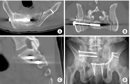
A B
C D
Fig. 2. Malposition of the S2 screw in the S1 foramen was identified in the axial (A), coronal (B), and sagittal (C) images of postoperative computed tomography.
(D) Postoperative radiograph of the pel- vic outlet view.
Table 2. Radiologic Results, Clinical Outcomes
Surgical outcome Number
Grade of reduction by Matta and Tornetta7)
Excellent (<5 mm) 5
Good (5-10 mm) 8
Fair (11-20 mm) 4
Poor (>20 mm) 0
Functional outcome by Majeed score16)
Excellent (≥85) 5
Good (70-84) 6
Fair (55-69) 4
Poor (<55) 2
A B
C D
Fig. 3. (A) A 35-year-old female patient fell from a height and sustained a type C pelvic ring injury. (B) Posterior pelvic stabilization using S1 and S2 screws was performed 1 day after the injury. No fur- ther procedure was performed for the anterior pelvic ring because acceptable reduction of the fractures in the pubic rami was identified after posterior fixa- tion. (C) No displacement occurred, and callus formation was observed at the 4-week follow-up. (D) The radiograph obtained at the 1-year follow-up showed complete bony union without complica- tions.
A B
C D
Fig. 4. (A) Emergent external fixation and pelvic packing were performed in a 17-year-old male patient with a vertically unstable (type C) pelvic ring injury with an open wound. (B) Posterior pelvic fixa- tion using S1 and S2 iliosacral screws and anterior fixation with rami screws were performed 2 days after the initial injury.
(C) Rami screws and the external fixator were removed, and debridement and the insertion of antibiotic-impregnated cement beads were conducted because of a deep infection 4 weeks after the initial surgery. Sacroiliac screw loosening was identified. (D) Anterior pelvic plating using the modified Stoppa approach and posterior pelvic screw exchange were performed after the infection had sub- sided completely.
의 S2 나사못 중 3개의 나사못에서 위치 이상 혹은 나사 이완 을 보였으나 만족스러운 방사선적, 임상적 결과를 보여 S1과 S2의 나사못 고정술은 효과적인 후방 골반 고정법이라고 보 고하였다. 본 연구에서는 1예(5.9%)에서 S2 나사못 위치 이상 이 수술 후 CT에서 확인되었으나 특별한 신경학적 증상이 없 어 추가적인 수술 없이 경과 관찰을 시행하였다. 천장 나사못 의 위치 이상을 방지하기 위해 신경 감시 체계, 3차원 영상증 폭기 및 항법 장치 등의 장비들이 소개되고, 그 효용성에 대 한 연구들이 보고되고 있어23,25-27) 의인성 신경 손상을 예방 하기 위한 유용한 방법들로 생각되나 이들은 모두 고가의 장 비로 사용이 제한되는 단점이 있다. 영상증폭기만으로도 안 전하게 S1과 S2에 천장 나사못 고정술을 시행할 수 있었다는 Osterhoff 등24)의 보고와 같이, 면밀한 술 전 계획,28) 골반골 에 대한 정확한 해부학적 이해와 영상증폭기의 주의 깊은 관 찰을 통해서도 나사못의 위치 이상을 예방할 수 있을 것으로 생각된다.
신경학적 손상은 수술 전 의식이 없거나 전신 상태 문제 및 통증으로 인해 검사가 제한되는 경우가 많아 정확하게 판 단하기는 어려웠으나 수술 후 CT를 통한 골절의 정복 정도와 나사못의 위치를 고려했을 때, 수술 후 5예에서 보인 신경학 적 증상은 의인성 손상으로 인한 것보다는 골반환 손상 시에 발생한 것으로 추측된다. Kim 등29)은 18예의 C형 골반환 손 상 중 근전도 검사상 5예(27.8%)에서 신경 손상을 보였으며 이들 중 4예가 요천골 신경총 병변이었다고 보고하였다. 본 연구에 포함된 대상들은 모두 C형 손상이었으며 17예 중 5예 (29.4%)에서 신경학적 증상을 보였고 이 중 4예가 요천골 신 경총 병변으로 비슷한 결과를 보였다.
대부분의 C형 골반환 손상은 후방 골반환 손상에 더해 전 방의 손상도 동반되며 적절한 안정성을 획득하기 위해 많은 경우에서 수술적 치료가 필수적이다. Sagi 등30)에 의하면 C 형 골반환 손상에서 후방 골반환 고정에 더해 전방 고정이 생 역학적 안정성에 중요한 역할을 하다고 보고하였다. C형 골 반환 손상은 전방 고정술도 필수적으로 본 연구에서도 금속 판 13예, 외고정 3예 등 1예를 제외하고 모든 경우에서 전방 고정을 시행하였다. 후방 고정술 후 전방 골반환이 해부학적 위치로 유지되었고, 입원 중 시행한 추시 관찰 중 정복 소실이 발생하지 않은 1예에서만 전방 고정을 시행하지 않았다(Fig.
3). 이는 치골 골절 부위의 전위가 심하지 않았고 전방 골반환 주위의 연부조직이 비교적 잘 보존되었던 상태로 회전 불안 정성이 거의 없어 후방 고정만으로 충분한 안정성을 얻을 수 있었던 드문 증례로 생각된다. 또한 본 연구에서 나사 이완이
발생하여 추가 고정을 시행했던 증례도 개방 골절과 이로 인 한 심부 감염으로 치골 결합부 파열과 치골 골절에 대해 외고 정 및 내고정을 일정 기간 동안 제대로 시행할 수 없었던 경 우(Fig. 4)로 C형 골반환 손상에서 전방 고정의 중요성을 확 인할 수 있었다.
본 연구는 의무 기록과 방사선 검사를 토대로 한 후향 적 검사이며 단일 센터에서 적은 증례 수를 대상으로 시행 된 연구라는 한계가 있다. 또한 많은 동반 손상과 다양한 환 자 상태로 후방 고정 방법을 제외하고는 증례들 간의 이종성 (heterogeneity)이 커 결과를 일반화시키는 데 제한이 있을 것 으로 생각된다. 추후 대규모의 다기관 연구를 통해 보다 많은 증례를 통한 연구가 필요할 것으로 생각된다.
결 론
수직 불안정 골반환 손상에서 나사못 이완 및 정복 소실 등의 수술적 결과를 고려할 때 S1과 S2의 후방 나사못 고정술 을 통해 만족스러운 결과를 얻을 수 있었다.
요 약
목적:
수직 불안정 골반환 손상에서 S1과 S2 후방 나사못 고 정술을 시행한 증례들에 대한 수술 결과를 분석해 보았다.대상 및 방법:
2011년 1월부터 2016년 4월까지 수직 불안정 골반환 손상을 동반한 환자들 중 S1과 S2에 나사못 고정술을 시행한 17명의 환자를 대상으로 하였다. 평균 나이는 43.9세 였으며 C1형 10예, C2형 6예, C3형이 1예였다. 방사선적 결과 와 나사못 이완, 신경 손상 등의 수술 후 합병증과 기능 평가 등의 임상적 결과를 영상 자료와 의무 기록을 통해 후향적으 로 조사하였다.결과:
전방 고정을 유지하지 못해 고정 실패를 보여 추가 고정 술을 시행한 1예를 제외하고는 나사못 이완 없이 평균 14.6주 에 골유합을 얻을 수 있었다. 1예에서 수술 후 심부 감염이 있 었으며, 수상 후 신경 증상을 보인 5예 중 3예에서 부분 회복 을 보였다. 임상적 결과는 우수 5예, 양호 6예, 보통 4예, 불량 2예였다. 방사선적 결과는 우수 5예, 양호 8예, 보통 4예였으 며, 1예에서 나사못 위치 이상을 보였다.결론:
수직 불안정 골반환 손상에서 나사못 이완 및 정복 소 실 등의 수술적 결과를 고려할 때 S1과 S2의 후방 나사못 고 정술을 통해 만족스러운 결과를 얻을 수 있었다.색인 단어:
수직 불안정 골반환 손상, 후방 골반환 고정술, S1,S2 나사못
ORCID
여광희, http://orcid.org/0000-0003-1392-9196 문남훈, http://orcid.org/0000-0001-9975-0992 안재민, http://orcid.org/0000-0002-8821-0891 정재윤, http://orcid.org/0000-0001-5291-4689 장재훈 http://orcid.org/0000-0002-2636-7957
References
1. Rommens PM, Hessmann MH: Staged reconstruction of pelvic ring disruption: differences in morbidity, mortality, radiologic results, and functional outcomes between B1, B2/B3, and C- type lesions. J Orthop Trauma, 16: 92-98, 2002.
2. Kabak S, Halici M, Tuncel M, Avsarogullari L, Baktir A, Bas- turk M: Functional outcome of open reduction and internal fixation for completely unstable pelvic ring fractures (type C): a report of 40 cases. J Orthop Trauma, 17: 555-562, 2003.
3. Lindahl J, Hirvensalo E, Böstman O, Santavirta S: Failure of reduction with an external fixator in the management of injuries of the pelvic ring. Long-term evaluation of 110 patients. J Bone Joint Surg Br, 81: 955-962, 1999.
4. Miranda MA, Riemer BL, Butterfield SL, Burke CJ 3rd: Pelvic ring injuries. A long term functional outcome study. Clin Orthop Relat Res, (329): 152-159, 1996.
5. Cole JD, Blum DA, Ansel LJ: Outcome after fixation of unstable posterior pelvic ring injuries. Clin Orthop Relat Res, (329): 160- 179, 1996.
6. Keating JF, Werier J, Blachut P, Broekhuyse H, Meek RN, O’
Brien PJ: Early fixation of the vertically unstable pelvis: the role of iliosacral screw fixation of the posterior lesion. J Orthop Trauma, 13: 107-113, 1999.
7. Tornetta P 3rd, Matta JM: Outcome of operatively treated un- stable posterior pelvic ring disruptions. Clin Orthop Relat Res, (329): 186-193, 1996.
8. Routt ML Jr, Kregor PJ, Simonian PT, Mayo KA: Early results of percutaneous iliosacral screws placed with the patient in the supine position. J Orthop Trauma, 9: 207-214, 1995.
9. Gorczyca JT, Varga E, Woodside T, Hearn T, Powell J, Tile M:
The strength of iliosacral lag screws and transiliac bars in the fixation of vertically unstable pelvic injuries with sacral fractures.
Injury, 27: 561-564, 1996.
10. Krappinger D, Larndorfer R, Struve P, Rosenberger R, Arora R, Blauth M: Minimally invasive transiliac plate osteosynthesis for type C injuries of the pelvic ring: a clinical and radiological
follow-up. J Orthop Trauma, 21: 595-602, 2007.
11. Sagi HC, Militano U, Caron T, Lindvall E: A comprehensive analysis with minimum 1-year follow-up of vertically unstable transforaminal sacral fractures treated with triangular osteosyn- thesis. J Orthop Trauma, 23: 313-319, 2009.
12. Shuler TE, Boone DC, Gruen GS, Peitzman AB: Percutaneous iliosacral screw fixation: early treatment for unstable posterior pelvic ring disruptions. J Trauma, 38: 453-458, 1995.
13. Griffin DR, Starr AJ, Reinert CM, Jones AL, Whitlock S: Verti- cally unstable pelvic fractures fixed with percutaneous iliosacral screws: does posterior injury pattern predict fixation failure? J Orthop Trauma, 17: 399-405, 2003.
14. Kim JW, Oh CW, Oh JK, et al: The incidence of and factors af- fecting iliosacral screw loosening in pelvic ring injury. Arch Or- thop Trauma Surg, 136: 921-927, 2016.
15. Matta JM, Tornetta P 3rd: Internal fixation of unstable pelvic ring injuries. Clin Orthop Relat Res, (329): 129-140, 1996.
16. Majeed SA: Grading the outcome of pelvic fractures. J Bone Joint Surg Br, 71: 304-306, 1989.
17. Routt ML Jr, Simonian PT: Closed reduction and percutaneous skeletal fixation of sacral fractures. Clin Orthop Relat Res, (329):
121-128, 1996.
18. Routt ML Jr, Nork SE, Mills WJ: Percutaneous fixation of pelvic ring disruptions. Clin Orthop Relat Res, (375): 15-29, 2000.
19. Zhang L, Peng Y, Du C, Tang P: Biomechanical study of four kinds of percutaneous screw fixation in two types of unilateral sacroiliac joint dislocation: a finite element analysis. Injury, 45:
2055-2059, 2014.
20. Gautier E, Bächler R, Heini PF, Nolte LP: Accuracy of com- puter-guided screw fixation of the sacroiliac joint. Clin Orthop Relat Res, (393): 310-317, 2001.
21. Carlson DA, Scheid DK, Maar DC, Baele JR, Kaehr DM: Safe placement of S1 and S2 iliosacral screws: the “vestibule” concept.
J Orthop Trauma, 14: 264-269, 2000.
22. Hinsche AF, Giannoudis PV, Smith RM: Fluoroscopy-based multiplanar image guidance for insertion of sacroiliac screws.
Clin Orthop Relat Res, (395): 135-144, 2002.
23. Moed BR, Geer BL: S2 iliosacral screw fixation for disruptions of the posterior pelvic ring: a report of 49 cases. J Orthop Trau- ma, 20: 378-383, 2006.
24. Osterhoff G, Ossendorf C, Wanner GA, Simmen HP, Werner CM: Percutaneous iliosacral screw fixation in S1 and S2 for posterior pelvic ring injuries: technique and perioperative com- plications. Arch Orthop Trauma Surg, 131: 809-813, 2011.
25. Moon SW, Kim JW: Usefulness of intraoperative three-dimen- sional imaging in fracture surgery: a prospective study. J Orthop Sci, 19: 125-131, 2014.
26. Zwingmann J, Konrad G, Kotter E, Südkamp NP, Oberst M:
Computer-navigated iliosacral screw insertion reduces malposi- tion rate and radiation exposure. Clin Orthop Relat Res, 467:
1833-1838, 2009.
27. Zwingmann J, Konrad G, Mehlhorn AT, Südkamp NP, Oberst M: Percutaneous iliosacral screw insertion: malpositioning and revision rate of screws with regards to application technique (navigated vs. conventional). J Trauma, 69: 1501-1506, 2010.
28. Lucas JF, Routt ML Jr, Eastman JG: A useful preoperative planning technique for transiliac-trassacral screws. J Orthop
Trauma, 31: e25-e31, 2017.
29. Kim JW, Bae DH, Kim JH, Kim YC: Neurologic injury within pelvic ring injuries. J Korean Fract Soc, 27: 17-22, 2014.
30. Sagi HC, Ordway NR, DiPasquale T: Biomechanical analysis of fixation for vertically unstable sacroiliac dislocations with iliosa- cral screws and symphyseal plating. J Orthop Trauma, 18: 138- 143, 2004.