251
Removal of a broken needle using three-dimensional computed tomography: a case report
Jin-Ha Kim1, Seong-Yong Moon2
1Dental Clinic, The Third Logistic Support Command, Incheon,
2Department of Oral and Maxillofacial Surgery, School of Dentistry, Chosun University, Gwangju, Korea
Abstract(J Korean Assoc Oral Maxillofac Surg 2013;39:251-253)
Inferior alveolar nerve block obtained maximum anesthetic effect using a small dose of local anesthetic agent, which also has low a complication inci- dence. Complications of an inferior alveolar nerve block include direct nerve damage, bleeding, trismus, temporary facial nerve palsy, and etc. Among them, the major iatrogenic complication is dental needle fracture. A fragment that disappears into the soft tissue would be hard to remove, giving rise to a legal problem. A 31-year-old woman was referred for the removal of a broken needle, following an inferior alveolar nerve block. Management in- volved the removal of the needle under local anesthesia with pre- and peri-operative computed tomography scans.
Key words: Broken dental needle, Inferior alveolar nerve block, Cone-beam computed tomography, Foreign body migration
[paper submitted 2013. 7. 8 / revised 2013. 8. 28 / accepted 2013. 9. 13]
structures (blood vessels and nerve). Still, it causes a lot of fear to both dentists and patients3,4. The prevalence rate is low since most dentists know the causes of these complications, such as weakness of the alloy, narrowness of the needle, re- usage of the needle, incorrect technique, sudden movement of the patient or practitioner, manufacturing defects, and bending1,5-7.
As to the development of material, disposable dental nee- dles are made of flexible stainless steel and are more durable than the previous ones8,9. Note, however, that the needle can be broken more easily when bent or improperly used with in- correct block anesthesia method7.
This is a case report of a broken dental needle due to sud- den movement during inferior alveolar nerve block anesthe- sia, which was positioned between the coronoid process and condyle neck area. Management involved the removal of the needle under local anesthesia with pre- and peri-operative computed tomography (CT) scans.
II. Case Report
A 31-year-old female patient was referred by a local clinic to the Department of Oral and Maxillofacial Surgery of the Chosun University Dental Hospital for the removal of a broken dental needle. When she underwent inferior alveolar block, she tilted her head quickly because of the shock sensa-
I. Introduction
Inferior alveolar nerve block anesthesia is one of the most popular methods, and most clinicians use it effectively with- out serious side effects. Despite the low incidence, however, various complications can develop. Complications related to inferior alveolar nerve block anesthesia can be divided into two large groups, i.e., during and after operation1. Complica- tions during operation are needle breakage, pain at injection, hypersensitivity or allergy, overdosage and toxicity, and lack of effect; those after operation include hematoma, trismus, postoperative paresthesia, or neuralgia1.
Among these complications during operation, needle brea- kage is a rare complication nowadays2. To date, however, a few cases, though not many, are continuously being reported.
Actually, needle breakage has not been reported to cause se- vere complications such as damage to important anatomical
CASE REPORT http://dx.doi.org/10.5125/jkaoms.2013.39.5.251 pISSN 2234-7550·eISSN 2234-5930
Seong-Yong Moon
Department of Oral and Maxillofacial Surgery, School of Dentistry, Chosun University, 309, Pilmun-daero, Dong-gu, Gwangju 501-759, Korea
TEL: +82-62-220-3817 FAX: +82-62-228-7316 E-mail: [email protected]
This is an open-access article distributed under the terms of the Creative Commons Attribution Non-Commercial License (http://creativecommons.org/licenses/by-nc/3.0/), which permits unrestricted non-commercial use, distribution, and reproduction in any medium, provided the original work is properly cited.
CC
Copyright Ⓒ 2013 The Korean Association of Oral and Maxillofacial Surgeons. All rights reserved.
J Korean Assoc Oral Maxillofac Surg 2013;39:251-253
252
as the suspected location of the broken needle based on the pre-operative CT scan. Afterward, CT scan was performed.
The location of the broken needle was identified in the peri- operative cone beam CT.(Fig. 3) Through blunt dissection around the peri-condylar area, we found the broken needle fragment and finally removed it. After removal, the patient took a panorama for the confirmation of the removal (Fig. 4), and the operation time was about 30 minutes.
III. Discussion
Needle breakages are well recognized to be more common with a small-diameter needle, often occurring at the hub5,8,9. In addition, bending the needle weakens it, as can any signifi- cant change in the direction of the needle located deep in the tissues. Finally, a 30-gauge needle often has to be inserted tion. The fragment disappeared into the tissues, and the dental
clinician was unable to retrieve it.
Firstly, she referred to another hospital for removal of the broken needle, and they tried to remove the needle using CT images but failed, and then she was recommended general anesthesia from the doctor before visiting our department.
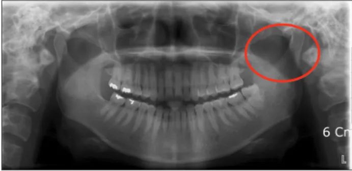
When she visited our department, she had a difficulty to open the mouth due to previous surgery. The fractured needle was found in the panoramic view (Fig. 1), which was located in the left condylar neck area. Likewise, cone beam CT was taken, and three-dimensional (3D) image was made.(Fig. 2)
Local anesthesia was performed via mandibular nerve block. We made about 6-7 cm vertical incision along the external oblique ridge, and then subperiosteal flap was el- evated to expose the medial and lateral aspects of the ramus.
The suture needle was positioned to the peri-condylar area
Fig. 1. The broken needle was shown in the left mandibular peri- condylar area in panoramic view.
Jin-Ha Kim et al: Removal of a broken needle using three-dimensional computed tomog- raphy: a case report. J Korean Assoc Oral Maxillofac Surg 2013
Fig. 2. The broken needle was shown in the left mandibular peri- condylar area in three-dimensional computed tomography view.
Jin-Ha Kim et al: Removal of a broken needle using three-dimensional computed tomog- raphy: a case report. J Korean Assoc Oral Maxillofac Surg 2013
Fig. 3. The broken needle was shown to be positioned in the left mandibular peri-condylar area in three-dimensional computed tomography view with the suture needle for localization during op- eration.
Jin-Ha Kim et al: Removal of a broken needle using three-dimensional computed tomog- raphy: a case report. J Korean Assoc Oral Maxillofac Surg 2013
Fig. 4. Panoramic view after the removal of the broken needle.
Jin-Ha Kim et al: Removal of a broken needle using three-dimensional computed tomog- raphy: a case report. J Korean Assoc Oral Maxillofac Surg 2013
Removal of a broken needle using three-dimensional computed tomography
253 dissection to identify the needle5-9. This case located the bro- ken needle in the peri-condylar area; we employed a vertical incision along the anterior border of the mandible, extending inferiorly to the external oblique ridge followed by subperios- teal dissection along the medial and lateral aspects of the ra- mus of the mandible. The initial subperiosteal dissection may help identify bony landmarks (such as lingula), which can be used as reference point during exploration; it also provide greater protection to the inferior alveolar and lingual nerves.
Focused extra periosteal blunt dissection based on CT scan information can be subsequently performed without inadver- tently damaging these nerves.
Therefore, in case of a broken dental needle during inferior alveolar nerve block anesthesia, prompt removal of the frag- ment with CT and guide-suture needle under local anesthesia make for an effective, successful method. This also prevents medicolegal issues from arising.
References
1. Säkkinen J, Huppunen M, Suuronen R. Complications following local anaesthesia. Nor Tannlegeforen Tid 2005;115:48-52.
2. Augello M, von Jackowski J, Grätz KW, Jacobsen C. Needle breakage during local anesthesia in the oral cavity--a retrospective of the last 50 years with guidelines for treatment and prevention.
Clin Oral Investig 2011;15:3-8.
3. Amies AB. Broken needles. Aust J Dent 1951;55:403-6.
4. Fraser-Moodie W. Location and localisation of metal in the tissues.
Br J Oral Surg 1966;4:99-105.
5. Zeltser R, Cohen C, Casap N. The implications of a broken needle in the pterygomandibular space: clinical guidelines for prevention and retrieval. Pediatr Dent 2002;24:153-6.
6. Faura-Solé M, Sánchez-Garcés MA, Berini-Aytes L, Gay-Escoda C. Broken anesthetic injection needles: report of 5 cases. Quintes- sence Int 1999;30:461-5.
7. Bhatia S, Bounds G. A broken needle in the pterygomandibular space: report of a case and review of the literature. Dent Update 1998;25:35-7.
8. Marks RB, Carlton DM, McDonald S. Management of a broken needle in the pterygomandibular space: report of case. J Am Dent Assoc 1984;109:263-4.
9. Bedrock RD, Skigen A, Dolwick MF. Retrieval of a broken needle in the pterygomandibular space. J Am Dent Assoc 1999;130:685-7.
10. Ethunandan M, Tran AL, Anand R, Bowden J, Seal MT, Brennan PA. Needle breakage following inferior alveolar nerve block: im- plications and management. Br Dent J 2007;202:395-7.
into the hilt to deposit the local anesthetic in the appropriate position. All of the above can contribute to needle breakage;
therefore, the use of a 30-gauge needle should be avoided when administering inferior alveolar nerve block1.
In case needle breakage does occur, every effort should be made to retrieve the needle immediately--if the tip is visible-- using fine hemostats. In the unfortunate event of the tip not being visible, the patient should be informed, and arrange- ments should be made for appropriate referral to the Depart- ment of Oral and Maxillofacial Surgery. Prompt retrieval is strongly recommended to minimize symptoms of pain, dys- phagia and trismus and to prevent the migration of the needle and potential damage to vital structures6. Note, however, that the retrieval of the needle in itself can lead to neurological and tissue damages during removal, and some suggest con- sidering removal only if the patient develops symptoms6. As reported by Ethunandan et al.10, the subject patient had trismus and pain upon leaving the clinic; thus, the initial decision was to leave the needle in situ, but it was subsequently removed six months later after the local symptoms persisted. A reason for the prompt removal of the needle is its possible migration and the development of severe complications; another factor is psychological. Therefore, removal of the fragment as soon as possible is recommended10.
Various methods have been described to find a broken nee- dle in the pterygomandibular space10. Plain radiographs are useful in confirming the broken needle and approximate posi- tion during the initial examination10. Nonetheless, they are unable to provide the accurate position of the broken needle and its relationship to adjacent structures10. 3D CT images can give us more precise information of the broken needle.
A site for incisions and exploration can be determined from the available information on the CT scan. It accurately shows the position of the needle with recognizable anatomical land- mark, especially with 3D reformatting10.
Most reports have suggested the use of vertical mucosal in- cision--often on the medial aspect of the mandible in the area penetrated by the needle--followed by blunt supra-periosteal