www.jpis.org
pISSN 2093-2278 eISSN 2093-2286 Copyright © 2011 Korean Academy of PeriodontologyThis is an Open Access article distributed under the terms of the Creative Commons Attribution Non-Commercial License (http://creativecommons.org/licenses/by-nc/3.0/).
Tissue reactions to suture materials in the oral mucosa of beagle dogs
Jae-Seok Kim1, Seung-Il Shin2, Yeek Herr2,3, Joon-Bong Park2,3, Young-Hyuk Kwon2,3, Jong-Hyuk Chung2,3,*
1Luden Dental Clinic, Seoul, Korea
2Department of Periodontology, 3Institute of Oral Biology, Kyung Hee University School of Dentistry, Seoul, Korea
Purpose: The objective of this study was to compare and evaluate the inflammatory responses of three widely used suture materials in the keratinized gingiva and buccal mucosa of beagle dogs.
Methods: Silk, polyglycolic acid, and nylon sutures were placed within the mandibular keratinized gingiva and maxillary buc- cal mucosa of four male beagle dogs. Biopsies were taken 3, 7, and 14 days after suturing. Specimens were prepared with hema- toxylin-eosin stain for evaluation under a light microscope.
Results: The suture materials placed in the oral mucosa elicited more inflammatory reactions than did those placed in the keratinized gingiva. The multifilament suture materials caused more inflammatory tissue reactions than did the monofilament suture materials in the oral mucosa.
Conclusions: If oral hygiene is well maintained and suture materials are placed in the keratinized gingiva, silk, nylon, and polyglycolic acid are considered to be proper suture materials for oral surgery. However, it is advisable to use monofilament suture materials if the suture site is within the oral mucosa.
Keywords: Gingiva, Histology, Inflammatory response, Oral mucosa, Suture materials.
INTRODUCTION
Suturing, which is the final procedure of a surgery, is used to reattach the removed tissue, to control bleeding, and allow for primary healing [1]. In addition to the physical require- ments for an ideal suture, such as maintaining proper form, ensuring the ease of sterilization and manipulation, and maintaining ligation and tension, the biological requirements of proper absorption time and minimum tissue response have also been suggested [2]. In intraoral surgeries, a variety of suture types have been applied [3]. Sutures can be divided into absorbable and non-absorbable ones and are classified as monofilament or multifilament types. Although microbial inflammation response due to the multifilament interstices
has been demonstrated, silk has been widely used in intra- oral surgeries because it has the advantages of easy manipu- lation, the capability of maintaining knot security, and less ir- ritation than nylon [4,5]. Polyglycolic acid, a polymer of lac- tide and glycolide, is used to manufacture an absorbable su- ture. After being extracted as a melted polymer, a thin fiber is manufactured into a braided form to improve its ease of ma- nipulation. Polyglycolic acid is absorbed in 60 to 90 days after insertion. It is hydrolyzed without any phagocytosis, which results in a weaker immune response than that of absorbable organic sutures [6]. One of the well-known non-absorbable monofilaments, nylon, is made from polyamide polymer and has been reported to show a high resistance to infection, high tension, and a low acute inflammation reaction [7].
Received: May 29, 2011; Accepted: Jun. 30, 2011
*Correspondence: Jong-Hyuk Chung
Department of Periodontology, Institute of Oral Biology, Kyung Hee University School of Dentistry, 1 Hoegi-dong, Dongdaemun-gu, Seoul 130-701, Korea E-mail: [email protected], Tel: +82-2-958-9380, Fax: +82-2-958-9387
infection by saliva, ingested food, microorganisms, etc., a su- ture placed in an oral cavity is affected differently than a su- ture in extraoral conditions [8]. In addition to the chronic in- flammation response, a suture placed in an infectious condi- tion is likely to show constant acute inflammation responses.
Furthermore, as a result of the inflammation response due to the suture itself, a suture can interfere with optimal wound healing and can result in the formation of excessive scar tis- sue, thereby reducing the tensile strength of the wound site.
This eventually negatively affects the immune response to infection. Therefore, the immune response to a suture is an important factor when choosing the proper suture type [9,10].
The suture type is one of the critical factors influencing in- fection occurrence post-surgery. It has been reported that braided sutures show a more severe inflammation reaction than do those without any braid, due to the potential for pen- etration and transfer by microorganisms [11,12]. On the other hand, it has been suggested that wound infection is not always associated with suture structure, but may also pertain to the number of fibers [13]. In addition, Gabrielli et al. [14] insisted that patient factors such as age and gender have an influence on the immune response and infection occurrence rate when they observed no significant differences in immune response among sutures.
Studies on suture-induced immune response according to oral mucosa tissue type have been conducted. Castelli et al.
[15] observed that the inflammation due to a suture located on the buccal mucosa was more severe than that of a suture located on the gingival mucosa. However, it was reported that there was no significant difference in immune response between the gingiva and alveolar mucosa in an adult dog [8].
The purpose of this study was to compare and evaluate the inflammatory responses induced by the three suture types widely used in clinical dentistry in the keratinized gingiva and buccal mucosa of beagle dogs.
MATERIALS AND METHODS
Surgical procedures
The sutures applied in this study were classified into mono- filament and multifilament types. The multifilament sutures included silk (size 5-0, reverse cutting needle FS-2, Ethicon Inc., Somerville, NJ, USA) and polyglycolic acid (coated Vicryl, size 5-0, reverse cutting needle FS-2, Ethicon Inc.), while the monofilament, nylon (size 5-0, reverse cutting needle FS-2, Ethicon Inc.) was also used.
Four one-year-old beagle dogs bred in isolated conditions and provided with a soft diet to protect the suture application site were used in this study. The experimental protocol
Animal Care and Use Committee of the Kyung Hee Univer- sity.
General anesthesia was conducted with the combination of 2 mL tiletamine and zolazepam (Zoletil, Virbac, Carros, France) and 0.5 mL xylazine (Rompun, Bayer Korea Ltd, Seoul, Korea) by intramuscular injection. The mandibular premolars (P1-P4) were extracted under local anesthesia of 2% lidocaine with 1:100,000 epinephrine (Lidocaine HCl, Huons Co., Seoul, Korea) by a sulcular incision and flap elevation approach. The extraction sites were sutured with an absorbable suture (plain gut, size 4-0, cutting needle P426, Ailee, Seoul, Korea). After a healing period of two months, the keratinized gingiva of the edentulous mandible and the buccal mucosa of the maxilla were sutured by single sutures of silk, polyglycolic acid, and nylon under general anesthesia. The sutures were inserted with minimal tissue tension, maintaining constant intervals and depth with a minimum suture gap of 7 mm. During the three days after single suturing, intramuscular injections of 2 mL gentamicin (Dongwha Pharm Co., Seoul, Korea) and 0.5 mL ketoprofene (Uni-Ketopro, UNIBiotech, Seoul, Korea) were administered. Three, seven, and 14 days subsequent to the single sutures, the tissue specimens were derived. During the entire experimental period, daily irrigation of normal sa- line and swabbing with 2% chlorhexidine were performed for plaque control.
Specimen preparation and observation
The sutures and surrounding tissues were retrieved en bloc, fixed with 10% neutrally buffered formalin solution, and subsequently embedded in paraffin. After the dissection of the specimens with a thickness of 8 µm, the specimens were double stained with hematoxylin and eosin for histo- pathological evaluation under a light microscope (BX-51, Olympus, Tokyo, Japan). Photography and analysis of the specimens were performed with a digital camera (DP-71, Olympus).
Statistical analysis
A modification of the method described by Racey et al. [16]
was used to evaluate the inflammatory reaction. The density of 3 cell-types (polymorphonuclear leukocytes, lymphocytes, macrophages) was counted and assigned a numerical grade from 1 (bare scattering) through 3 (dense aggregation).
A two-sample t-test was used to determine the inflamma- tory grade difference between the groups in the keratinized gingiva and buccal mucosa. To measure the statistical signif- icance of the inflammatory grade according to time point, one way analysis of variance and Duncan’s method for post- hoc testing were performed (P-value<0.05).
Immune response to silk sutures
Seven days after suturing, severe infiltration of inflamma- tory cells was noticed around the sutures (Fig. 1). Fourteen days post-suturing, a moderate degree of inflammatory cell infiltration and epitheloid formation were observed around the sutures. In the epithelium surrounding the sutures, a low degree of inflammatory cell infiltration was observed (Fig. 2).
For the suture located on the buccal mucosa, a more signifi- cant inflammation response was noted (Fig. 3). During the entire study, the group sutured on the mandibular keratin- ized gingiva showed only a slight inflammation response, compared with the sutures on the buccal mucosa. There was
as time progressed.
Immune response to polyglycolic acid sutures
Until 14 days after suturing, absorption of the suture was not observed, and the suture interstices were maintained.
Three days after suture, the inflammatory cell infiltration and immune responses within the surrounding epithelium and connective tissue of the mandibular keratinized gingival suture group were weak (Fig. 4). Seven days post-suturing, extensive inflammatory cell infiltration was observed sur- rounding the sutures and within the connective tissues of both groups. In addition, neutrophils and leukocytes were observed, along with the formation of bacterial plaque (Fig. 5).
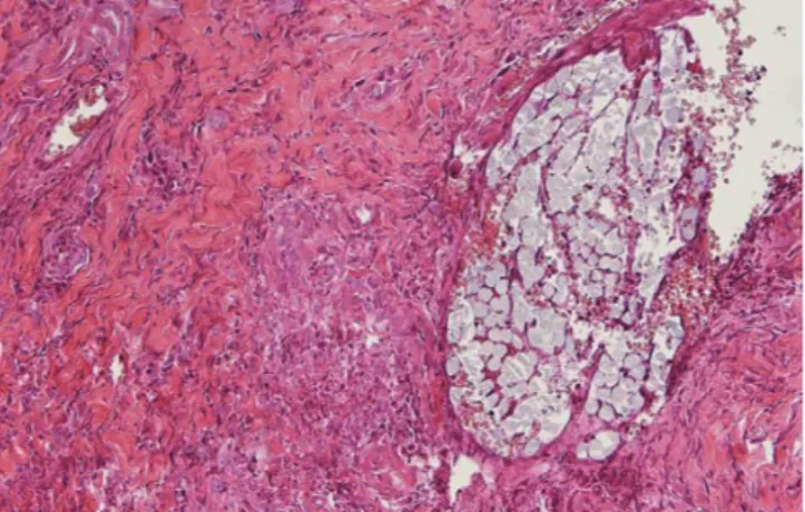
Figure 1. Silk suture at seven days in buccal oral mucosa. The su- ture loop is surrounded by dense inflammatory cell infiltrate and a zone of inflammatory reaction (H&E staining, original magnifica- tion ×40).
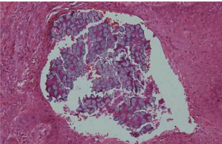
Figure 2. Silk suture at 14 days in keratinized gingiva. Bacterial plaque and inflammatory cell infiltration in the suture filaments is observed. Epithelial invagination is well advanced, and the suture material is lined by epithelium (H&E staining, original magnifica- tion ×200).
Figure 3. Silk suture at 14 days in buccal oral mucosa. Numerous polymorphonuclear leucocytes and lymphocytes are evident. Epi- thelial cells are observed around the suture material (H&E staining, original magnification ×400).
Figure 4. Polyglycolic acid suture at three days in keratinized gingi- va. A few lymphocytes and moderate polymorphonuclear leucocyte infiltration are observed around the suture material. Very few lym- phocytes and macrophages are observed in the interstitium be- tween the collagen bundles (H&E staining, original magnification
×200).
The immune response after 14 days had similar histologic features to that after seven days. While the tissues surround- ing the sutures showed a moderately high inflammation re- action, the inflammation reaction of the submucosa was slight with both sutures remaining unabsorbed. Infiltration of in- flammatory cells was found within the suture interstices (Fig.
6).
Immune response to nylon sutures
Infiltration of various inflammatory cells was examined from the tissues surrounding the sutures. While polymor- phonuclear leukocytes (PMNL) were mainly observed during the early stage, the proportion of macrophages present com- pared to other cell types increased as time proceeded. Al- though infiltration of inflammatory cells, such as PMNL,
neutrophils, and macrophages, could be observed around the suture, infiltration within the connective tissue was low. As for nylon suture located on the keratinized gingiva, seven days after suturing the number of PMNLs, lymphocytes, and macrophages decreased, from the surrounding tissue of the suture toward the submucosa tissue (Fig. 7). Fourteen days post-suturing, the infiltration of inflammatory cells was not significant. In particular, the infiltration of inflammatory cells within the connective tissue was very rare (Figs. 8 and 9).
The difference in inflammation reaction between the kera- tinized gingiva and the buccal mucosa was not significant.
Moreover, there was no difference in the inflammation reac- tion between the tissues surrounding the suture as time pro- ceeded.
Figure 5. Polyglycolic acid suture at seven days in buccal oral mu- cosa. Inflammatory cells are present within the suture, as well as in the adjacent submucosal area. The suture material is still intact (H&E staining, original magnification ×200).
Figure 6. Polyglycolic acid suture at 14 days in keratinized gingiva.
There is migration of monocytes into the interstices. The perisutur- al granulation tissue contains a few lymphocytes and macrophages, as well as elongated collagen fibers (H&E staining, original magni- fication ×200).
Figure 8. Nylon suture at 14 days in keratinized gingiva. The mi- croscopic findings around the suture reveal some neutrophils and lymphocyte infiltration. Perisutural granulation tissue containing many capillaries and collagen fibrils is visible. Moderate inflamma- tory reaction can be observed (H&E staining, original magnification
×400).
Figure 7. Nylon suture at seven days in keratinized gingiva. The perisutural tissue contains predominantly polymorphonuclear leu- cocytes and lymphocytes. Newly formed collagen fibrils can be ob- served (H&E staining, original magnification ×400).
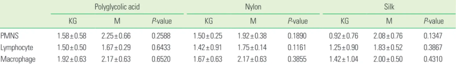
The inflammatory reaction grade was more severe in the buccal mucosa than keratinized gingiva in all groups, but there was no statistical significance (Table 1). The inflamma- tory reaction grade according to the point in time showed that lymphocyte and macrophage infiltration were signifi- cantly low in the keratinized gingiva after three days, where- as macrophage infiltration was significantly high in the buc- cal mucosa after one week (Table 2).
DISCUSSION
Adaptation and maintenance of the wound site by sutures are critical events for the success of a surgical procedure.
can increase the susceptibility to tissue infection [17]. Infec- tion caused by the incision of a wound site, spread of infec- tion during suturing and suture removal, and the delay of healing after surgery can all lead to a serious situation for a patient with a chronic disease, such as diabetes mellitus or cardiovascular disease. It has been reported that the risk of infection is increased by the presence of a suture within a wound [18].
Therefore, the importance of yielding the minimum im- mune response, which is an ideal result of suturing, should be considered with great attention when selecting suture type. Accordingly, this study was conducted to compare and evaluate the function of three suture types, all of which are widely used in intraoral surgeries, through the comparison of their tissue affinities. One of the most important functions of a suture is to maintain the stability of the wound edges until they can sustain themselves. The removal of oral sur- gery sutures is routinely performed one week post-surgery.
However, in the case of regeneration therapy, maintenance of the suture for at least two weeks is frequently allowed in clinical practice for the enhancement of tissue maturity [19].
Therefore, in this study, a long term evaluation was used to examine the early- and late-stage immune responses. The experiment was divided into three time periods for the ob- servation of acute inflammation, the common period of su- ture removal, and the maximum tensile force of the incision:
three days post suturing, seven days post suturing, and 14 days post suturing. As for the sutures in the experiment, silk, polyglycolic acid, and nylon sutures were selected based on their wide utilization in clinical practice.
Figure 9. Nylon suture at 14 days in buccal oral mucosa. Significant inflammatory cell infiltration--polymorphonuclear leucocytes, mac- rophages, and lymphocytes--is observed along the suture track (H&E staining, original magnification ×200).
Table 1. Inflammatory reaction grade in the keratinized gingiva and buccal mucosa of suture materials (two-sample t-test, mean±SD).
Polyglycolic acid Nylon Silk
KG M P-value KG M P-value KG M P-value
PMNS 1.58±0.58 2.25±0.66 0.2588 1.50±0.25 1.92±0.38 0.1890 0.92±0.76 2.08±0.76 0.1347
Lymphocyte 1.50±0.50 1.67±0.29 0.6433 1.42±0.91 1.75±0.14 0.1161 1.25±0.90 1.83±0.52 0.3867 Macrophage 1.92±0.63 2.17±0.63 0.6520 1.67±0.63 2.17±0.63 0.3855 1.42±1.04 2.00±0.50 0.4310 N=4, KG: keratinized gingiva, M: buccal mucosa; PMNS, polymorphonuclear leucocyte.
Table 2. Result of inflammatory reaction (ANOVA and post-hoc test [Duncan’s method], mean±SD) at 3 days, 1 week, and 2 weeks.
Keratinized gingiva Buccal mucosa
3 days 1 week 2 weeks P-value 3 days 1 week 2 weeks P-value
PMNS 1.00±0.66 1.92±0.29 1.08±0.29 0.0848 1.75±0.43 2.50±0.25 2.00±0.75 0.2746
Lymphocyte 0.91±0.38a) 1.92±0.38 1.33±0.29 0.0364 1.75±0.25 1.91±0.38 1.58±0.38 0.5305
Macrophage 0.83±0.52a) 2.17±0.38 2.00±0.25 0.0125 1.67±0.29 2.67±0.14b) 2.00±0.43 0.0203
a)P<0.05 significant low. b)P<0.05 significant high.
N=4, PMNS, polymorphonuclear leucocyte.
the soft tissue in the extraction site included highly organized fibrous connective tissues at 30 days post-extraction, while the isolation of the newly formed hard tissue was mainly composed of woven bone and marginal mucosa at 60 days post-extraction. In this study, the suturing was conducted two months after the extraction to rule out the influence of the immune response caused by the soft tissue healing pro- cess and the woven bone formation.
The keratinized gingiva of the mandibular premolar re- gions and the buccal mucosa of the maxillary premolar re- gions were selected as the suture application sites for this study. Castelli et al. [15] reported that the mucosa is suscepti- ble to the infiltration of inflammatory cells due to its compo- sition of muscle fibers, minor saliva glands, and common connective tissues. Romanos et al. [21] immunohistologically identified type V collagen as containing the largest portion of lamina propria in sound keratinized gingiva. They also showed that type V collagen has great resistance to collage- nase and acts as a mechanical barrier to bacterial penetra- tion. It was also shown in this study that the sutures located on the buccal mucosa resulted in a higher infiltration of in- flammatory cells into the internal tissue of the sutures than did those on the keratinized gingiva. However, the difference in the inflammation reaction between suture locations at 14 days post-suturing was insignificant.
In addition, it has been suggested that the physical form of a suture determines the degree of the inflammation reaction [15]. In many studies, the cause of the severe inflammation reaction observed in multifilament sutures, compared with monofilament sutures, has been referred to as ‘the wicking effect’ and results in the spread of the infection among the multifilament sutures and the presence of bacteria within the suture interstices [8,12,22]. However, Grigg et al. [4] found that silk did not produce more fluid movement by capillary action than coated Vicryl, and produced less than a polymer suture. According to a clinical study on the influence of su- ture absorption in two types of multifilament sutures and the resulting complication occurrence of wound healing and the failure of dental implants by Ivanoff and Windmark, neither type of suture showed any infection or implant failure [5].
Sortino et al. [23] counted the number of microorganisms with a light microscope in order to evaluate the inflamma- tion reactions after removal of silk and polymer sutures from an intraoral environment eight days post-suturing. They re- ported that there was no significant difference between the treatment outcomes with regard to suture type, due to simi- lar distributions of aerobic and anaerobic bacteria within the sutures. Also, it was reported that comparisons of infection, dehiscence, redness, scar hyperplasia, and patient satisfaction
weeks and six months after suturing did not show any signif- icant differences [24]. In our study, blood clots from the trau- ma of suturing and infiltration of inflammatory cells around the suture and within the connective tissue due to acute in- flammation reaction were observed around the suture and within the submucosa during the entire period of observa- tion. However, there were no significant differences in the degree of inflammation among the three types of suture lo- cated on the keratinized gingiva. As for the silk and polygly- colic acid sutures, the existence of neutrophils within suture interstices and the migration of leukocytes were identified throughout the study.
In this study, intramuscular injection was administered with 2 mL of gentamicin for three days after suturing, and plaque control was performed with normal saline and 2% chlorhexi- dine. According to the study of Leknes et al. [25] on immune responses due to the use of silk and expanded polytetrafluo- roethylene sutured onto the mandibular keratinized gingiva of a beagle, the administration of 2% chlorhexidine and sys- temic broad-spectrum antibiotics reduced the formation of biofilm and inflammation within the suture path. On the other hand, it was reported that the use of chlorhexidine had no effect on the species or existence of microorganisms, re- gardless of the kind of suture utilized [23]. However, because it used human participants, the latter study was considered to be incapable of strict control. Moreover, considering that the surgery sites were at the mandibular right angle area, the maintenance of oral hygiene was regarded as unmanageable.
In this study, post suturing oral disinfection was conducted daily with 2% chlorhexidine, and unlike the study of Sortino et al. [23], the oral hygiene control of the suture sites in the mandibular premolar region (P1-P4) was possible, resulting in a definite outcome.
In conclusion, the nylon monofilament suture resulted in a favorable treatment outcome when placed in the alveolar mucosa, lesions arising from acute inflammation or an envi- ronment in which oral hygiene maintenance was unavailable.
On the other hand, the structural difference in the monofila- ment and the multifilament sutures, with regard to immune response, did not affect the treatment outcome when placed in histologically dense keratinized gingiva with good oral hygiene.
CONFLICT OF INTEREST
No potential conflict of interest relevant to this article was reported.
1. Shaw RJ, Negus TW, Mellor TK. A prospective clinical evaluation of the longevity of resorbable sutures in oral mucosa. Br J Oral Maxillofac Surg 1996;34:252-4.
2. Swanson NA, Tromovitch TA. Suture materials, 1980s:
properties, uses, and abuses. Int J Dermatol 1982;21:373-8.
3. Cohen ES. Atlas of cosmetic and reconstructive periodon- tal surgery. 3rd ed. Hamilton: BC Decker; 2007.
4. Grigg TR, Liewehr FR, Patton WR, Buxton TB, McPherson JC. Effect of the wicking behavior of multifilament sutures.
J Endod 2004;30:649-52.
5. Ivanoff CJ, Widmark G. Nonresorbable versus resorbable sutures in oral implant surgery: a prospective clinical study.
Clin Implant Dent Relat Res 2001;3:57-60.
6. Craig PH, Williams JA, Davis KW, Magoun AD, Levy AJ, Bogdansky S, et al. A biologic comparison of polyglactin 910 and polyglycolic acid synthetic absorbable sutures.
Surg Gynecol Obstet 1975;141:1-10.
7. Silverstein LH. Principles of dental suturing: the complete guide to surgical closure. New Jersey: Montage Media Co.; 1999.
8. Selvig KA, Biagiotti GR, Leknes KN, Wikesjo UM. Oral tis- sue reactions to suture materials. Int J Periodontics Re- storative Dent 1998;18:474-87.
9. Sanz L, Smith S. Mechanisms of wound healing, suture material, and wound closure, strategies in gynecologic surgery. New York: Springer; 1986.
10. Yaltirik M, Dedeoglu K, Bilgic B, Koray M, Ersev H, Issever H, et al. Comparison of four different suture materials in soft tissues of rats. Oral Dis 2003;9:284-6.
11. Leknes KN, Røynstrand IT, Selvig KA. Human gingival tissue reactions to silk and expanded polytetrafluoroeth- ylene sutures. J Periodontol 2005;76:34-42.
12. Lilly GE, Armstrong JH, Salem JE, Cutcher JL. Reaction of oral tissues to suture materials. II. Oral Surg Oral Med Oral Pathol 1968;26:592-9.
13. Rothenburger S, Spangler D, Bhende S, Burkley D. In vitro antimicrobial evaluation of Coated VICRYL* Plus Anti- bacterial Suture (coated polyglactin 910 with triclosan) us- ing zone of inhibition assays. Surg Infect (Larchmt) 2002;3 Suppl 1:S79-87.
Suture materials and other factors associated with tissue reactivity, infection, and wound dehiscence among plastic surgery outpatients. Plast Reconstr Surg 2001;107:38-45.
15. Castelli WA, Nasjleti CF, Diaz-Perez R, Caffesse RG. Cheek mucosa response to silk, cotton, and nylon suture materi- als. Oral Surg Oral Med Oral Pathol 1978;45:186-9.
16. Racey GL, Wallace WR, Cavalaris CJ, Marguard JV. Com- parison of a polyglycolic-polylactic acid suture to black silk and plain catgut in human oral tissues. J Oral Surg 1978;36:766-70.
17. Blomstedt B, Osterberg B, Bergstrand A. Suture material and bacterial transport. An experimental study. Acta Chir Scand 1977;143:71-3.
18. King RC, Crawford JJ, Small EW. Bacteremia following in- traoral suture removal. Oral Surg Oral Med Oral Pathol 1988;65:23-8.
19. Blumenthal NM. A clinical comparison of collagen mem- branes with e-PTFE membranes in the treatment of hu- man mandibular buccal class II furcation defects. J Peri- odontol 1993;64:925-33.
20. Cardaropoli G, Araújo M, Lindhe J. Dynamics of bone tis- sue formation in tooth extraction sites. An experimental study in dogs. J Clin Periodontol 2003;30:809-18.
21. Romanos GE, Schröter-Kermani C, Weingart D, Strub JR.
Health human periodontal versus peri-implant gingival tissues: an immunohistochemical differentiation of the extracellular matrix. Int J Oral Maxillofac Implants 1995;
10:750-8.
22. Lilly GE. Reaction of oral tissues to suture materials. Oral Surg Oral Med Oral Pathol 1968;26:128-33.
23. Sortino F, Lombardo C, Sciacca A. Silk and polyglycolic acid in oral surgery: a comparative study. Oral Surg Oral Med Oral Pathol Oral Radiol Endod 2008;105:e15-8.
24. Gabel EA, Jimenez GP, Eaglstein WH, Kerdel FA, Falanga V.
Performance comparison of nylon and an absorbable su- ture material (Polyglactin 910) in the closure of punch bi- opsy sites. Dermatol Surg 2000;26:750-2.
25. Leknes KN, Selvig KA, Bøe OE, Wikesjö UM. Tissue reac- tions to sutures in the presence and absence of anti-infec- tive therapy. J Clin Periodontol 2005;32:130-8.