J Lung Cancer 2010;9(2):106-109
106
Tumor Lysis Syndrome Induced by Radiotherapy in Non- Small Cell Lung Cancer
Tumor lysis syndrome (TLS) is an oncologic emergency that is characterized by numerous metabolic abnormalities, including hyperuricemic nephropathy, hyperphosphatemia, hypocalcemia, hyperkalemia and increased serum creatinine. This syndrome is common for tumors with rapid cell turnover and growth rates, and for bulky tumors with high sensitivity to anti-neoplastic treatments. Hence, TLS is a well-recognized clinical problem in hematologic malignancies. TLS is rarely observed to be induced in solid tumors by chemo- therapy. Herein we present the second case of TLS that developed during radiotherapy in a patient with non-small cell lung cancer. (J Lung Cancer 2010;9(2):106 109)
Key Words: Tumor lysis syndrome, Radiotherapy, Non-small cell lung carcinoma
Dong-Hyo Noh, M.D.1 Ki-Eun Hwang, M.D.1 Jeong-Hyun Shin, M.D.1 Dong Kim, M.D.1 Kyung-Hwa Cho, M.D.1 Keum-Ha Choi, M.D.2 Seong-Hoon Park, M.D.3 Eun-Taik Jeong, M.D.1 and Hak-Ryul Kim, M.D.1
Departments of 1Internal Medicine,
2Pathology, 3Radiology, Institute of Wonkwang Medical Science, Won- kwang University School of Medicine, Iksan, Korea
Received: July 14, 2010 Revised: September 14, 2010 Accepted: September 27, 2010 Address for correspondence Hak-Ryul Kim, M.D.
Department of Internal Medicine, Won- kwang University School of Medicine, 344-2, Shinyong-dong, Iksan 570-749, Korea
Tel: 82-63-859-2583 Fax: 82-63-855-2025
E-mail: [email protected] This study was supported by grant from Wonkwang University in 2009.
Tumor lysis syndrome (TLS) is an oncologic emergency. It is characterized by a group of metabolic derangements that are caused by the massive and abrupt release of cellular components into the blood after the rapid lysis of malignant cells (1). The release of intracellular metabolites, including nucleic acids, proteins, phosphorus and potassium, can overwhelm the normal homeostatic mechanisms and this potentially leads to hyperuricemia, hyperkalemia, hyperphos- phatemia, hypocalcemia and uremia (2-5). TLS is most frequently observed in patients with hematologic malignancies such as myeloproliferative disease, acute leukemia and high- grade non-Hodgkin’s lymphoma, and especially Burkitt’s
lymphoma after the initiation of cytotoxic therapy, although TLS may also occur spontaneously and/or in other types of tumor that have a high proliferative rate, a large tumor burden or high sensitivity to cytotoxic therapy (6,7). The occurrence of TLS in solid tumors is relatively rare, but this malady has a high mortality rate. TLS is likely a consequence of less pre-emptive prophylaxis and reduced awareness of the occurrence of this problem in solid tumors. We report here on a case of TLS in a patient with non-small cell lung cancer and it occurred during radiotherapy. Sadly, the patient died despite undergoing hemodialysis.
TLS by Radiotherapy in Non-small Cell Lung Cancer 107
Fig. 1. Chest computed tomography shows a 3.3 cm mass with irregular margins and heterogenous density in the apical segment of the right upper lobe.
Fig. 2. Microscopic findings show the acinar type of adeno- carcinoma with malignant glands infiltrating the collagenous stroma (H&E stain, ×200).
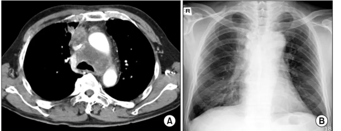
Fig. 3. (A) Chest computed tomo- graphy shows mediastinal lymph node enlargement after the third cycle of docetaxel. (B) The chest X-ray shows no significant changes after the 6th fraction of radiotherapy.
CASE REPORT
A 67-year-old male was diagnosed with stage IV non-small cell lung cancer 8 months previously. He was previously healthy and he had smoked a pack a day for 20 years. A chest computed tomography (CT) scan obtained for a health check-up revealed a 3.3-cm mass that was heterogenous with irregular margins, and this was in the right upper lobe apical segment (Fig. 1). Among the other CT findings were multiple enlarged lymph nodes, including a right supraclavicular lymph node, and a pulmonary nodule in the left lower lobe. He underwent a percutaneous needle aspiration biopsy. The histologic findings were consistent with a moderately differentiated adenocarci- noma. Immunohistochemical staining was performed for TTF-1 and the result was positive (Fig. 2). The laboratory tests, including the complete blood cell count and blood chemistry,
were within the normal ranges. He was treated with six cycles of gemcitabine and cisplatin chemotherapy. A post-treatment CT scan showed progressive disease with an increased size of both the lung mass in the right upper lobe and the mediastinal lymph nodes, and an increased number of bilateral pulmonary nodules. He subsequently began second-line chemotherapy with docetaxel. After the third cycle of docetaxel, a CT scan showed progressive disease (Fig. 3). After 2 weeks, his dysphagia, dyspnea and hoarseness worsened. After presentation of the case at a multidisciplinary meeting, we made the decision to offer palliative radiotherapy. A total radiation dose of 30 Gy divided into 10 fractions was planned to relieve the obstruction of the superior vena cava. Before the radiotherapy, the laboratory tests were normal as follows: blood urea nitrogen, 18.5 mg/dL (normal range, 8∼20 mg/dL); creatinine, 0.94 mg/dL (normal range, 0.5∼1.3 mg/dL); potassium, 4.5 mEg/L
108 J Lung Cancer 2010;9(2):106-109
(normal range, 3.5∼5.5 mEg/L); uric acid, 3.6 mg/dL (normal range, 3.5∼7.2 mg/dL); calcium, 8.8 mg/dL (normal range, 8.4
∼10.2 mg/dL); and lactate dehydrogenase, 250 IU/L (normal range, 100∼450 IU/L).
After the 6th fraction of radiotherapy with a total of 18 Gy, the urine volume decreased to 500 mL/day. The laboratory tests showed the following: blood urea nitrogen, 69.1 mg/dL;
creatinine, 2.53 mg/dL; potassium, 5.9 mEg/L; uric acid, 10.2 mg/dL; calcium, 8.8 mg/dL; lactate dehydrogenase, 450 IU/L.
There were no significant changes seen on the chest X-ray during radiotherapy. We did not perform a CT scan due to the short interval between the 1st day of radiotherapy and the time of symptom aggravation. Despite intensive management, including vigorous hydration with normal saline, concomitant use of furosemide and allopurinol (a xanthine oxidase inhibitor for the management of hyperuricemia), he became anuric with worsening of the laboratory findings the next day, as follows:
blood urea nitrogen, 80.2 mg/dL; creatinine, 2.8 mg/dL;
potassium, 6.5 mEg/L; phosphorus, 6.7 mg/dL; uric acid, 11.1 mg/dL; calcium, 8.4 mg/dL; lactate dehydrogenase, 1,513 IU/L.
Although emergency hemodialysis was started, he passed away that day due to uncontrolled metabolic acidosis.
DISCUSSION
TLS is rarely observed in solid tumors. In one review, there were only 45 cases of TLS in patients with solid tumor cases from the first report in 1977 to 2002 (7). The incidence of TLS is more common in highly responsive tumors with a large tumor burden. Small cell lung cancer, breast cancer, germ cell tumors and malignant melanoma are the most common solid tumors reported to be associated with TLS (8,9). Although diverse causes for TLS in solid tumors have been reported, most cases of TLS cases in solid tumors are induced by chemotherapy (9). The incidence of TLS in association with radiotherapy is rare.
In this case, we thought the decrease in urine volume and the lab findings of TLS were caused by radiotherapy, but not by tumor progression itself because the tumor’s size wasn’t bulky or growing rapid enough to cause TLS. Further, these findings of TLS were seen immediately after radiotherapy, so we concluded that the cause of TLS was radiotherapy. The present case is the 2nd reported case of TLS that developed
during radiotherapy in a patient with non-small cell lung cancer. The first case was described in 2008 in a patient with non-small cell lung cancer after receiving radiotherapy of 6 Gy (10).
The potential risk factors for TLS in solid tumors include a high turnover burden with a metastatic presentation, elevated levels of serum lactate dehydrogenase and uric acid, the response to anti-neoplastic treatment, pre-existing renal insuffi- ciency, treatment with nephrotoxic agents and underlying problems, such as infections and intravascular volume depletion (7,9,11-13). Because renal clearance is the primary method of eliminating most toxic products released into the circulation, aggressive hydration before and during treatment in high-risk patients is the mainstay of prevention.
The mortality rate for patients with solid tumors and TLS appears to be higher than that reported for TLS following treatment of hematologic malignances. This high mortality rate of solid tumor patients with TLS may be explained by the lack of prophylactic measures that are usually implemented before the start of chemotherapy for hematologic malignancies.
Contrary to hematologic malignancies, TLS in solid tumors can occur several days to weeks after treatment. Thus, awareness of the possibility of TLS in solid tumors, alertness and timely laboratory work-ups are essential for high-risk patients.
Frequent laboratory evaluations with a complete blood count and assessing the serum sodium, potassium, chloride, bicar- bonate, calcium, phosphorus, uric acid and creatinine, and urine analysis before and during treatment are important for the early detection of TLS.
We have presented a rare case of TLS that was induced by radiotherapy in a patient with non-small cell lung cancer.
Whether or not a patient has a solid tumor and/or is treated by radiotherapy, this type of patient should be carefully observed because of the substantial risk for TLS.
REFERENCES
1. Arrambide K, Toto RD. Tumor lysis syndrome. Semin Nephrol 1993;13:273-280.
2. Cohen LF, Balow JE, Magrath IT, Poplack DG, Ziegler JL.
Acute tumor lysis syndrome: a review of 37 patients with Burkitt's lymphoma. Am J Med 1980;68:486-491.
3. Brereton HD, Anderson T, Johnson RE, Schein PS. Hyper- phosphatemia and hypocalcemia in Burkitt lymophoma:
TLS by Radiotherapy in Non-small Cell Lung Cancer 109
complications of chemotherapy. Arch Intern Med 1975;135:
307-309.
4. Drakos P, Bar-Ziv J, Catane R. Tumor lysis syndrome in nonhematologic malignancies: report of a case and review of the literature. Am J Clin Oncol 1994;17:502-505.
5. Kjellstrand CM, Cambell DC 2nd, von Hartitzsch B, Buselmeier TJ. Hyperuricemic acute renal failure. Arch Intern Med 1974;133:349-359.
6. Hande KR, Garrow GC. Acute tumor lysis syndrome in patients with high-grade non-Hodgkin's lymphoma. Am J Med 1993;94:133-139.
7. Baeksgaard L, Sørensen JB. Acute tumor lysis syndrome in solid tumors: a case report and review of the literature. Cancer Chemother Pharmacol 2003;51:187-192.
8. Crittenden DR, Ackerman GL. Hyperuricemic acute renal failure in disseminated carcinoma. Arch Intern Med 1977;137:
97-99.
9. Gemici C. Tumour lysis syndrome in solid tumours. Clin Oncol (R Coll Radiol) 2006;18:773-780.
10. Noh GY, Choe DH, Kim CH, Lee JC. Fatal tumor lysis syndrome during radiotherapy for non-small-cell lung cancer.
J Clin Oncol 2008;26:6005-6006.
11. Mott FE, Esana A, Chakmakjian C, Herrington JD. Tumor lysis syndrome in solid tumors. Support Cancer Ther 2005;2:
188-191.
12. Coiffier B, Altman A, Pui CH, Younes A, Cairo MS.
Guidelines for the management of pediatric and adult tumor lysis syndrome: an evidence-based review. J Clin Oncol 2008;26:2767-2778.
13. Yuh YJ, Kim SR. Lactate dehydrogenase (LDH) as a tumor marker for non-small cell lung cancer. Cancer Res Treat 2002;34:339-344.