www.krspine.org
Radiological and Clinical Outcome after Simple Discectomy of Central Massive Lumbar Disc Herniation
Young Do Koh, M.D., Seung Jun Rhee, M.D., Dong Jun Kim, M.D.
J Korean Soc Spine Surg 2010 Dec;17(4):169-176.
Originally published online December 31, 2010;
doi: 10.4184/jkss.2010.17.4.169
Korean Society of Spine Surgery
Department of Orthopaedic Surgery, Ewha Womans University Collge of Medicine
#911-1 Mok-dong, Yangcheon-gu, Seoul, 158-710, Korea Tel: 82-2-2646-6808 Fax: 82-2-2646-6804
©Copyright 2010 Korean Society of Sping Surgery pISSN 2093-4378 eISSN 2093-4386
The online version of this article, along with updated information and services, is located on the World Wide Web at:
http://www.krspine.org/DOIx.php?id=10.4184/jkss.2010.17.4.169
This is an Open Access article distributed under the terms of the Creative Commons Attribution Non-Commercial License (http://
creativecommons.org/licenses/by-nc/3.0) which permits unrestricted non-commercial use, distribution, and reproduction in any medium, provided the original work is properly cited.
Journal of Korean Society of
Spine Surgery
Radiological and Clinical Outcome after Simple Discectomy of Central Massive Lumbar Disc Herniation
Young Do Koh, M.D., Seung Jun Rhee, M.D., Dong Jun Kim, M.D.
Department of Orthopedic Surgery, School of Medicine, Ewha Womans University, Seoul, Korea
Study Design: This is a retrospective case control study.
Objectives: To analyze our results following simple discectomy of central massive disc herniation focusing on instability for the usefulness of intervertebral fusion.
Summary of Literature Review: Lumbar instability is a complication of central massive disc herniation. However, there is limited evidence on the correlation between lumbar instability and loss of disc material.
Materials and Methods: A total of 25 patients who had undergone discectomy for a single-level lumbar disc herniation were followed up for two years. The clinical group (group A) included 12 patients that had a compromised canal with greater than 50% of the herniated disc, while the central axis of the herniated disc was less than 20% deviated from the center axis of the spinal canal, as seen on MRI.
The control group (group B) had 13 patients that had a compromised canal with less than 50% of the herniated disc while their axis was more than 20% deviated from the center axis of the spinal canal. Clinical and radiologic instability, pain and functional disability were compared between the two groups.
Results: No differences was found between the two groups in clinical instability, radiological instability, visual analogue scale (VAS), and the Oswestry disability index (ODI).
Conclusions: Central massive disc herniation after discectomy did not show a significant difference in clinical or radiological instability from that of other herniation types.
Key Words: Discectomy, Central massive disc herniation, Instability
Received: July 16, 2010 Revised: December 10, 2010 Accepted: December 10, 2010 Published Online: December 31, 2010 Corresponding author: Dong Jun Kim, M.D.
Department of Orthopedic Surgery, Mokdong Hospital, Ewha Womans University, School of Medicine, 911-1, mok 6-dong, Yangcheon-gu, Seoul 158- 710, Seoul, Korea
TEL: 82-2-2650-2873, FAX : 82-2-2642-0349 E-mail: [email protected]
“This is an Open Access article distributed under the terms of the Creative Commons Attribution Non-Commercial License (http://
creativecommons.org/licenses/by-nc/3.0/) which permits unrestricted non-commercial use, distribution, and reproduction in any medium, provided the original work is properly cited.”
본 논문의 요지는 대한척추외과학회 제27차 춘계학술대회에서 발표되었음.
INTRODUCTION
The intervertebral discs distribute external loads in radial form and the fibrous rings surrounding the intervertebral discs connect each segment firmly, contributing to the stability of spinal segments. A disc herniation damages these structures and contributes to instability of each segment. Since Knutsson et al.1) reported in 1944 that change in intervertebral discs caused instability of spinal segments, numerous studies have been conducted on measuring the correlation between degenerative change (loss of the intervertebral disc) and instability of spinal segments. However, studies on the long-term effect of the loss of intervertebral discs on stability of the spinal segments is quite limited.
There has been a constant debate on the necessity of spinal fusion after discectomy because of difficulties in intervertebral instability assessments as a result of anatomical position of disc
Young Do Koh et al Volume 17 • Number 4 • December 2010
www.krspine.org 170
herniation and loss of the intervertebral disc. Therefore, this study assessed the instability based on the anatomical position of disc herniation and the extent of loss of the intervertebral disc.
Patients with a compromised canal with more than a 50% of the herniated disc, while the axis was less than 20% deviated from the central axis of the spinal canal, as seen on MRI, were categorized as a central massive disc herniation group and were expected to have a high risk of developing lumbar instability after surgery. Short-term and long-term follow-up observations were carried out. In this research, we wanted to estimate the correlations between the extent of disc loss, clinical outcome and development of lumbar instability, and gather evidence that would help decision making in diagnosis and treatment for lumbar instability following disectomy.
MATERIAS AND METHODS
1. Materials
This study included patients who had been diagnosed as having a lumbar disc herniation and undergone simple discectomy by the same surgeon from February 2005 through November 2007. In lumbar spine lateral radiographs taken in standing, flexion, extension, horizontal and angular displacements were measured for radiological lumbar instability.
Patients with spondylolysis, destructive spodylolisthesis and instability or more than 2 segments, as well as subjects with a history of surgery or trauma were excluded from this study.
Only subjects with intervertebral disc herniation that developed at the level of L4-5 or L5-S1, at which spinal segment instability largely occurred, were included. Among all subjects, patients with a compromised canal with more than 50% of the herniated disc, while the axis was less than 20% deviated from the center axis of the spinal canal on MRI, were categorized as group A.
Patients with a compromised canal with less than 50% of the herniated disc whose axis was more than 20% deviated from the center axis of the spinal canal on MRI, were categorized as the control group. A total of 107 surgery cases were performed during the period of investigation. Seventeen patients (15.8%) satisfied the inclusion criteria of the clinical group and 12 of them with two-year follow-up observations were selected. The clinical group included 3 males and 9 females with a mean age of 39.7 years old (17~64). Twenty-seven (25.2%) patients satisfied the criteria for the control group and thirteen patients who had undergone two-year follow-up observations were included.
The control group consisted of 11 males and 2 females with a mean age of 41.6 years old. (22-62)
2. Methods
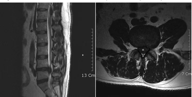
The scale of herniation in the spinal canal to that of the normal spinal canal of the axial image, in which an intervertebral disc herniation was verified by MRI images, was expressed in percentile. The percentile was obtained by measuring the encroached area three times by two surgeons. Among the measured values of the total of six instances, the mean value of four, after subtracting the highest and lowest value, was used to determine the extent of spinal canal encroachment (Fig. 1). The herniation in the spinal canal of the clinical group was 71.06%
on average while that of the control group was 27.47% on average (Table 1).
A condition where the central axis of the herniated disc is displaced less than 20% from the central axis of the spinal canal on the same axial MRI was defined as central intervertebral disc herniation (Fig. 2). The axial lateralization of disc herniation was 7.83% in the clinical group while that of the control group was 59.77%.
On the lumbar spine lateral radiographs taken twice in Table 1. Constitution of the clinical group and control group
Group A GroupB
Case (no.) 12 13
Sex (M:F) 3 : 9 11:2
Age (years) 39.75 41.69
Canal compromised (%) 71.06 27.47
Axis lateralization rate (%) 7.83 59.77
Level L4-L5 L5-S1 L4-L5 L5-S1
11 1 4 9
*Group A; clinical group with canal compromised more than 50% and with axis lateralization rate less than 20% in MRI
*Group B; control group with canal compromised less than 50% and with axis lateralization rate more than 20% in MRI
standing flexion and extension, one month and two years after surgery, horizontal and angular displacement were measured by using Dupuis’s method.2), and radiological instability was assessed. Intervertebral disc segment with horizontal displacement of 3 mm or longer, or angular instability of 15 degrees or greater, was classified as unstable (Fig. 3). In all cases, radiological focus was on the L3-L4 lumbar intervertebral space, and the distance from the x-ray tube to the film was unified at 100 cm.
Clinical instability was examined by investigating “instability catch”, “painful catch” and “apprehension.” It is considered as significantly unstable when all three tests yield positive results.
Instability catch is a test to induce acute pain in the lower lumbar vertebra when a patient is asked to gradually stand up straight from the maximally flexed standing position.
Painful catch is a test to determine if one feels lumbar pain when a raised leg is slowly lowered. Apprehension was assessed with a questionnaire on the experience of acute pain in the lower lumbar region. Visual analogue scale (VAS) and Oswestry disability index (KODI) were used to make comparative analyses of the extent of pre-and-post operative pain and that of functional disorder. Chi-Squared test and independent t-test were performed for statistical verification. Paired t- test was used
to compare the clinical and radiological result of the clinical and control groups.
RESULTS
1. Clinical outcomes
1) Visual analogue scale (VAS)
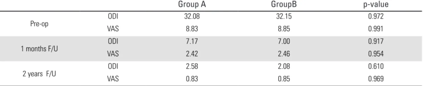
The VAS score of the clinical group (Group A) before surgery was 8.83 which reduced to 2.42 one month after surgery, showing 72.59% improvement. Two years after surgery, the VAS score was 0.83, revealing an improvement of 91.95%.
The VAS score of the control group (Group B) before surgery was 8.85, which reduced to 2.46 one month after surgery, showing 72.20% improvement. However, the VAS score measured during two years of follow ups after surgery was 0.85, showing 90.30% improvement. There was no significant difference between these two groups (p>0.05) (Table 2).
2) Oswestry disability index (ODI)
The ODI score of Group A before surgery was 32.08, and reduced to 7.17 one month after surgery, showing 77.81%
improvement. Two years after surgery, the ODI score of Group A improved to 2.58, showing a 90.60% improvement. The ODI
Fig. 1. MRI finding of disc herniation and the method of canal compromised area.
Canal compromised (%) = A+AB x 100 (A + B : spinal canal area , A : involved canal area )
Young Do Koh et al Volume 17 • Number 4 • December 2010
www.krspine.org 172
score of Group B before surgery was 32.15, and reduced to 7.00 one month after surgery, showing a 78.23% improvement. The ODI score taken during two years of follow ups after surgery was 2.08, showing a 93.53% improvement. There was no significant difference between these two groups (p>0.05) (Table 2).
3) Follow-up observations on clinical instability
“Instability catch”, “painful catch” and “apprehension” tests were investigated to verify clinical instability and conducted during the follow-up observations at one month. It revealed clinical instability complaints in three patients (25.00%) of Group A, and two patients (15.38%) of Group B. In final follow-up observations, clinical lumbar instability was evident in
two patients (16.67%) of Group A and in one patients (7.69%) of Group B. Nevertheless, three patients who had shown clinical instability did not exhibit radiological instability.
2. Radiological outcomes 1) Horizontal displacement
During one month follow up after surgery the lumbar spine lateral radiographs were taken in standing flexion and extension.
On radiographs the mean horizontal displacement of the central massive disc herniation group was 2.54mm. There were four cases (33.33%) that had a horizontal displacement more than 3mm. However, the two-year follow-ups showed the mean horizontal displacement was 1.17 mm in the central massive disc herniation group and there was only one case (8.33%) that was
Fig. 2. Axial lateralization of disc herniation in MRI Axial lateralization (%) = AB x 100
(A : radius of spinal canal, B : length from canal center to axis of herniated disc) Table 2. Clinical result of clinical group and control group
Group A GroupB p-value
Pre-op ODI 32.08 32.15 0.972
VAS 8.83 8.85 0.991
1 months F/U ODI 7.17 7.00 0.917
VAS 2.42 2.46 0.954
2 years F/U ODI 2.58 2.08 0.610
VAS 0.83 0.85 0.969
*Group A; clinical group with canal compromised more than 50% and with axis lateralization rate less than 20% in MRI
*Group B; control group with canal compromised less than 50% and with axis lateralization rate more than 20% in MRI
†.ODI : Oswestry disability index
‡.VAS : Visual analogue scale
more than 3mm.
The mean horizontal displacement distance of Group B at one-month follow-up was 1.80 mm as shown in the lumbar spine lateral radiographs taken in standing flexion and extension. There were two cases (15.38%) that exceeded the level of instability criteria. The mean horizontal displacement was 1.08 mm and one case (7.69%) showed displacement more than 3mm. This showed that there was no significant difference between these two groups (p>0.05) (Table 3). Two patients, who had shown radiological instability of horizontal displacement for each group in the two-year follow-ups did not show clinical instability.
2) Angular displacement
The mean angular displacement of the central massive disc herniation group (Group A) was 11.96° as shown in the lumbar spine lateral radiographs taken in standing, flexion, and extension, at one month follow up. There were four cases (33.33%) that showed an angular displacement more than 15 degrees. In two-year follow-ups, the mean angular displacement
was 6.29° and only one case (8.33%) showed an angular displacement greater than 15 degrees.
Among the control group, the mean angular displacement was 10.23° and two cases (15.38%) were shown to have greater angular displacement than 15 degrees. The two-year follow-up observations showed a mean angular displacement of 7.50° and one case (7.69%) showed a greater displacement than 15 degrees. With respect to angular displacement, there was no significant difference between these two groups (p>0.05) (Table 3).
Two patients who had shown radiological instability in angular displacement in the two-year follow-up of each group did not manifest clinical instability. Patients that showed an angular displacement in the control group also showed instability in horizontal displacement.
DISCUSSION
Surgical treatment of patients with intervertebral hernia has been under debate over the need and indication of spinal fusion for some time. Nachlas3) reported that there had been no clinically significant difference between groups with discectomy and interbody fusion, and groups with discectomy alone in the surgical treatment of intervertebral disc herniation. Later, several authors reported sufficiently satisfactory results on discectomy alone.4-6) On the other hand, other reports insisted on the necessity of spinal fusion due to pain recurrence and the possibility of re-surgery due to segmental instability of the spine and repeated intervertebral disc herniation.7-10)
Vaughan et al.8) insisted on the need for interbody fusion by presenting the following evidence: 85% clinical satisfaction of spinal fusion for a patient group with L4-L5 intervertebral disc hernia, as opposed to 39% clinical satisfaction for a group with no spinal fusion. Young et al.9) reported clinical satisfaction in a patient group with spinal fusion through examining lumbago Table 3. Radiological result of clinical group and control group.
Group A Group B p-value
1month F/U Horizontal displacement(mm) 2.54mm 1.80mm 0.276
Angular displacement(°) 11.96° 10.23° 0.351
2year F/U Horizontal displacement(mm) 1.17mm 1.08mm 0.829
Angular displacement(°) 6.29° 7.50° 0.368
*Group A; clinical group with canal compromised more than 50% and with axis lateralization rate less than 20% in MRI
*Group B; control group with canal compromised less than 50% and with axis lateralization rate more than 20% in MRI Fig. 3. Measurement of angular difference and horizontal displacement
on flexion/extension radiogram (Radiographic method of Dupuis and co- workers)
Horizontal displacement = AO – RO Angular displacement = θ”-θ’
Young Do Koh et al Volume 17 • Number 4 • December 2010
www.krspine.org 174
and sciatic neuritis after surgery. Also, Takesima et al.10) reported significant improvement of lower lumbar pain in patients who had undergone spinal fusion regardless of interbody instability and several reports insisted on the need of spinal fusion for patients with intervertebral disc herniation. In addition, Satoh et al.11) reported massive intervertebral disc hernia and intervertebral disc herniation accompanied by spinal segmental instability as an indication for interbody fusion.
On the other hand, LaMont et al.6) reported that performing interbody fusion for intervertebral disc hernia patients does not show significant clinical benefits compared to performing a simple discectomy. Therefore, the need for spinal fusion has continuously been debated. Several authors repeatedly reported comparative studies between spinal fusion and simple discectomy for the surgical treatment of patients with intervertebral disc herniation.12-19)
This study was intended to examine an additional need for spinal fusion, in cases of discectomy, by making follow-up clinical and radiological observations of the central massive disc hernia group with respect to instability of surgical segments. The spinal fusion was performed on central massive disc herniation patients suffering from lumbar instability at the same time.
Cribb et al.20) performed long-term follow up observations of patients with massive intervertebral disc hernia and diagnosed by referring to the defined standard. The massive intervertebral disc group with 50% or more displacement of the spinal canal as seen on MRI tends to have central or paramedian lumbar disc herniation (CLDH).
Barlocher et al.21) defined such cases as central lumbar mass prolapse accompanied by injuries of the posterior longitudinal ligament. Knop-Jergas et al.22) reported that central massive intervertebral disc herniation had a tendency of developing segmental instability, as opposed to other intervertebral herniation, as well as having an poor prognosis.
Walker et al.23) suggested there was a high possibility of segmental instability since central massive intervertebral herniation had often been accompanied by posterior longitudinal ligament injury.
Central massive intervertebral disc herniation does not have a good long-term prognosis due to heavy loss of the intervertebral disc and injury of the posterior longitudinal ligament. This group of patients may have intersegmental instability. In the
treatment of an intervertebral disc herniation, this is a typical basis for interbody fusion.24-26) However, from the result of this investigation, central massive lumbar disc herniation did not exhibit many occurrences of a long-term spinal instability as opposed to that of the control group. There was no significant difference in the clinical result either. In contrast to the simple discectomy, interbody fusion is a surgical procedure with several disavantages such as increased extent of surgery, blood loss in surgery, longer time of operation and hospital stay, instability of adjacent spinal segment, development of pseudoarthrosis, and economic aspects. Clinicians should give a detailed explanation regarding the treatment to patients with intervertebral disc herniation.
A minimum 2-years follow-up observation period for this study is thought to be a one limitation of this study. In addition, the lack of intervertebral disc herniation cases experiencing segmental instability before surgery led to limited research methodology and needs to be addressed in future investigations.
CONCLUSIONS
Central massive intervertebral disc herniation treated with simple discectomy did not show significant differences in clinical or radiological instability from that of other herniation types.
REFERENCES
1. Knutsson F. The instability associated with disk degeneration of the lumbar spine. Acta Radiol Scand. 1944;25:593-609.
2. Dupis PR, Yong-Hing K, Cassidy JD, Kirkaldy Willis WH.
Radiologic diagnosis of lumbar spinal instability. Spine.
1985;10:262-76.
3. Nachlas IW. End-results study of the treatment of herniated nucleus pulposus by excision with fusion and without fusion. J Bone Joint Surg Am. 1952;34:981–8.
4. Barr JS, Kubik GS, Molloy MK, et al. Evaluation of end results in treatment of ruptured lumbar intervertebral discs with protrusion of nucleus pulposus. Surg Gynecol Obstet.
1967;125:250–6.
5. Frymoyer JW, Hanley E, Howe J, Kuhlmann D, Matteri R. Disc excision and spine fusion in the management of lumbar disc disease : A minimum ten-year follow-up.
Spine. 1978;3:1–6.
6. LaMont RL, Morawa LG, Pederson HE. Comparison of disc excision with combined disc excision and spinal fusion for lumbar disc ruptures. Clin Orthop. 1976;121:212–6.
7. Rish BL. A comparative evaluation of posterior lumbar interbody fusion for disc disease. Spine. 1985;10:855–7.
8. Vaughan PA, Malcolm BW, Maistelli GL. Results of L4–
L5 disc excision alone versus disc excision and fusion. Spine.
1988;13:690–5.
9. Young HH, Love GJ. End results of removal of protruded lumbar intervertebral discs with and without fusion. Am Acad Orthop Surg Inst Course Lecture. 1959;16:213–6.
10. Takeshima T, Kambara K, Miyata S, Ueda Y, Tamai S.
Clinical and radiographic evaluation of disc excision for lumbar disc herniation with and without posterolateral fusion. Spine. 2000;25:450–6.
11. Satoh I, Yonenobu K, Hosono N, Ohwada T, Fuji T, Yoshikawa H. Indication of posterior lumbar interbody fusion for lumbar disc herniation. J Spinal Disord Tech.
2006;19:104-8.
12. Tominaga S, Date K, Ouchi K, et al. Comparison between non-fusion and fusion group for lumbar discopathy—
Effects of intervertebral fusion. Rinshou Seikei Geka.
1988;23:236–46.
13. White AH, von Rogov P, Zucherman J, Heiden D.
Lumbar laminectomy for herniated disc; a prospective controlled comparison with internal fixationfusion. Spine.
1987;12:305–7.
14. Eie N. Comparison of the results in patients operated upon for ruptured lumbar discs with and without spinal fusion.
Acta Neurochir (Wien). 1978;41:107–13.
15. Barr JS. Low back and sciatic pain: Result of treatment . J Bone Joint Surg Am. 1951;33:633-49.
16. Burns BH, Young RH. Results of surgery in sciatica and low back pain. Lancet. 1951;257:245-9.
17. Decker HG, Shapiro SW. Herniated lumbar intervertebral disks ; results of surgical treatment without the routine use of spinal fusion. AMA Arch Surg. 1957;75:77-84.
18. Frymoyer JW, Hanley EN Jr, Howe J, Kuhlmann D, Matteri RE. A comparison of radiographic findings in fusion and nonfusion patients ten or more years following lumbar disc surgery. Spine. 1979;4:435-40.
19. Weber H. Lumbar disc herniation : a controlled, prospective study with ten years of observation. Spine. 1983;8:131-40.
20. GL Cribb, DC Jaffray, VN Cassar pullicino. Observation on the natural history of massive lumbar disc herniation. J Bone Joint Surg Br. 2007;89:782-4.
21. Barlocher CB, Krauss JK, Seiler RW. Central lumbar disc herniation. Acta Neurochir (wien). 2000;142:1369-75.
22. Knop-Jergas BM, Zucherman JF, Hsu KY, DeLong B.
Anatomic position of a herniated nucleus pulposus predicts the outcome of lumbar discectomy. J Spinal Disord.
1996;9:246-50.
23. Walker JL, Schulak D, Murtagh R. Midline disc herniation of the lumbar spine. Southern Med J. 1993;86:13-7.
24. Ki SC, Choi YS, Kim KS, Kuk WJ. Lumbar Discectomy Using Tubular Retractor and Microendoscopy. J Korean Soc Spine Surg. 2008;15:265-71.
25. Chung YK, Yoo JH, Chung KJ, Wang JS. Clinical results of percutaneous endoscopic discectomy in herniated intervertebral disc of lumbar spine. J Korean Soc Spine Surg.
2005;12:224-8.
26. Lee KY, Sohn SK, Lee MJ, Chung IK. The influence of the degree of lum-bar disc degeneration on MRI and the amount of removed disc on clinical outcomes. J Korean Soc Spine Surg. 2005;12:184-91.
Young Do Koh et al Volume 17 • Number 4 • December 2010
www.krspine.org 176
중심성 거대 추간판 탈출증 환자의 단순 추간판 절제술 후 영상 및 임상적 결과
고영도 • 이승준 • 김동준
이화여자대학교 의과대학 정형외과학교실
연구 계획 : 본 연구는 중심성 거대 추간판 탈출증 환자의 단순 추간판 절제술 후 추시결과에 대한 후향적 대조군 연구이다.
목적: 거대 추간판 탈출증 환자에서 단순 추간판 절제술 후의 예상되는 해부학적 추간판 탈출의 위치 및 추간판 소실 양과 요추부 불안정성의 발생의 상 관관계를 밝혀 추간판 절제술에 따른 요추 불안정성의 진단과 치료 방향의 결정에 도움을 얻고자 하였다.
선행문헌의 요약: 요추 불안정성은 중심성 거대 추간판 탈출증의 합병증으로 고려되지만 요추 불안정성과 추간판 절제와의 연관성에 대한 증거는 제한 적이다
대상 및 방법: 한 분절의 요추부 추간판 탈출증이 있는 16세에서 64세 총 25명의 환자를 2년까지 추시하였으며, 그 중 수술 전 자기공명영상을 통해 추 간판 탈출이 척추관을 50%이상 침범하며 추간판 탈출의 중심 축이 척추관 중심 축에서 20% 이내에 위치한 12명의 환자를 실험군으로 정하였다. 대조 군은 추간판이 척추관을 50% 미만으로 침범하며 추간판 탈출의 중심 축이 척추관 중심 축에서 20%를 벗어난 MRI 소견을 보이는 13명의 환자를 대상 으로 하였으며, 수평전위, 각 전위를 측정하여 방사선학적 불안정성을, 그외 임상적 불안정성과 VAS, ODI를 비교하였다.
결과: 두군간의 임상적, 방사선학적 불안정성 및 VAS 와 ODI 로 측정한 임상적 결과 역시 두군간의 유의한 차이를 보이지 않았다.
결론: 본 연구에 따르면 술 전 예상되는 추간판 탈출의 해부학적 위치 및 추간판 소실 정도는 단순 추간판 절제술 후 요추부의 장기적 불안정성과 의미 있는 상관관계가 없는 것으로 생각된다.
색인 단어: 추간판 절제술 , 거대 추간판 탈출증, 요추부 불안정성.
약칭 제목: 거대 추간판 탈출증의 수술 후 추시