λ ~!i.1IJ. ~‘t Mtl샘셰에 J‘ 꺼\22 양 짜 1 쩌It pp. 124 - 130, 1986 Journal of Korean Radiological Society, Vo1.22, No.l, 1986
下大靜服 中總뾰에 對한 放射線學的 考察
서 울大學校 廣科大學 放射線科學敎室
崔然않·李東鎬·金榮九·朴在亨·延敬模·韓萬춤 - Abstract -
Infrahepatic Interruption of Inferior Vena Cava
Yeon Hyeon Choe, M.D., Dong Ho Lee, M.D., Young Goo Kim, M.D.,
Jae Hyung Park, M.D., Kyung Mo Yeon, M.D., Man Chung Han, M.D.
Department of Radiology, College of Medicine, Seoul National University
Congenital anomaly 01 IVC is rare, but understanding of this anoll1aly is important in radiological diagnosis, angiographic procedures and major retroperitoneal and thoracic surgery
We analysed 23 cases of IVC interruption diagnosed by cardiac angiography at Seoul National University Hospital
The results were as lollows
1. The incidence of infrahepatic interruption of IVC was 0.45% oi lhe patients having cardiac angiography and most patients showed cyanosis (91%)
2. The 1l10st cOll1mon associated cardiac anomaly was right ventricular outflow lract obstruction (60%). Other associated cardiac delects were VSD, ASD, valvular anomalies in 9 cases (39'Y,) respectively; double outlet right ventricle, bilateral superior vena cava, single ventricle in 6 cases (26‘l(,) respectively; single atrium. PDA in 5 cases (22%) respectively
6 cases of situs inversus, 3 cases of situs ambiguus, 2 cases of visceral heterotaxia and one case of asplenia were observed als。
7 cases 01 lelt-sided IVC were associated with IVC interruption in normal situs
1. 績 은ιi a빼
下大靜服의 先天性 略形에 대 한 解 !}IJ學的 지 식 은 心 導子術 및 心血管造影術 또는 뽑靜服 레닌 채취, 副賢 靜服造影術등에서의 카테타 조작이나 복부와 흉부
CT
상 종양의 임파선 轉移 여부의 결정에 도움을 줄 뿐 아 니라 흉부 수술이나 후복강 수술에 중대한 영향을 미칠
이 논문은 1986년 l윌 18일에 접수하여 1986 년 1월 31일에 채택되었음.
수 있으므로 임상적으로나 방사선학적으로 중요하다 1-17)
저자들은 서울대학교병원 진단방사선과에서 경험한 23 例의 下大靜服 中絡lJE(Infrahepatic interruption of
i nf er ior vena cava ) 을 대 상으로 방사선 학적 소견을 분 석하였다
2. 對象고} 方法
서울대학교병윈 진딘방사선과에서 1978 년 2월 부터 1985년 6월 까지 7 년 4 개월 동안 심도자술 및 심장
一}참然;tt 外: 下大靜뼈 中*~1il':에 對한 放射짧쩡的 j양察 -
조영숭을 시행하여 진단한 23 例의 하대정액 中絡효을 대상으로 하였으며 이중 22 例는 심 장영화조영술을 시 행하였다. 심 장영화조영숨은 GE MSI-1250 양면촬영 기로 35rnm 펼름에 妙當 30 또는 60 frame 속도로촬
영하였고 조영제는 자동주엽기
(Medrad @) 로
meglu-mine iothalamate ( Telebrix@
38) 를 kg 당 2~4
cc주입하였다.
3.
結 果하대정맥 중절증 23 例中 22 例는 심 장영화조영숨을 시행한 예로서 4903명중 약 0.45% 를 차지하였다. 대상 의 연령은 生後 3 개월에서 29 세의 분포를 보였고. 男 女比는 8 : 15로 여자가 훨씬 많았다 2 名을 제외한 대다수에서 좁色j[을 동반하였으며 ( 91 %) , 內驗遊位가
6 例 situs ambi guus 가 3 例였다 si t us ambi guus환 자 중 I 例는 방사선옹위 원소 주사상 無牌職뾰을 보였 고 임상적으로 多數의 비장이 있을 것으로 의심되는 1 例는 확인되지 않았으며 또 한 예에서는 비장의 상태가 결정되지 않았다 2 例에서는 內魔異常位置뾰( viscer-
a 1 heterotaxi a ) 이 관찰되 었다.
內騙正位인 14例中 7 例는 우측의 하대정맥에서 奇靜 服( azygos vein ) 으로 연속되 었고 ( Fig. 1) , 5 例는
左測性 下大靜服( Lt. postrenal ! VC) 에서 半奇靜服 ( hemiazygos vein) 으로 연속되 어 副半奇靜服( acces- sory hemiazygos vei n) 을 거쳐 左뼈ij에 있는 t大靜服 으로 流入하였으며 CFig. 2), 2 例는 左測性 下大靜服 에서 반기정맥요로 연속되어 下部 뼈堆骨 수준에서 正中 線을 가로칠러 右測으로 자리를 바꾼 후 기정맥, 右測 t大靜服으로 유엽하는 경로를 취하였다( Fig. 3).
Si tus amb iguus 3 例에서는 모두 右測 下大靜服에서 右測에 위치한 기정맥을 지나 우측 상대정맥으로 유입하 였으며, 內魔뺑位에서는 全例가 左 f則 下大靜服에서 左測 에 위치한 기정맥으로 유입하였다. 內魔正{fJ로 左없ij 下 大靜服에서 左測 上大靜眼으로 연속되 는 5 例중 1 例에 서는 冠狀靜服洞으로 유입하는 대신 좌섬방으로 직접 유 입하였다 ( Table 1 ).
Tab!e 1. Summary of 23 Cases of IVC Interruption
Classification
1. Situs solitus
a. Rt. IVC to azygos cont b. Lt. IVC to hemiazygos to
accessory hemiazygos cont.
c. Lt. IVC to hemiazygos to azygos cont
2. Situs inversus with Lt. IVC to azygos cont
3. Situs ambiguus with Rt. IVC to azygos cont
No
14 7
5
2
6
3
동반된 심장기형으로는 폐동맥 유출로 폐쇄 ( pulmon- ary outflow tract obstruction) 가 14 例 ( 60 %)로 가 장 많았고, 心室中隔缺협, 心房中隔缺慣, 었뿔멍윷略形。 l 各
9 i꺼u
(
39 %), 兩大血管右心室起始효, 양측성上大靜眼,單一心室이 各 6 例( 26 %) , 單一心房, 開存{生 動眼管 이 各 5 例( 22 %) 가 있었고 心房室中隔缺협 ( endoca-
rd ia 1 cushi on def ect ), 右位心( dext ro cardi a ) , 심 장역위의 左位心, 大血管뺑位j[ ( t ransposition of gre-
at arteri es ) 이 各 4 例( 17 % ) 에서 관찰되 었다 ( Ta- ble 2 ).
4.
考 쩔Fig. 1. Infrahepatic interruption of IVC with azygos con
tinuation in situs solitus. Azygos vein was 下大靜服 뼈形을 이해하는 데 있어 중요한 뼈生學을
enlarged 살펴 보띤 다음과 같다 10,18
’.
後題용靜眼系는 세 쌍의- 125 -
-大짧放射線醫學會誌‘ · 第22卷 第l 號 1986 -
R읍兒靜!w系인 後主靜!W(posterior cardinal vein), 下 主靜服(subcardinal vein), 上主靜服(s pracardinal vein) 으로 부터 발달하며 이들은 각기 태생 6, 7, 8 주의 주된 정맥계를 이룬다. 後主靜服은 최초의 횡경막 하 태아정맥계로서 이들의 內題뼈~에 下主靜服이 발달하 면서 점차 퇴화하게 되는데 퇴화하지 않고 賢後分節를 형 성하는 경 우 右뼈~ 수뇨관을 內없IJ으로 끌어 大靜服後民 管(retrocaval ureter ) 을 초래하는 것으로 생각되고 있 다. 태생 7 주에 하주정액은 후주정맥과 外명~문합을 이 루고 정 상 題測左賢靜眼이 될 하주정 맥 간문합 ( inter-
A
A
subcardinal anastomosis) 을 이 루고, 우측 하주정 액 과 우측 간정 액 이 府下主靜服間物슴 ( hepato -subcardinal
channel) 을 이루어 府分節을 형성하게 되는데 이 後者 의 문합이 이 루어 지지 않으연 下大靜服 中組훈이 발생 하게 된다. 또한 우측 뽑陽間題靜服 (omphalo mesen.
teric vein) 이 커져서 府과 右心房 사이를 연결하는 府 上分節(suprahepatic portion) 을 형성한다. 세 번째 의 정 액 계 인 上主靜服은 下主靜服의 背뼈~, 後主靜뼈의 內없~에 발달하여 아래쪽으로는 賢後分節(post renal se.
gment) 을 형성하고 위쪽으로는 奇靜服系플 형성한다.
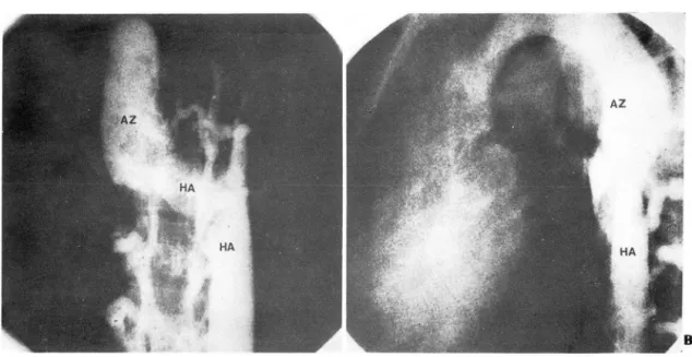
Fig. 2. Left postrenal IVC with interruption. Left IVC continued to hemiazygos (HA), accessory hemiazygos vein (AH) and left superior vena cava (LSVC) and finally drained into coronary smus
On lateral view (B), this anomaly showed so called ‘candy cane' or ‘shepherd crook' ap' pearance‘
B
- 126-
- 않然섰 外: 下大뽑服 中*용'ifÉ에 對한 放射線學的 :; 察-
A
Fig. 3. Left postrenal IVC with interruption in AP (A) and lateral view (B).
Hemiazygos vein (HA) crossed midline at low thoracic vertebral level (T7-8) to join azygos vein (AZ)
상주정맥과 하주정맥 사이의 문합이 양측에 이루어 져서 賢分節 (renal segment) 를 형성하며 대동액 뒤의 正中 線 문합이 잔존하는 경우 大動服後左賢靜服( retroaor-
tic left renal vein) 을 형 성하고 題f則의 左賢靜服이 함께 있는 경 우 소위 大動服周圍賢靜服環( rena 1 venous
collar) 을 형성한다.
이와같이 정상적인 右뼈ij 하대정맥은 右測 상주정맥이 賢後分節을, 우흑 下上主靜願間物合( sub-supracardin-
al anastomosis) 이 賢分節을, 우측 하주정액이 賢前分 節을, 우측 간정액이 府分節을, 우측 l價陽$禮靜服이 府上 分節을 이루어서 구성된다(Fig. 4). 양측성 하대정맥
(double IVC) 은 좌우측 상주정맥에 의해 형성되며,좌 측성 하대정맥 ( 1 eft post renal 1 VC )은 좌측 상주정 맥의 잔존에 의해 형성된다. 이 밖에 우측 후주정액과 우측 상주정액 이 共存하여 수뇨관 주위에 정맥의 고리를 만드는 經靜服性 llJ(管( transcaval ureter) 이나 양측 후 주정맥의 잔존에 의한 양측성 대정맥후뇨관( bilateral
retrocaval ureter) 도 보고된 바 있으며 19’, 賢前分節의 기형과 賢後分節의 기형의 組合이 있다 16 )
Huntington과 Mc Clure는 태생학적 근거에 의하여 우측 후주정맥, 우측 상주정맥, 좌측 상주정맥, 좌측 후 주정맥을 각각 A , B , C, 0 로 지칭한 도표를 제안하였 으며 신후분절기형을 15型으로 분류하였다 2) 그후 Chuang등 2 ,이 이를 보완하여 가설적이거나 동물에서만
발견되는 유형을 제외하고 방사선학적, 임상적으로 중 요한 A , B , C , BC 의 4 형과 뽑前, 賢後分節기형을 추 가한 단순화된 분류표를 제안한 바 있다. 그러나 이 밖 에도 하대정맥 중절증에 기정맥 또는 반기정맥 연속이 아 닌 맑門服 연속이나 20, 上行體靜服, 홉堆靜服覆과의 연 결만 있는 例가 있고 2l) , 복부에서 척추의 어느 한쪽에 위치한 하대정맥이 횡격막 수준에서 정중션을 가로질러 반대쪽으로 경로를 바꾼 후 우심방으로 유입하는府分節 略形들이 있£며 22 -엉 하대정맥이 좌심방이나 관상정 맥동으로 유입하는 경우, 하대정맥이 비정상적으로높은 위치에서 우심방과 연락되는 경우와 R주上分節이 폐쇄되 는 경우 등의 뺨上分節기형들이 보고되어 26’ 저자들은
Table 3 과 같이 정리하여 보았다( Table 3 ).
문헌에 의하면 하대정맥 중절증은 심 장조영술 시행 껴j中 O.2~1.3%의 빈도를 보이고 선천 성 심 장기형이 85 %에서 관찰된다고 하였 S며 1,2) 이는 저자들의 例 에서와 비슷한 빈도이다. 하대정맥 중절증에 섬장기형 이 동반되었을 때는 多牌魔효과 연관된 복잡심장기형이 많다고 알려져 왔~나 27 , 28) 저자들의 경우에 다비장증S 로 확인된 例는 없었다.
최근에 CT 나 혈관조영술의 이용이 증가하연서 하대 정액의 기형을 더욱 자주 발견하게 되었다. 썩*모睡을 위시한 각종 악성종양 환자의 病期 결정에 있어 확장된 기정액, 반기정맥을 橫l隔體R왜後 빼巴R용陣大( retrocru r- - 127-
limb) 에서 시행하는 것이 신장으로 부터오는 혈류의 희석이 적어 유리하며 부신정맥초영술시에는 앞쪽 좌신 정맥 (preaortic limb)에서 부산정맥으로 선택진입하여
야 한다 2,5,6,8) •
대 동액 A造物( prosthesis) 대 치 술이 나 간문맥고혈 압 환자에 대한 短絡수솔, 體部 교감신경절제솔, 신장이식 수술 등의 후복강 수슐에 있어서 하대정액의 기형이 존 재하는 경우에 해부학적 이해가 필요하며 兩測性 下大靜 服의 下大靜服間連絡靜服 ( i ntercaval communi cat íng veín) 의 손상이나 대동맥주위 정백환의 背測정맥 파열 이 생기지 않도록 주의하여야 한다, 血桂효의 예땅을 위한 하대정맥 결찰 또는 umbrella 설치 시에도 양측성 Table 2. Assocíated Cardiac Anomalies of IVC Inter.
* Situs inversus in 6 cases and situs all1biguus in 3 cases out of all 23 cases
* * One case of sitlls all1biguus sh。、vecl aspleni~ ancl another one case \\’as sllspectecl to have 1l1111tiple spleens but was not confirll1ed by scintigraphy
(N=23)
M 9 9 9 7 6 6 6 5 5 4 4 4 4 3 2 1
--- No.
PlIlmonary olltflow tract obstruction VSD
ASD. secllndull1
Valvlllar anomalies
Left-siclecl IVC (sitlls solitus) DOllble outlet RV
Bilateral superior vena cava Single ventricle
Single atrillll1
PDA
Enclocarclial cllshion defect Dextrocarcl ia
Isolated levocardia
Transposition of great arteries Right aortic arch
Coronary artery anoll1aly TOF
Criss-cross heart
Total anOll1alOllS pulll10nry venolls return Left sllperior 、ena cava to LA
Polysplenia
As히p비〕끼le잉l1! a
Anoll1alies - 大합放껴,t짧醫챙會誌 . 第22卷 第l 號 1986
ruptlon
al lymphadenopathy) 로 오인할 수 있으며 11-14) 좌측 성 하대정맥 또는 양측성 하대정액도 대동맥 주위 임파 선 종대와 구별하여야 한다16 , CT상 연속적인 管狀 구조가 彈한 均質 造彩增彈을 보이거나, 冠狀切片에서 管 狀으로 보이연 정맥임을 획언 할 수 있다 9, 11 )
하대정맥 중절증이 생기면 기정맥의 확장이 단순 흉부 촬영像에서 관찰되므로 據見할 수 있다 1,3) 심도자숭 ( cardiac catheterization) 시행시 카테타 끝이 우심방 에 있는데 좌우 움직임이 제한되고 지속적우로 카테타가 후방에 위치하는 것£로 생각되면 하대정맥 중절증을 의 섬하여야 하며 1
,
카테타가 반기정맥, 좌측 상대정맥을 지나 관상정액동으로 가는 경로를 취하여도 특정적인 카 테타 경로를 알연 술자가 당황하지 않고 검사를 진행할 수 있다 초음파검사를 시행하면 정상적으로 쉽게 관찰 되는 밤分節이 안 보이는 것과 대동맥 주위로 늘어난 기 정맥, 반기정맥을 관찰함£로써 비교적 쉽게, 비침습적 무로 하대정맥 중절증을 진단할 수 있다 29,30’-
또한 신정맥에서 rell1 n을 채취할 경우에 大動服周圍 뽑靜服環이 있으면 分技가 적 은 윗 쪽 좌신정 맥 Cinferior
ζ。-ω-〉-(〕-mrαZ’φ」α도。-이-〉-〔〕 -어ζφ∞,gm。ι
PARS RENALIS
PARS SUPRA CARDINALlS
PARS SUB- CARDINALlS L. Adrenal
야
-
뼈 /
L
니” e
PARS HEPATICA L. Subclav
Fig. 4. Adult inferior vena cava (Adaptecl from Ferris'" ')
Iliac Renal R. Renal
Azygos
~k.然 n 外· 下大해'ìlll1it cp쩌자에 훈f한 i.&M*.m&용“] 考察- Table 3. Classification of Anomalies of IVC
1. Posternal segment 1. Rt. retrocaval ureter 2. Transcaval ureter 3. Lt. postrenal IVC 4. Double IVC
5. Bilateral retrocaval ureter 6. Lt. retrocaval ureter 11. Renal segment
1. Circumaortic renal venous ring 2. Retroaortic left renal vein IJI. Prerenal segment
1. Infrahepatic interruption of IVC with azygos con- tmuatlOn
2. lnfrahepatic interruption of IVC with hemiazygos cont1l1Uatlon
3. Infrahepatic interruption of IVC with portal con- tinuation
4. Infrahepatic interruption of IVC with vertebral plexus and ascending lumbar vein connection IV. Hepatic segment
Lt. (Rt.) IVC shifting of sicJe at liver to enter rt. atrium V. Suprahepatic segment
1. Anomalous connection of IVC to LA
2. Anomalous connection of IVC to coronary sinus 3. Anomalous high insertion of IVC to RA 4. Congenital obliteration of suprahepatic IVC
하대정액의 연결부위 하부에서 시행하여야 효과적인 수 숭이 된다. 하대정맥 중절증에 기정맥으로 연속된 하대 정맥을 가졌던 환자에서 펴1 암 수술시의 기정맥결찰로 인 한 사밍에 보고 된 바 있으므로 흉부 수울시에도하대정 액 중절증 유무를 아는 것이 중요하다 17)
5.
結 論저자들은 서울대학교병원 진단방사선과에서 심혈관조 영솔로 진단한 23 例의 하대정액 중절증을 분석하여 다 음과 같은 결과를 얻었다.
1. 下大靜服 中絡효 23 例는 최근 심 장영화초영술을 시행한 4903 名中 약 0.45 %를 차지하였고 91% 에서 청 색증을 보였다
2. 동반 심 장기 형 으로는 右心室流出路 閒짧가 14fJIJ
- 129
( 60 %)로 가장 많았고, 心室中隔缺협, 心房中隔缺협,
離體쐐形이 各 9 例(39 %)이었으며, 그 밖에도兩大血 管右心室起始료, 兩測性 上大靜服, 單-ι、室 등이 있었 다’
또한 內魔뺑位가 6 例( 26 %), situs arnbiguus 가 3例 였고, 內魔異常位置효 ( vi sceral heterotaxia) 이 2 例,
확인된 無牌魔뾰이 I例였다. 內魔正位 14 ØlJ 中 7 例의 좌측성 하대정맥이 관칠되어 齊後分節略形이 下大靜뼈 中絡효에 많이 연관된 것을 알 수 있었다.
3. 하대정맥 중절증에 대한 지식은 心훨子術 및 섬 장 영화조영술시의 카테타 조직이나 복부, 흉부 CT 상임파 선전이 유무 판별 등에 방사선학적으로 유용하며• n힘部 수술사에도 이에 대한 熟知기 요구된다.
REFERENCES
1. Anderson RC, Adams PA, Burke B: Anomalous inferior vena cava with azygos continuation (infrahepatic inter- ruption of lhe in{erior vena cava). J Pediat 59:370-383, 1967
2. Chuang VP, Mena CE, Hoskins PA: Congenital anomalies of the inferior vena cava - review of embryogenesis and presentation of a simplified classification. Br J Radiol -17:206-213, 1974
3. Milledge RD: Absence of inferior vena cava. Radiology 85:860-865, 1965
-1. Mitty HA: Circumaortic renal collar, a potentially hazar dous anomaly of the left renal vein. AJR 125;307-310, 1975 5. Kottra JJ, Castellino RA: The circumaortic left renal vein,
angiographic appearance. Radiology 95;141-143, 1970 6. Chuang VP, Mena CE, Hoskins PA: Congenital anomalies
of the left renal vein - angiographic consideration. Br J RadioI47;274-278, 1974
7. Bosnik MA. Madayag M: Angiographic appearance of the circumaortic left renal vein. J Urol 108;18-20, 1972 8. Field S, Chir B, Saxton H: Venous anomalies complicating
left adrenal catheterization. Br J Radiol of anomalies of the inferior vena cava and le{t renal vein. AJR 732:759-763, 1979
9. Royal SA, Callen PW: CT evaluation o( anomalies o( the inferior vena cava and left renal vein. AJR 732:759-763, 1979
10. Mayo J. Gray R, Louis ES: Anomalies o( the in(erior vena cava, review. AJR 1-10;339-345, 7983
11. Jasinski RW, Yang CF, Rubin JM: Vena cava anomalies
- 大韓放射線醫學會誌‘• 第22卷 第l 號 1986-
simulating adenopathy on computed tomography. J Com- only at the iliac anastomosis. J Urol 91;478-481, 1964 put Assist Tomogr 5;921-924, 1981. 22. Freedom RM, Treves 5: Splenic scintigraphy and ra- 12. Ginaldi 5, Chuang VP, Wallace S: Absence of hepatic seg- dionuclide venography in heterotaxy syndrome
ment of the inferior vena cava with azygos continuation. 23. Freedom RM, Fellows KE: Radiographic visceral patterns J Comput Assist Tomogr 4;112-114, 1980. in the asplenia syndrome. Radiology 106;387-391, 1973.
13. Breckenridge jW, Kinlaw WB: Azygos continuation of in- 24 유시준, 김승협, 한만청 : 선천성 심장질환을 동반 ferior vena cava: CT appearance. J Comput Assist Tomogr 한 무비 장증 3 례보고. 대 방의지 VoI.XVl; 51 9-528
,
4;392-397, 1980. 1980
14. Churchill Rj, Wesby 111 G, Marsan RE et al: Computed 25 유시준, 임정기, 연경모 · 섬장의 이상위치. 대방의 tomographic demonstration of anomalous inferior vena 지 VoI. XV ; 86-93
,
1979cava with azygos continuation. J Comput Assist Tomogr 26. Feedom RM, Culham jAG, Moes CAF: Angiocardiography 4;398-402, 1980. of congenital heartdisease. 58-62, Macmilliaπ New York, 15. Kumar D, Kumar 5: Circumaortic left renal vein. J Com- 1984
put Assiçt Tomogr 5;914-916, 1981. 27. Adams FH: Heart disease in infants, children and 16. Faer Mj, Lynch RD, Evans HO, et al: Inferior vena cava adolescents. 3rd Ed, 448-451, 씨lilliams & 싸lilkins,
duplication: Demonstration by computed tomography. 8altimore, 1983
Radiology 130;707-709, 1979. 28. Vaughan
n
Hawkins IF, Elliott LP: Diagnosis of polysplenia 17. Effler DB, Greer AE, 5ifers EC: Anomaly of the vena cava syndrome. Radiology 101;511-518, 1971inferio ι report of fatality after ligation. JAMA 29. Garris jB, Kangaroo H, 5ample WF: Ultrasonic diagnosis 146;1321-1322, 1951. of infrahepatic interruption of the inferior vena cava with 18. Clemente CD: Anatomy of the human body. 29th Ed, azygos continuation. Radiology 134;179-183, 1980
788.792, Lea & Febiger, 1985. 30. Train T5, Henderson ι1R, 5mith AP: Sonographic 19. Abrahams HL: Angiography. 3rd Ed, 895-921, Little, 8rown demonstration of left-sided inferior vena cava with
and Company, 8oston, 1983. hemiazygos continuation. AJR 134;1057-1059, 1980 20. Bercoff E, Colin R, Benozio M, et al.: Infrahepatic infer- 31. Ferris Ej, Hipona FA, Kahn PC, et al.: Venography of in-
ruption of the inferior vena cava with portal continuation. ferior vena cava and its branches; 1-20, 이lilliams and
Radiology 154;771-772, 1985. Wilkinε 8altimore, 1969.
21. Colborn GL: A case of bilateral inferior vena cava joined