대한치주과학회지 : Vol. 37, No. 3, 2007
Full-mouth disinfection의 단기간의 임상적 효과
이신화*, 김옥수, 김영준, 정현주
전남대학교 치의학전문대학원 치주과학교실, 전남대학교 치의학연구소
I. 서론
치주 질환은 치은연하 치태 내에 존재하는 치주 원인균에 의해 발생하는 혼합 감염이며 치주낭 내 세균의 부하가 국소적, 전신적인 숙주 방어 기전의 균형을 깨뜨리는 경우 치주 조직 파괴가 일어난다.
치주 치료의 성공은 구강 내의 ecological niche와 치아에 관련된 치태 내에 존재하는 병원성 세균 감 소에 의존하며1), 치주 감염의 치료에 주로 추천되는 방법은 기계적인 접근법으로 치석제거술과 치근활택 술(SRP)을 들 수 있다2). 기계적인 기구조작 후 치은 연하 세균의 부하는 1000배 정도 감소되지만3), 사분 악 또는 육분악으로 나누어 시행하는 conventional SRP의 경우 치료되지 않은 치주낭, 혀, 점막, 편도, 인두, 타액 등 구강 내 다른 부위에서 유래한 병원성 세균이 1주일 이내에 치료된 치주낭 내로 쉽게 재집 락화될 수 있다4). 따라서 치료되지 않은 감염된 치 주낭으로부터 치료된 치주낭으로의 교차 감염을 줄 이기 위하여 Quirynen5)은 full-mouth SRP와 함께 시술 전에 클로르헥시딘 젤(1%)로 1분간 tongue brushing, 클로르헥시딘 용액(0.2%)으로 1분간 구강
세정, 시술 동안 클로르헥시딘 젤(1%)로 10분 이내에 3번씩 치은연하 세척, 시술 후 2주 동안 클로르헥시 딘 용액(0.2%)으로 1분간 구강 세정하는 방법을 소개 하였고 이 술식을 full-mouth disinfection (Fdis)이 라고 하였다. 이후 여러 연구에서 Fdis가 임상적, 미 생물학적으로 유의한 효과가 있다고 보고되었다
1,6-11). 그러나 Fdis가 conventional SRP와 비교하여 임상적 효과에 있어서 유의한 차이가 없는 것으로 보고되기도 하였다12-15).1)
본 연구에서는 중등도 이상의 전반적 만성 치주염 환자 치료 시 치석제거술 후 임상에서 쉽게 구할 수 있는 클로르헥시딘 용액(0.1%)을 사용하여 실제 적 용하기 쉽도록 변형시킨 full-mouth disinfection (Fdis)을 시행하여 단기간의 임상적 효과를 con- ventional SRP와 비교하였다.
II. 연구 대상 및 방법
전신적으로 건강한 전반적 만성 치주염 환자 10명 (남자 6명, 여자 4명, 평균 47.3세, 비흡연자)을 대 상으로 하여 환자의 선호도에 따라 Fdis 군(5명) 또
* 교신저자 : 김옥수, 광주광역시 동구 학동 8번지 전남대학교 치의학전문대학원 치주과학교실, 501-757 (전자우편 : [email protected])
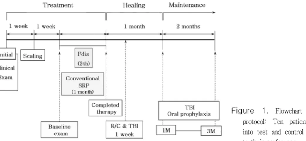
Figure 1. Flowchart of experimental protocol: Ten patients were divided into test and control groups according to their preferences.
는 conventional SRP 군(5명)을 결정하였으며 Fdis 를 시행한 군을 실험군으로, conventional SRP를 시행한 군을 대조군으로 하였다. 임상 지수는 치석 제거술 시행 1주 후 subgingival SRP 전(baseline) 과 Fdis/conventional SRP 시행 후 1, 3개월에 측 정하였다(Figure 1). 반복 측정에 대한 접근성을 고 려하여 상악 우측 사분악 치아 협설면의 근원심 및 중앙부를 포함한 6 부위 즉, 환자당 42 부위에서 치 은열구 출혈 지수(SBI), 치태 지수(PI), 치은 퇴축 (GR), 치주낭 탐침 깊이(PD), 임상적 부착수준(CAL) 을 측정하였다. 치은열구 출혈 지수는 탐침 후 30초 이내의 출혈을 관찰하여 출혈이 없는 경우 0, 치은 출혈이 있으나 치은 변색과 부종이 없는 경우 1, 치 은 출혈과 변색이 있으나 부종이 없는 경우 2, 치은 출혈, 변색, 부종을 수반한 경우 3, 치은 출혈, 변 색, 부종 및 궤양을 수반하는 경우 4, 치은 출혈이 저절로 되고, 변색이 있으며 현저한 부종 및 궤양이 있는 경우 5로 표시하였다. 치태 지수는 Silness &
Lӧe Index를 사용하였으며, 치태가 부착되어 있지 않은 상태를 0, 치은 변연에 부착된 치태로서 탐침 소자로 치면을 긁어보아 확인할 수 있는 엷은 상태 를 1, 치은 변연을 따라 육안적으로 확인될 수 있을 정도로 과량의 치태가 부착되어 있고 치간부에는 치 태가 없는 상태를 2, 치은 변연에 많은 양의 치태가 침착되어 있고 치간부에도 치태로 채워져 있는 상태 를 3으로 나타내었다. 치은퇴축은 Williams probe
(23W, Hu-Friedy, USA)를 사용하여 백악법랑경계 부에서 유리치은 변연까지 1mm 단위로 측정하였고, 치주낭 탐침 깊이는 동일한 기구로 유리치은 변연에 서 치주낭 기저부까지 1mm 단위로 측정하였다. 임 상적 부착수준은 백악법랑경계부로부터 치주낭 기저 부까지의 거리로 치은퇴축과 치주낭 탐침 깊이의 합 (mm)으로 나타내었다.
실험군(Fdis group)에서는 치석제거술 후 1주일에 국소마취 하에서 subgingival full-mouth SRP를 시행하였다. Quirynen의 원래의 프로토콜5)을 변경 하여 클로르헥시딘 용액(0.1%)으로 시술 동안 치은 연하 세척을 하였고, 환자에게 시술 전 40초간 구강 세정, 시술 직후 20초간 tongue brushing, 시술 후 2주 동안 1일 2회 40초간 구강 세정을 지시하였고 구강 세정 시 마지막 10초간은 클로르헥시딘 용액이 편도에 머무를 수 있게 하였다. 대조군에서는 치석 제거술 시행 1주 후부터 1주일 간격으로 사분악씩 국소마취 하에서 subgingival SRP를 시행하였다.
칫솔질 교육과 치면 세마는 두 군 모두에서 시술 1 개월과 3개월 후 필요시 시행하였다.
III. 임상 증례
1. 증례 I
환자는 칫솔질할 때 가끔 잇몸에서 피가 난다는
B
SBI 222 222 222 222 111 213 303
PI 212 212 212 211 202 111 100
CAL 535 536 624 523 323 324 323
GR 000 000 000 000 000 000 000
PD 535 536 624 523 323 324 323
#17 #16 #15 #14 #13 #12 #11
P
PD 534 436 635 533 323 323 323
GR 001 010 000 000 000 000 000
CAL 535 446 635 533 323 323 323
PI 212 212 202 202 201 102 201
SBI 222 222 222 222 202 223 323
B: buccal, P: palatal, SBI: sulcus bleeding index, PI: Silness & Lӧe plaque index, CAL: clinical attachment level (mm), GR:
gingival recession (mm), PD: probing pocket depth (mm).
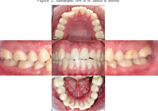
Table 1. Clinical measurements in sampled sites of case I at baseline Figure 2. Radiographic view of Rt. maxilla at baseline
Figure 3. Clinical view at baseline: generalized gingival inflammation and redness and swelling of interdental papillae were seen.
Figure 4. Clinical view at one month after Fdis: severe gingival recessions of interdental papillae between #15-16 and
#25-26 were observed.
Figure 5. Clinical view at 3 months after Fdis: it maintained the healthy state, but gingival recession was seen especially at interdental papillae in anteriors.
Site
Time #17 #16 #15 #14 #13 #12 #11
PD
M 0 B 535 536 624 523 323 324 323
P 534 436 635 533 323 323 323
M 3 B 333 323 323 222 222 223 222
P 423 324 323 322 222 222 222
CAL
M 0 B 535 536 624 523 323 324 323
P 535 446 635 533 323 323 323
M 3 B 333 434 423 223 222 223 222
P 434 336 423 322 222 222 222
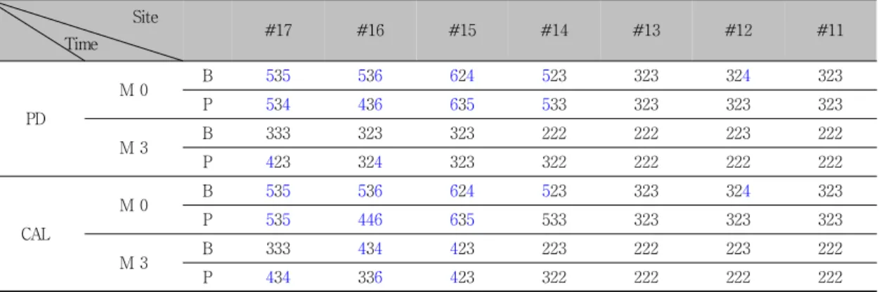
PD: probing pocket depth (mm), CAL: clinical attachment level (mm), M 0: at baseline, M 3: 3 months after Fdis, B: buccal, P: palatal.
Table 2. Initial and final measurements of probing pocket depth (mm) and clinical attachment level (mm) in sampled sites
주소로 내원한 38세 남자 환자였으며 구강 검사 시 전반적인 치태 및 치석의 침착과 치은 염증을 보였 다(Figure 2, 3). 초진 시 우측 사분악 구치부에서 가시적 치은 퇴축은 없었으며, 치주낭은 중등도 깊 이로 측정되었다(Table 1). Fdis 시행 1개월 후 치은 염증은 전반적으로 개선되었으며, 특히 #15~#16,
#25~#26 사이의 치간유두가 소실되어 black tri- angle 양상을 보였다(Figure 4). 구강 위생은 양호
한 편이었으며 Fdis 시행 3개월 후 상하악 전치부가 더욱 개선된 양상을 보였고, black triangle 부위의 치간 유두가 일부 재생되었다(Figure 5). 4~6mm의 중등도 치주낭 깊이는 초진 시 15 부위에서 측정되 었으나 시술 3개월 후에 2 부위에서만 측정되었고, 4~6mm의 임상적 부착수준은 초진 시 16 부위에서 시술 3개월 후 7 부위로 감소하였다(Table 2).
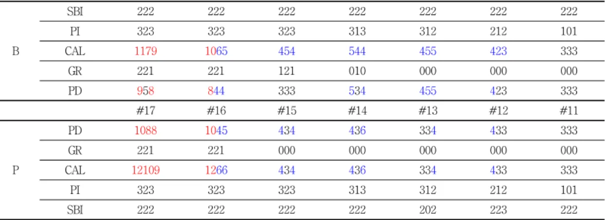
B
SBI 222 222 222 222 222 222 222
PI 323 323 323 313 312 212 101
CAL 1179 1065 454 544 455 423 333
GR 221 221 121 010 000 000 000
PD 958 844 333 534 455 423 333
#17 #16 #15 #14 #13 #12 #11
P
PD 1088 1045 434 436 334 433 333
GR 221 221 000 000 000 000 000
CAL 12109 1266 434 436 334 433 333
PI 323 323 323 313 312 212 101
SBI 222 222 222 222 202 223 222
B: buccal, P: palatal, SBI: sulcus bleeding index, PI: Silness & Lӧe plaque index, CAL: clinical attachment level (mm), GR:
gingival recession (mm), PD: probing pocket depth (mm).
Table 3. Clinical measurements in sampled sites of case II at baseline
Figure 6. Radiographic view of Rt. maxilla at baseline. Severe bony destruction was seen at #16, 17.
Figure 7. Clinical view at baseline: gingival inflammation and recession were seen.
2. 증례 II
Figure 8. Clinical facial view. A. at baseline: generalized gingival inflammation was seen. B. one month after Fdis: the state of gingiva was improved looking coral pink. C. three months after Fdis: the patient controlled plaque better and maintained the healthy gingiva well.
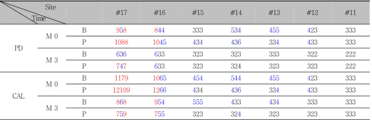
Site
Time #17 #16 #15 #14 #13 #12 #11
PD
M 0 B 958 844 333 534 455 423 333
P 1088 1045 434 436 334 433 333
M 3 B 636 633 323 323 333 322 222
P 747 633 323 324 323 323 222
CAL
M 0 B 1179 1065 454 544 455 423 333
P 12109 1266 434 436 334 433 333
M 3 B 868 954 555 433 434 333 333
P 759 755 323 324 323 323 333
PD: probing pocket depth (mm), CAL: clinical attachment level (mm), M 0: at baseline, M 3: 3 months after Fdis, B: buccal, P: palatal.
Table 4. Initial and final measurements of probing pocket depth (mm) and clinical attachment level (mm) in sampled sites
씹을 때 양쪽 어금니가 힘이 없고, 특히 오른쪽 위가 시큰거린다는 주소로 내원한 52세 여자 환자로 전반적인 치은 염증과 치태 및 치석의 침착을 보였 고, 중등도에서 심도의 전반적 만성 치주염으로 진 단되었다. 초진 시 우측 사분악 구치부는 치은 퇴축
을 동반하며 치주낭은 10mm 정도로 깊게 측정되었 다(Figure 6, 7, Table 3). Fdis 시행 1개월 후 치은 은 전반적으로 산호빛 분홍색의 건강한 상태로 개선 되었으며(Figure 8), Fdis 시행 3개월 후 중등도 이 상의 치주낭 깊이는 초진 시 24 부위에서 측정되었
A
B
C
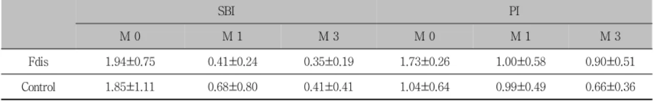
SBI PI
M 0 M 1 M 3 M 0 M 1 M 3
Fdis 1.94±0.75 0.41±0.24 0.35±0.19 1.73±0.26 1.00±0.58 0.90±0.51
Control 1.85±1.11 0.68±0.80 0.41±0.41 1.04±0.64 0.99±0.49 0.66±0.36
M 0: at baseline, M 1: one month after Fdis, M 3: 3 months after Fdis.
Data shown are mean±SD.
Table 5. The measurements of sulcus bleeding index (SBI) and plaque index (PI) in sampled sites
GR CAL
M 0 M 1 M 3 M 0 M 1 M 3
Fdis 0.55±0.32 0.80±0.33 0.90±0.34 4.40±0.56 3.70±062 3.58±0.55
Control 0.38±0.33 0.70±0.55 0.80±0.62 4.22±1.18 3.56±0.65 3.47±0.73
M 0: at baseline, M 1: one month after Fdis, M 3: 3 months after Fdis.
Data shown are mean±SD.
Table 6. The measurements of gingival recession (GR) and clinical attachment level (CAL) in sampled sites
Figure 9. Changes of probing depth by tooth type. Figure 10. Changes of probing depth according to initial probing depth.
으나 시술 3개월 후 8 부위로 감소하였다(Table 4).
4mm 이상의 임상적 부착수준은 초진 시 28 부위에 서 시술 3개월 후 19 부위로 감소하였고, #17은 시 술 3개월 후 2.5mm의 부착획득을 보였다(Table 4).
IV. 연구결과
치석제거술 시행 1주일 후 Fdis를 시행한 실험군 5 명과 conventional SRP를 시행한 5명에서 기준선, 시 술 1, 3개월 후에 임상지수의 평균을 내어 비교하였다.
1. 실험군과 대조군의 임상지수 비교
치은열구 출혈 지수는 시술 1개월 후 실험군에서 더 많이 감소하는 경향이 있었으며, 시술 3개월 후 에는 치료 방식 간의 차이는 작아졌다. 치태 지수는 시술 1개월 후 실험군에서 현저한 감소를 보였고, 시 술 3개월 후에는 대조군에서 더 많이 감소하였다 (Table 5).
2. 치주낭 깊이, 치은 퇴축과 임상적 부착 수준의 변화량 비교
치주낭 깊이의 변화를 단근치/다근치 그리고 기준 선에서의 치주낭 깊이에 따라 분류하여 비교하였다.
단근치에서는 시술 1개월 후 실험군에서 대조군보다
더 많은 감소를 보였으나, 다근치에서는 두 군 모두 비슷하게 감소하였다(Figure 9). 치주낭을 심도별로 분석한 경우 중등도의 치주낭은 두 군에서 유사하게 감소하였으나, 초진 시 7mm 이상인 치주낭은 시술 1개월 후 실험군에서 3.75mm, 대조군에서 2.77mm 감소하였고, 초진 시 3mm 이하인 치주낭은 시술 1 개월 후 실험군에서 0.65mm, 대조군에서 0.18mm 감소하였으며 두 군 모두에서 시술 3개월 후에 치주 낭 깊이 감소율은 시술 1개월 후보다 감소하였다 (Figure 10). 치은퇴축은 시술 3개월 후 실험군과 대 조군에서 각각 0.35mm, 0.42mm 증가하였고, 임상 적 부착수준은 각각 0.82mm, 0.75mm의 부착획득 을 보였다(Table 6).
V. 토의
치주 치료 후 구강 내에서 병원성 세균은 구강 내 의 치주낭, 이외에 혀의 배면, 점막, 편도, 타액에서 재집락화할 수 있으며, 이러한 세균은 타액의 흐름
16), 치주탐침기17), 익스플로러18), 심지어 구강 위생 기구19) 등을 통해서 전파될 수 있다. 따라서 기계적 치주 치료 시 부분적인 접근법보다 구강 전체적으로 접근하는 방법이 더욱 합리적일 수 있으며, 이러한 접근법은 1995년 Quirynen이 Fdis를 도입한 이후 여러 연구에서 기존의 부분악으로 접근하는 방법에 비해 임상적, 미생물학적으로 유의한 효과가 있는 것으로 보고되었다. 그러나 Fdis와 관련하여 이전에 보고된 연구에서는 대부분 사분악을 2주 간격으로 나누어 치료하였으며, 클로르헥시딘 젤, 스프레이, 용액(0.2%) 등은 시중에서 구하기가 쉽지 않다. 따 라서 이번 연구에서는 중등도 이상의 전반적 만성 치주염 환자 치료 시 임상에서 쉽게 구할 수 있는 클로르헥시딘 용액(0.1%)을 사용하였으며 치석제거 술 후 실제 적용하기 쉽도록 변형시킨 Fdis를 시행 하여 단기간의 임상적 효과를 conventional SRP와 비교하였다.
이번 연구에서 치은열구 출혈 지수와 치태 지수가 기존의 방법에 비해 Fdis 시행 1개월 후 현저히 감
소되었으나 시술 3개월 후 개선율이 줄었다. 그래서 Fdis는 치유 초기에 치은 염증과 치태 지수를 더욱 효율적으로 감소시킬 수 있음을 알 수 있었다. 치태 지수는 시술 1개월과 3개월 사이에 실험군에 비해 대조군에서 더욱 감소하였는데 이것은 대조군의 경 우 보다 빈번한 내원을 통해 구강 위생 관리에 대한 동기 유발이 지속된 것과 관련시킬 수 있다. 단근치 의 치주낭 깊이는 실험군에서 약간 더 많이 감소하 였으나 다근치에서의 치주낭 깊이 감소는 실험군과 대조군에서 비슷하였다. Bollen 등1)은 심한 치주염 환자에서 구강 내 소독을 강화하기 위하여 구강 세 정 기간을 2개월로 연장하고 편도에 클로르헥시딘 스프레이를 병용하여 변경된 Fdis를 시술한 결과 시 술 4개월 후 대조군에 비해 7mm 이상의 깊은 치주 낭은 다근치에서 1.4mm, 단근치에서 2.3mm의 부가 적인 치주낭 깊이 감소를 보고하여 Fdis가 깊은 치 주낭을 가진 단근치에서 더욱 큰 효과가 있음을 보 여주었다. 이번 연구에서 초진 시 4~6mm인 치주낭 은 1.77mm 감소하였고 초진 시 7mm 이상인 치주낭 은 Fdis 시술 3개월 후 4.13mm 감소하여 깊은 치주 낭에서 더 큰 효과를 보였으며, 이전의 연구20,21)에서 7mm 이상 치주낭은 3.4mm의 감소를 보인 것과 비 교해 보면 더욱 큰 개선을 보였다. 그리고 Fdis 시행 후 치은 퇴축은 더욱 적었으나 임상적 부착수준이 약 간 더 개선된 점을 고려해 보면 부착 획득 측면에서 Fdis가 더욱 유리한 것으로 해석할 수 있다.
지금까지 보고된 연구 결과에서 Fdis의 효과는 일 관되지 않았으나 대체적으로 Fdis의 효과가 더 큰 것으로 보고되었다. 이러한 차이는 실험 프로토콜의 차이, 특히 세균의 교차감염 위험성 관점에서의 주 요한 차이에 기인할 수 있다22). 클로르헥시딘의 적 용 방법(농도, 적용 형태, 적용 부위) 차이, 치료 간 격, 구강 위생, 질환의 심도, 술전 치석의 양, 술후 관리 등의 요인이 교차감염 가능성과 관련되어 있다.
이번 연구에서는 클로르헥시딘의 부가적인 사용 효 과를 배제할 수 없겠지만 그 효과를 비교하기 위해 클로르헥시딘을 사용하지 않고 24시간 이내에 SRP 만 시행하는 full-mouth planing (Frp) 군을 포함
시키지 않았는데 이전의 연구에서 Frp는 Fdis와 임 상적, 미생물학적인 차이가 크지 않은 것으로 보고 되었다23). Fdis의 주요한 효과는 24시간 이내에 기 구조작을 완료한 것과 관련되며6), 이것은 치료되지 않은 치주낭이 병원성 세균의 저장소로서 큰 비중을 가지는 것을 의미한다24). 이외에 Fdis의 효과는 lo- cal Schwartzman reaction25) 즉, 1회의 기구조작 후 혈류 내로 다량의 항원이 방출되어 항체 생산을 유도하는 것으로 해석되기도 하지만 Apatzidou 등12) 은 이러한 백신 효과가 유의하지 않다고 하였다.
Fdis에서 클로르헥시딘 사용 시 치태가 현저히 감소 되는 효과를 얻었지만 사용을 중단한 경우 치태 축 적이 많아진 것을 고려해 볼 때 효율적인 구강 위생 관리를 강조할 필요가 있다.
일부의 연구에서 Fdis 술식의 부작용으로 시술 후 체온 증가, herpes labialis 등의 부작용이 드물게 보고되었으나 이번 연구에서는 herpes labialis 병력 이 있는 1명의 여자 환자에서만 보고되어 부작용은 거의 없었다. 또한 체온 증가는 전투약을 통해 감소 시킬 수 있으며26), 시술 시 의인성 외상을 줄이도록 노력한다면 이러한 부작용은 더욱 줄일 수 있을 것 이다.
이상의 내용을 고려해 볼 때 Fdis는 치태 조절이 부적절한 경우, 중증 치주염 환자, 치료되지 않은 부 위에 치태와 치석이 다량 침착되어 교차감염 위험이 큰 경우에 특히 유용하므로 시간, 비용 측면에서 여 러 번 내원하여 치료받기 어려운 경우나 전신 질환 을 가진 경우, 특히 방사선 치료가 예정되어 조기 치 유가 필요한 경우에 적절히 적용한다면 보다 향상된 치료 결과를 얻을 수 있을 것이다.
VI. 결론
전반적 만성 치주염 환자에 실제 적용 가능하도록 변형된 Fdis를 시행하고 3개월 간의 임상적 효과를 관찰한 결과 기존의 방법에 비하여 치은 염증 및 치태 감소, 단근치의 치주낭 및 깊은 치주낭 깊이 감소 등 에서 더욱 많은 임상적 개선을 치유 초기에 얻었다.
따라서 전반적 만성 치주염 치료 시 변형된 Fdis 는 효과적인 비외과적 술식이며 장기간의 효과를 확 인하기 위해서는 더욱 많은 환자를 대상으로 한 좀 더 긴 기간의 연구가 필요할 것이다.
VII. 참고문헌
1. Bollen CML, Mongardini C, Papaioannou W, Van Steenberghe D, Quirynen M. The effect of a one-stage full-mouth disinfection on different intra-oral niches. Clinical and microbiological observations. J Clin Periodontol 1998;25:56-66.
2. Cobb CM. Non-surgical pocket therapy.
Ann Periodontol 1996;1:443-490.
3. Goodson JM, Tanner A, McArdle S, Dix K, Watanabe SM. Multicenter evaluation of tetracycline fiber therapy. III. Microbio- logical response. J Periodont Res 1991;26:
440-451.
4. Harper DS, Robinson PJ. Correlation of histometric, microbial, and clinical in- dicators of periodontal disease status be- fore and after root planing. J Clin Periodontol 1987;14:190-196.
5. Quirynen M, Bollen CML, Vandekerckhove BN, Dekeyser C, Papaioannou W, Essen H.
Full- versus partial-mouth disinfection in the treatment of periodontal infections:
short-term clinical and microbiological observations. J Dent Res 1995;74:1459-1467.
6. Quirynen M, Mongardini C, De Soete M, Pauwels M, Coucke W, Van Eldere J, Van Steenberghe D. The role of chlohexidine in the one-stage full-mouth disinfection treatment of patients with advanced adult periodontitis. Long-term clinical and micro- biological observations. J Clin Periodontol 2000;27:578-589.
7. Bollen CML, Vandekerckhove BN, Papaioa- nnou W, Van Eldere J, Quirynen M. Full- versus partial-mouth disinfection in the treatment of periodontal infections. A pilot study: long-term microbiological observa- tions. J Clin Periodontol 1996;23:960-970.
8. Vandekerckhove BN, Bollen CML, Dekeyser C, Darius PL, Quirynen M. Full- versus partial-mouth disinfection in the treatment of periodontal infections. Long-term clin- ical observations of a pilot study. J Periodontol 1996;67:1251-1259.
9. Mongardini C, van Steenberghe D, Dekeyser C, Quirynen M. One stage full- versus par- tial-mouth disinfection in the treatment of chronic adult or early-onset periodontitis.
I. Long-term clinical observations. J Clin Periodontol 1999;70:632-645.
10. De Soete M, Mongardini C, Pauwels M, Haffajee AD, Socransky SS, Van Steenberghe D, Quirynen M. One stage full-mouth disinfection. Long-term micro- biological results analyzed by checkerboard DNA-DNA hybridization. J Periodontol 2001;72:374-382.
11. 조익현, 정의원, 차정헌, 김중수, 이대실, 김창 성, 김종관, 최성호. Full mouth disinfection therapy의 단기간 임상 효과 연구. 대한치주과 학회지 2005;35(3):597-608.
12. Apatzidou DA, Kinane DF. Quadrant root planing versus same-day full-mouth root planing. J Clin Periodontol 2004;31:152- 159.
13. Koshy G, Kawashima Y, Kiji M, Nitta H, Umeda M, Nagasawa T, Ishikawa I. Effects of single-visit full-mouth ultrasonic de- bridement versus quadrant-wise ultrasonic debridement. J Clin Periodontol 2005;32:
734-743.
14. WennstrÖm JL, Tomasi C, Bertelle A, Dellasega E. Full-mouth ultrasonic de- bridement versus quadrant scaling and root planing as an initial approach in the treatment of chronic periodontitis. J Clin Periodontol 2005;32:851-859.
15. Jervoe-Storm PM, Semaan E, AlAhdab H, Engel S, Fimmers R, Jepsen S. Clinical outcomes of quadrant root planing versus full-mouth root planing. J Clin Periodontol 2006;33:209-215.
16. Van Winkelhoff AJ, Van der Velden U, De Graaff J. Microbial succession in recolonis- ing deep periodontal pockets after a single course of supra- and subgingival debridement.
J Clin Periodontol 1988;15:116-122.
17. Papaioannou W, Bollen CML, Van Eldere J, Quirynen M. The adherence of perio- dontopathogens to periodontal probes. A possible factor in intra-oral transmissions?
J Periodontol 1996;67:1164-1169.
18. Loesche WJ, Svanberg ML, Pape HR.
Intra-oral transmission of Streptococcus mutans by a dental explorer. J Dent Res 1979;58:1765-1770.
19. Preus HR, Lassen J, Ass A, Christersson LA. Prevention of transperiodontal sites during subgingival application of antibio- tics. J Clin Periodontol 1993;20:299-303.
20. Nordland P, Garrett S, Kiger R, Vanooteghem R, Hutchens LH, Egelberg J.
The effect of plaque control and root de- bridement in molar teeth. J Clin Periodontol 1987;14:231-236.
21. Loos B, Claffey N, Egelberg J. Clinical and microbiological effects of root debridement in periodontal furcation pockets. J Clin Periodontol 1988;15:453-463.
22. Quirynen M, Teughels W, Van Steenberghe
D. Impact of antiseptics on one-stage, full-mouth disinfection. Letter to the editor. J Clin Periodontol 2006;33:49-52.
23. Quirynen M, Mongardini C, Pauwels M, Bollen CML, Van Eldere J, Van Steenberghe D. One stage full- versus partial-mouth disinfection in the treatment of chronic adult or generalized early-onset periodontitis. II.
Long-term impact on microbial load. J Periodontol 1999;70:646-656.
24. Danser MM, Van Winkelhoff AJ, De Graaff J, Loos BG, Van der Velden U. Short-term effect of full-mouth extraction on perio
dontal pathogens colonising the oral mu- cous membranes. J Clin Periodontol 1994;
21:484-489.
25. Aguillon JC, Ferreira V, Nunez E, Paredes L, Molina MC, Colombo A. Hermosilla T, Ferreira A. Immunomodulation of LPS abil- ity to induce the local Schwartzman reaction.
Scandinavian J Immuno 1996;44:551-555.
26. Gomi K, Yashima A, Nagano T, Kanazashi M, Maeda N, Arai T. Effects of full-mouth and root planing in conjunction with sys- temically administered Azithomycin. J Periodontol 2007;3:422-429.
- Abstract -
Clinical short-term effects of full-mouth disinfection
Shin-Hwa Lee, Ok-Su Kim*, Young-Joon Kim, Hyun-Ju Chung
Department of Periodontology, School of Dentistry, Dental Science Research Institute, Chonnam National University
Full-mouth disinfection (Fdis) completes the entire scaling and root planing (SRP) in one stage within 24 hours for the prevention of microbial recolonization from untreated sites and ecological niches. The aim of this study is to compare the clinical short-term effects of modified Fdis with those of the conventional SRP in the therapy of moderate and severe chronic periodontitis. Modified Fdis group (5 patients) received the entire SRP within 24 hours using chlorhexidine solution (0.1%) and conventional SRP group (5 patients) received SRP per quadrant at one-week intervals. Clinical parameters were measured at baseline, one month and three months after both therapies.
The results of this case report were as follows:
1. There were considerable decreases in sulcus bleeding index and plaque index one month after Fdis.
2. The mean probing depth of single-rooted teeth decreased more in Fdis group than conventional SRP group after therapy and, that of multi-rooted teeth decreased similarly in both groups.
3. The mean probing depth decreased 1.77mm in case of initial probing depth of 4-6mm and it decreased 4.13mm in case of initial probing depth of ≥ 7mm three months after Fdis.
4. There were the smaller increases in gingival recession together with the larger gains in attach- ment in Fdis group than conventional SRP group after three months.
Within the limitations of this study, one could conclude that Fdis has beneficial clinical effects in the treatment of moderate and severe chronic periodontitis and further research would be helpful in- cluding more subjects during a longer period to confirm the beneficial long-term effects of Fdis.2)
Key words : full-mouth disinfection, fdis, chlorhexidine, SRP