http://dx.doi.org/10.5125/jkaoms.2011.37.5.403
403
Ⅰ. Introduction
Midline dermoid cysts, although rare, typically present as glabellar or nasal masses with potential sinus tract extension to the skin. Clinical features of Craniofacial dermoid cysts pre- sent in varied ways, including infection, asymptomatic puncti, or seizure secondary to intracranial invasion.
The following case report is a rare example of dermoid cyst that appeared in the upper lip midline area.
Ⅱ. Case report
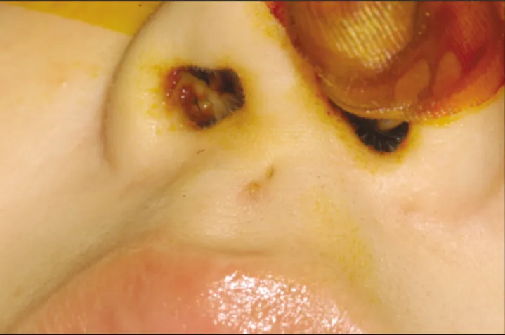
A 17-years-old girl had a conspicuous swelling of the upper lip. She had pain on the upper lip area where it is fistular and pus formation. The lesion had a minute sinus tract extending from the frenulum to a midline punctum at the base of the col- umella.(Figs. 1, 2) Radiographs of MRI documented 12×10 mm well defined cyst in midline upper lip. This lesion showed fluid signal on T1W1 and T2W1 with relatively thick rim enhancement. Therefore it is suggestive of dermoid cyst with combined inflammation. Under general anesthesia, she was treated with conservative surgical excision through a horizon- tal incision in the upper lip mucosa. At surgery, dissection of the sinus tract at the base of the columella revealed continuity
with a cyst in the labial frenulum.(Figs. 3-5) Complete exci- sion was performed without complication and acceptable cosmesis was achieved at the site of surgical excision.
Microscopic findings supported the diagnosis of a dermoid cyst.(Fig. 6) At present, she has not pain on the upper lip area and not swelling. There is not recurrence and prognosis is favorable.(Fig. 7)
Ⅲ. Discussion
Dermoid are benign tumors of neuroectodermal and meso- dermal origin presenting as cysts filled with keratinized debris that may lie along the path or at the end of a sinus tract lined by squamous epitheilum.1-3According to Cauchois et al.4, a midline nasal pit at the base of the columella as described in this report is pathognomonic for a nasal dermoid. This nasal dermoid, however, is significant in terms of the embryologic development of its unique location.
In 1936, New and Erich5described two report of nasal der- moid cysts (NDCs) with upper lip involvement and pits at the base of the columella. There was no mention of cyst location specific to the labial frenulum of the upper lip. they theorized that these cases of soft palate cysts arose during the abnormal midline fusion of palatine plates that grow from the maxillary processes. Similiary, in this case, we propose that the labial frenulum dermoid sinus arose between the 6th and 10weeks of fetal life. During this time the nasal placodes invaginate to form the medial nasal processes, which in turn form the inter- maxillary processes. Fusion of the intermaxillary processes by the 10th week of gestation and entrapment of neuroextoderm at the midline of these processes may have resulted in the frenu- lum dermoid as reported in this case.
고 세 욱
435-040 경기도 군포시 산본동1142
원광대학교 치과대학 산본 치과병원 구강악안면외과 Sewook Koh
Department of Oral and Maxillofacial Surgery, School of dentistry, wonkwang university
Sanbon-dong 1142, Gunpo, 435-040, Korea TEL: +85-31-390-2569 FAX: +82-31-390-2777 E-mail: [email protected]
Midline dermoid cyst of the upper lip: case report
Sewook Koh
Department of Oral and Maxillofacial Surgery, School of Dentistry, Wonkwang University, Gunpo, Korea
Midline craniofacial dermoids are rare lesions resulting from the abnormal fusion of embryologic structures. The clinical features of craniofacial der- moid cysts show a range of presentations, including infection, asymptomatic puncti, or seizure secondary to intracranial invasion. Appropriate man- agement involves an accurate diagnosis of the dermoid cyst and a complete resection.
Key words: Craniofacial dermoid cyst, Upper lip
[paper submitted 2011. 5. 2 / revised 2011. 10. 3 / accepted 2011. 9. 29]
Abstract (J Korean Assoc Oral Maxillofac Surg 2011;37:403-5)
*This paper was supported by wonkwang university in 2010.
J Korean Assoc Oral Maxillofac Surg 2011;37:403-5
404
Fig. 1.Preoperative view of the upper lip sinus.
Sewook Koh: Midline dermoid cyst of the upper lip: case report. J Korean Assoc Oral Maxillofac Surg 2011
Fig. 2.Preoperative view of the frenulum.
Sewook Koh: Midline dermoid cyst of the upper lip: case report. J Korean Assoc Oral Maxillofac Surg 2011
Fig. 3.Dermoid sinus dissected from intraoral exposure.
Sewook Koh: Midline dermoid cyst of the upper lip: case report. J Korean Assoc Oral Maxillofac Surg 2011
Fig. 4.Postoperative cutaneous defect.
Sewook Koh: Midline dermoid cyst of the upper lip: case report. J Korean Assoc Oral Maxillofac Surg 2011
Fig. 5.Resected dermoid cyst and sinus.
Sewook Koh: Midline dermoid cyst of the upper lip: case report. J Korean Assoc Oral Maxillofac Surg 2011
Fig. 6.H&E (×40) cyst-like structure.
Sewook Koh: Midline dermoid cyst of the upper lip: case report. J Korean Assoc Oral Maxillofac Surg 2011
Midline dermoid cyst of the upper lip: case report
405 Nasal dermoids typically present during childhood with one
case study by McCaffrey et al.1showing 70% NDCs diagnosed in the first month of life by a pediatrician or family physician.
Therefore, although NDCs are rare, suspicion should be high for NDCs in a child presenting with a midline mass, especially associated with discharge not resolved by antibiotic therapy.6 Presentation of dermoid cysts ranges from a cutaneous mass with or without a sinus opening to a hair tuft to a cheesy or frothy discharge. As in this report, dermoid tissue presenting as a formulalike discharge was forced from the frenulum cyst and columellar sinus during feeding, most likely as pressure within the mouth transferred pressure to the base of the dermoid.
The presentation of a midline dermoid cyst should prompt the physician to investigate not only for sinus extension but also for associated congenital anomalies. Data regarding the incidence and correlation of midline dermoids with other anomalies are rare, with no clear association with a specific syndrome. A case study of 36 patients with dermoids by Deyonelle et al.,2however, found that 3 of the 36 patients had cysts presenting in conjunction with anomalies such as bilater- al aural atresia, bilateral pinna abnomalities, or nasal cavity
agenesia. Wardinsky et al.6found a 41% association of NDCs with other craniofacial abnormalities. But our case is not asso- ciated with craniofacial abnormality.
Treatment of NDCs requires complete surgical excision of the cyst and sinus tract. Any remaining dermoid, neuroectoder- mal, or epithelial tissue after surgery represents incomplete resection. McCaffrey et al.1concluded that of 21 patients with dermoid cysts, 15 with complete excision as determined at the time of surgery had a recurrence rate of 7%. Comparatively, three cases of irrigation and debridement as well as three cases of incomplete resection resulted in a 100% recurrence rate.
Although dermoid cysts are benign lesion, incomplete resec- tion increases surgical complication of recurrence, infection, decreased cosmesis, and possible malignant transformation.
Ⅳ. Conclusion
Midline craniofacial dermoids represent rare lesions resulting from abnormal fusion of embryologic structures. This previ- ously unreported location of a dermoid retains similar etiology, epidemiology, histology, and basic embryology of a nasal der- moid but is unique in its location outside of the nose and its extension beyond the upper lip into the frenulum. Proper man- agement relies on both the primary care physician who diag- noses the dermoid and the surgeon who resects it.
References
1. McCaffrey T, McDonald T, Gorenstein A. Dermoid cysts of the nose: review of 21 cases. Otolaryngology Head Neck Surg 1979;
81:52-59.
2. Deyonelle F, Ducroz V, Roger G, et al. Nasal dermoid sinus cysts in children. Laryngoscope 1997;107:795-800.
3. Hughes GB, Sharpino G, Hunt W, et al. Management of the con- genital midline nasal mass: a review. Head Neck Surg 1980;2:
222-233.
4. Cauchois R, Laccourreye O, Bremond D, et al. Nasal dermoid si- nus cyst. Ann Otol Rhinol Laryngol 1994;103:615-618.
5. New G, Erich J. Dermoid cysts of the head and neck. Surg Gynecol Obstet 1937;65:48-55.
6. Wardinsky TD, Pagon RA, Kropp RJ, et al. Nasal dermoid sinus cysts: association with intracranial extension and multiple mal- formations. Cleft Palate Craniofac J 1991;28:87-95.
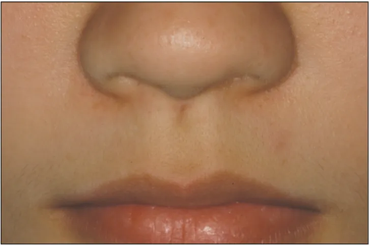
Fig. 7.Postoperative 22months.
Sewook Koh: Midline dermoid cyst of the upper lip: case report. J Korean Assoc Oral Maxillofac Surg 2011