대한소화기학회지 2006;48:351-354
접수: 2006년 10월 26일, 승인: 2006년 3월 20일
연락처: 류지곤, 110-744, 서울시 종로구 연건동 28번지 서울대학교 의과대학 내과학교실
Tel: (02) 2072-1962, Fax: (02) 762-9662 E-mail: [email protected]
Correspondence to: Ji Kon Ryu, M.D.
Department of Internal Medicine, Seoul National University College of Medicine, 28, Yeongeon-dong, Jongno-gu, Seoul 110-744, Korea
Tel: +82-2-2072-1962, Fax: +82-2-762-9662 E-mail: [email protected]
췌장의 Somatostatinoma 1예
서울대학교 의과대학 내과학교실
최유식․박주경․이상협․윤원재․이준규․류지곤․김용태․윤용범
A Case of Pancreatic Somatostatinoma
You Sik Choi, M.D., Joo Kyung Park, M.D., Sang Hyub Lee, M.D., Won Jae Yoon, M.D., Jun Kyu Lee, M.D., Ji Kon Ryu, M.D., Yong-Tae Kim, M.D., and Yong Bum Yoon, M.D.
Department of Internal Medicine, Seoul National University College of Medicine, Seoul, Korea
Somatostatinoma is a rare neoplasm usually arising from the pancreas and duodenum which typically presents with indolent, nonspecific symptoms in the absence of systemic neuroendocrine manifestations that characterize somatostatinoma syndrome. It accounts for less than 1% of all gastrointestinal endocrine tumors with an annual incidence of 1 per 40 million. It is often associated with regional and/or portal metastasis at the time of diagnosis, and complete tumor resection is possible only in 60% to 70% of cases. We experienced a case of pancreatic somatostatinoma recently. A 51-year-old woman presented with right upper quadrant abdominal pain and loose stool for one month. A hypermetabolic lesion in the pancreatic head was detected on positron emission tomography-CT (PET-CT) scan. The tumor was resected by pylorus preserving pancreaticoduodenectomy. Immuno- histochemical staining of the tumor tissue exhibited diffuse positivity for somatostatin, but was negative for insu- lin and glucagon. Herein, we report a case of pancreatic somatostatinoma diagnosed postoperatively. (Korean J Gastroenterol 2006;48:351-354)
Key Words: Somatostatinoma; Pancreas; Islet cell tumor; Pancreaticoduodenectomy
서 론
Somatostatinoma는 췌장 또는 십이지장에서 발생하는 매우 드문 내분비 종양으로1,2 당뇨, 지방변, 담석증 등의 전형적인 somatostatinoma 증후군의3,4 양상으로 발현할 수 있지만, 대부 분의 환자에서는 모호한 복부통증, 체중감소, 배변 습관의 변 화 등 비특이적인 증상만이 나타난다.5 전형적인 증례의 경우 방사선 검사에서 크기가 크고 고립성인 췌장 두부 종양을 보 이며 진단 또는 수술 시점에 림프절이나 간전이가 되어 있는 경우가 흔하다.6 1977년 Larsson 등이7 처음 보고한 이래 지금
까지 전 세계적으로 150예가8 보고되어 있으며 국내에서는 췌 장에서 발생하여 간 및 자궁에 전이된 1예와9 십이지장, 바터 팽대부에 각각 1예씩 보고되어 있으나,10,11 이번 증례는 비특 이적인 증상을 주소로 내원하여 조기에 수술로 치료한 췌장에 국한된 somatostatinoma로서 국내에서 매우 희귀한 예이다.
저자들은 우상복부 통증과 무른 변을 주소로 내원한 환자 에서 positron emission tomography (PET) 스캔과 컴퓨터단층 촬영으로 췌장 소도세포 종양 의심하에 수술 절제를 통해 somatostatinoma로 확진한 1예를 경험하였기에 문헌 고찰과 함께 보고한다.
352 대한소화기학회지: 제48권 제5호, 2006
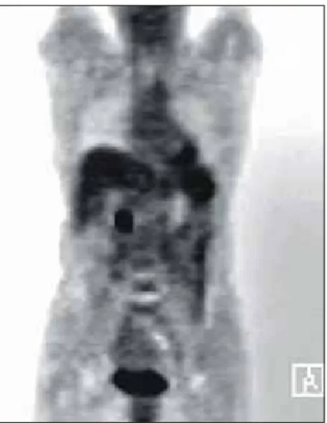
Fig. 1. F-18 FDG PET scan. It shows a focal hypermetabolic lesion on the ampulla of Vater.
PET, positron emission tomography.
Fig. 2. CT scan of the abdomen. It shows about a 2 cm-sized well-enhancing mass on the pancreatic head at early arterial phase.
There is no dilatation of the pancreatic duct.
Fig. 3. Endoscopic retrograde cholangiopancreatography. There is no abnormal finding.
증 례
65세 여자가 2개월 전부터 시작된 간헐적인 우상복부 통 증과 하루 3-4회의 무른 변을 주소로 인근 검진센터 방문하 였고, PET에서 바터 팽대부에 국소 과대사 병변이 관찰되어 (Fig. 1) 바터 팽대부암 의심하에 전원되었다. 과거력, 사회 력, 가족력에서 특이사항은 없었다. 신체 검사에서 전신상태 는 양호하였으며 혈압 140/90 mmHg, 호흡 14회/분, 맥박 66 회/분, 체온 36.5oC였다. 말초혈액검사에서 백혈구 8,800/mm3, BUN 11.8 mg/dL, 크레아티닌 0.7 mg/dL, 총 단백 6.4 g/dL, 알부민 3.9 g/dL, 총 빌리루빈 0.9 g/dL, AST 18 IU/L, ALT
20 IU/L이었으며, 알칼리 포스파타제 58 IU/L,γGT 25 IU/L 이었다. CEA는 1 ng/mL 미만이었고, CA 19-9는 5 U/mL이 었다. 복부 전산화단층촬영의 동맥기 영상에서 매우 강한 조영증강 소견을 보이는 2 cm의 결절이 췌장 두부에서 관 찰되었고, 췌관이나 담관의 확장 소견은 없어 췌장의 내분 비 종양이 의심되었다(Fig. 2).
내시경역행담췌관조영술에서는 특이 소견이 없었다(Fig.
3). 호르몬 검사에서 혈청 글루카곤은 24.3 pg/mL (정상 범 위; 40-130)이었고, 인슐린은 9.3 IU/mL (정상 범위; 2-25)이 었다. 이상의 소견을 종합하여 췌장 두부의 내분비 종양으 로 의심하고 조직학적인 확진과 근치 치료를 위하여 수술을 시행하였다. 수술 소견은 췌장 두부에서 고형 종괴가 발견 되었고 유문보전췌십이지장절제술을 시행하였다.
병리 소견에서 종양 크기는 2.2×1.5×1.3 cm이며 원위부 총담관 바로 아래쪽의 췌장두부에 위치하고 있었다. 종괴는 피막에 잘 둘러싸여 있어 주변 조직으로 침윤하는 부분은 관찰되지 않았으며, 유착, 림프절 전이 등도 없었다. 광학현 미경 소견은 저배율에서 선(gland) 구조를 형성하고 있는 전 형적인 종양세포가 관찰되었고, 고배율에서는 타원 또는 원 형의 핵과 미세한 과립의 세포질이 관찰되었다(Fig. 4).
감별 진단을 위해 시행한 면역조직화학염색에서 somatos- tatin은 양성이었고(Fig. 5), synaptophysin과 chromogranin도 양성이었으며 인슐린, 글루카곤은 음성이었다. 최종적으로 췌장의 somatostatinoma로 진단되었다. 수술 후 환자의 증상 은 소실되었고 5개월이 지난 현재 특이 소견 없이 외래 추 적관찰 중이다.
최유식 외 7인. 췌장의 Somatostatinoma 1예 353
Fig. 4. Microscopic finding. Tumor cells have relatively uniform round to oval nuclei with fine granular cytoplasm (H&E stain, × 400).
Fig. 5. Somatostatin immunohistochemical stain. The tumor cells show diffuse positivity on somatostatin stain (×400).
고 찰
Somatostatinoma는 매우 드문 내분비 종양으로1,2 주로 췌 장 두부에서 큰 크기의 고형 악성종양 형태로 나타난다.6 이 전에는 당뇨, 지방변, 담석증 등을 주 증상으로 하는 soma- tostatinoma 증후군의 발현이 특징적으로 알려졌으나,3,4 최근 연구에서는 173예의 somatostatinoma 환자 중에서 단지 17명 만이 somatostatinoma 증후군을 일으켰다.8
Somatostatinoma는 췌장 두부에서 46-75%가 발생하며 그 밖에 십이지장, 바터팽대부, 공장, 담낭관에서 발견된 예들 이 보고되었다.8,12 90-96%가 단일성이며 크기는 1.5-10 cm였 고 췌장에서 발생된 somatostatinoma의 평균 크기가 십이지 장에서 발생된 경우보다 크다.8 53-84%의 종양이 전이를 일 으키며 호발 부위는 간이며 그 밖에 림프절, 뼈 등에 전이된
다.8,12 병리 소견에서 섬유 중격을 가진 잘 분화된 종양으로
나타나며12 십이지장 somatostatinoma의 경우에는 특징적으 로 사종체(psammoma body)를 동반한다.8,13 전자현미경 소견 에서는 D세포에서 전형적인 분비성 과립이 관찰된다.8,12 면 역조직화학염색에서는 somatostatin 면역 반응이 모든 예에 서 관찰된다.8,11 전형적인 somatostatinoma 증후군의 경우에 는 종양에서 분비된 somatostatin이 다양한 위장관 호르몬과 펩타이드들(인슐린, 글루카곤, 성장 호르몬, 세크레틴, 가스 트린, 콜레시스토키닌, glucose- dependent insulin releasing peptide)과 중탄산염 및 다른 췌장의 외분비 기능을 억제하 여 고혈당, 담석증, 지방변, 저염산증 등의 증상을 유발한 다.1,3 Somatostatinoma의 호발 연령은 40-60세이며 남녀 발생 률의 차이는 확실치 않다.8 전술한 대로 전형적인 somato- statinoma 증후군은 173예 중 단지 17예에서만 발생하였다는 보고가 있는 반면 복부 통증(40%), 체중 감소(26%), 황달
(23%), 설사(18%), 오심 또는 구토(16%), 복부 종양이나 간 비대의 발견(22%) 등이 주 증상이다.8 55%의 환자에서 당뇨 가 동반되며 대개의 경우 경증이어서 경구용 혈당강하제나 소량의 인슐린으로 혈당 조절이 가능하며, 담낭 또는 담도 계 질환은 65%에서 발생하고 35%의 환자에서 담석이 발견 된다.12 설사나 지방변은 전체적으로는 18- 35%의 환자에서 발생하나, 췌장 종양의 경우에는 설사와 지방변이 각각 83%, 92%에서 동반된다.8,12 설사는 하루 3-10회 정도 발생 하며 일부 환자에서는 설사의 기간이나 정도가 질병 진행과 관련이 있으며 전이가 발생하면 악화되고 수술 후 호전된 다. 저염산증은 70%에서 발견되었고 췌장 종양 환자의 32-33%에서 9-21 kg의 체중감소가 관찰되며,8 15-67%에서 경증의 빈혈이 발견된다.8 전형적인 somatostatinoma 증후군 은 췌장종양에서 빈도가 더 높으며,8 somatostatinoma 환자의 절반은 multiple endocrine neoplasm (MEN I) 또는 MEN II와 동반되어 발생한다.12
대부분의 somatostatinoma는 담낭 절제를 위한 개복술 시 또는 복통이나 설사의 증세 등 비특이 증세로 위장관계 영 상 검사를 시행하였을 때 발견된다.1,8,12 Somatostatinoma가 의심될 때에는 혈중 somatostatin의 상승이 진단 의미를 지니 지만 수술 전 진단은 어려운 경우가 많으며 대부분의 경우 에서 췌장 종양의 수술 후 조직병리 소견으로 진단된다.14 전산화단층촬영과 자기공명영상이 췌장 종양의 발견과 간 전이 등 병기 결정에 효과적이며15 재발 유무, 이차 위치 추 적, 잠재 파종성 전이의 진단에는 somatostatin 수용체에 대 한 핵의학 검사가 유용하다.16
Somatostatinoma는 발생률이 낮고 장기 추적관찰의 자료 가 부족하여 최선의 치료방법이 정립되어 있지 않다. 그러 나 일반적으로 2 cm 미만의 종양은 국소 절제만으로 충분 하고, 그 이상의 크기에서는 췌십이지장절제술을 추천한다.5
354 The Korean Journal of Gastroenterology: Vol. 48, No. 5, 2006
Streptozocin, 5-fluorouracil과 doxorubicin을 이용한 항암 치료 는 일부 환자에서 부분 반응을 보인다. Somatostatinoma 증 후군을 동반한 수술이 불가능한 환자에서 octreotide 주입이 효과적이다.12,17,18 생존율에 대한 자료는 제한되어 있지만 국소 종양에서 성공적인 절제가 이루어질 경우 장기 생존이 가능하고,8 전이암의 경우에는 수술 절제와 수술 후 항암치 료를 통해 60%에서 6개월에서 5년 생존을 기록했다는 연구 가 있다.18
이번 증례를 요약하면 전형적인 증상인 somatostatinoma 증후군 없이 2개월간 지속된 우상복부 통증과 무른 변을 주 소로 내원하여 PET 검사를 통해 췌장 종양이 발견되었고, 수술을 통해 매우 드문 종양인 somatostatinoma가 진단되었 다. 이에 저자들은 문헌 고찰과 함께 보고한다.
참고문헌
1. Green BT, Rockey DC. Duodenal somatostatinoma presenting with complete somatostatinoma syndrome. J Clin Gastroen- terol 2001;33:415-417.
2. Mozell E, Stenzel P, Woltering EA, Rosch J, O'Dorisio TM.
Functional endocrine tumors of the pancreas: clinical presen- tation, diagnosis and treatment. Curr Probl Surg 1990;27:
301-386.
3. Krejs GJ, Orci L, Conlon JM, et al. Somatostatinoma syn- drome: biochemical, morphologic and clinical features. N Engl J Med 1979;301:285-292.
4. Tanaka S, Yamasaki S, Matsushita H, et al. Duodenal soma- tostatinoma: a case report and review of 31 cases with special reference to the relationship between tumor size and meta- stasis. Pathol Int 2000;50:146-152.
5. O'Brien TD, Chejfec G, Prinz RA. Clinical features of duo- denal somatostatinomas. Surgery 1993;114:1144-1147.
6. House MG, Yeo CJ, Schulick RD. Periampullary pancreatic somatostatinoma. Ann Surg Oncol 2002;9:869-874.
7. Larsson LI, Hirsch MA, Holst JJ, et al. Pancreatic soma-
tostatinoma: clinical features and physiological implications.
Lancet 1997;26:666-668.
8. Soga J, Yakuwa Y. Somatostatinoma /inhibitory syndrome: a statistical evaluation of 173 reported cases as compared to other pancreatic endocrinoma. J Exp Clin Cancer Res 1999;
18:13-22.
9. Kim TY, Park YI, Im YH, et al. A case of pancreatic somatostatinoma with metastasis to the liver and uterus.
Korean J Med 1993;44:689-696.
10. Nam MS, Lee EJ, Cho JH, et al. A case of duodenal somatostatinoma presenting with diabetic ketoacidosis. Korean J Med 1995;48:822-827.
11. Lee HW, Kim HC, Song OP, et al. Somatostatinoma of the ampulla of vater. Korean J Surg 2004;66:251-255.
12. Vinik AI, Strodel WE, Eckhauser FE, Moattar AR, Lloyd R.
Somatostatinoma, PPomas, neurotensinomas. Semin Oncol 1987;14:263-281.
13. Mao C, Shah A, Hanson DJ, Howard JM. Von Reckling- hausen's disease associated with doudenal somatostatinoma:
contrast of duodenal versus pancreatic somatostatinomas. J Surg Oncol 1995;59:67-73.
14. Marakis G, Ballas K, Rafailidis S, Alatsakis M, Patsiaoura K, Sakadamis A. Somatostatin-producing pancreatic endocrine carcinoma presented as relapsing cholangitis-a case report.
Pancreatology 2005;5:295-299.
15. Tjon A Tham RT, Jansen JB, Falke TH, Lamers CB. Imaging features of somatostatinoma: MR, CT, US and angiography. J Comput Assist Tomogr 1994;18:427-431.
16. Krausz Y, Bar-Ziv J, de Jong RB, et al. Somatostatin recep- tor scintigraphy in the management of gastroenteropancreatic tumors. Am J Gastroenterol 1998;93:66-70.
17. Harris GJ, Tio F, Cruz AB Jr. Somatostatinoma: a case report and review of the literature. J Surg Oncol 1987;36:8-16.
18. Fulfaro F, Quagliuolo V, De Conno F, Ripamonti C. Car- cinoid somatostatinoma of the duodenum. Eur J Surg Oncol 1998;24:601-604.