S L E는 인구 1 0만 명당 약 6 - 3 5명에서 볼 수 있는 질환으로 주로 젊은 여성에서 발병하고 특히, 아프리카나 아시아에서 유병율이 높은 것으로 알려져 있다. SLE는 만성염증성질환으 로 원인은 정확히 알려져 있지 않으나 피부, 관절, 신장, 신경 계, 장막, 그 밖에 다른 여러 장기를 침범하는 전신질환이다.
염증병변은 주로 면역복합체나 항체에 의해 작은 혈관의 침 범으로 발생하나, 병의 치료에 쓰이는 스테로이드 제재에 의 한 의인성 변화도 볼 수 있다. 병의 진행은 호전과 악화를 반 복하는 임상양상을 보이고, 임상증상은 침범장기에 따라 다양 하게 나타난다. 복부를 침범하는 경우 급성복통, 구토, 설사, 혈뇨, 신기능저하 등을 일으키고 간혹 급성복증을 감별하기 어려운 경우가 있다(1, 2).
S L E는 병변 자체의 다양한 임상소견 및 합병증에 따르는 비특이적 양상 때문에 영상진단이 요구되지만, 이에 대한 보 고는 드물다. SLE로 확진되고 복부영상진단에서 이상 소견이 관찰된 2 0명의 환자(전례 여성, 나이 1 3 - 4 8세)를 대상으로 하 여, SLE의 다양한 복부침범 소견을 소개하고자 한다.
소화기계
위장관침범의 기본병리는 작은 혈관의 면역복합체와 항체 에 의한 염증변화와 허혈변화로 장간막과 장벽의 변화 그리 고 동반된 복수를 볼 수 있다. 장간막의 혈관들은 C T상 염주 양 변화를 보이고 말단에서는 정상적인 점감( t a p e r i n g )을 보 이지 않으며, 혈관주위의 장간막지방조직에 침윤을 보인다 (Fig. 1)(3). 소장과 대장의 변화는 장벽비후로 잘 알려져 있
고 심한 경우 허혈장염을 일으켜 장벽 내에 공기를 볼 수 있 다. 바륨검사상 소장 주름의 비후와 연축( s p a s m )을 볼 수 있 으며(Fig. 2A), 초음파검사와 C T상 미만성 장벽비후 및 이중 표적 징후(double-target sign)를 보인다(Fig. 2B). 주 침범부위 는 상장간막동맥의 공급을 받는 공장과 회장이며 십이지장동 맥과 췌십이지장 동맥궁의 공급을 받는 십이지장의 비후도 종종 볼 수 있는데 이는 주 공급동맥의 혈관염 때문으로 추측 된다(Fig. 3). 장벽 비후의 분포는 한 동맥 영역에 국한되거나 (Fig. 4) 여러 동맥이 동시에 침범하므로 다양하게 나타난다.
간침범의 경우는 간종대로 나타나는데 드물지 않게 보이는 소견으로 발생기전은 밝혀진 바 없다. 간종대와 더불어 스테 로이드 치료에 의한 이차적인 소견으로 지방간과 선종이 발 생할 수 있다(Fig. 5).
췌장염의 빈도는 정확히 알려져 있지 않으나, 혈관염과 스테 로이드치료의 이차적변화로 여겨지고, 다양한 췌장염의 합병증 을 볼 수 있다. 간혹 SLE 환자에서 췌장의 미만성 혹은 국소적 인 비대가 나타나며(Fig. 6), 임상적으로 췌장염의 증상과 아밀 라제( a m y l a s e )의 증가 없이 췌장 비대를 보이는 경우가 있다.
장막
S L E환자의 약 3 0 %에서 흉막염, 심막염 등이 나타나고 복 막염의 빈도는 그 보다 적은 것으로 알려져 있다(6). 복막을 비롯한 장벽, 담낭벽, 방광벽은 장막으로 둘러싸여 있으며, S L E에서 염증변화로 장막비후를 초래하게 된다(4). Hoffman 등( 5 )은 부검을 시행한 S L E환자 중 약 6 0 %에서 복막염이 있 었다고 보고하였을 정도로 드문 소견이 아니다. 복막의 비후 는 전반적으로 미만변화를 보이고 C T상 조영증강을 보인다 전신성홍반성루프스(Systemic lupus erythematosus, 이하 S L E로 약함)은 원인이 정확히 밝
혀져 있지 않은 전신성면역질환으로 기본 병리는 면역복합체나 항체에 의한 혈관염이나 장 막염이다. 대부분은 비특이적인 소견을 보이고 다양한 형태의 복부침범을 하는데 소장과 대 장의 장벽 비후, 간 및 췌장종대, 장막염, 림프절종창과 비종대, 신장염과 간질성 방광염, 그 리고 혈전정맥내막염 등을 볼 수 있다. 임상적으로 미 류마티즘협회(American Rheumatism Associa- tion)의 진단기준 및 조직검사에 의하여 S L E를 확진 받은 환자를 대상으로 영상진 단을 시행하고 위에서 언급한 복부장기침범을 중심으로 방사선학적 소견을 알아보았다. SLE 환자에서 복부장기침범의 다양한 방사선학적소견을 숙지함은 S L E의 복부침범 및 합병증을 조기 진단 하는데 도움이 되리라 생각된다.
1한양대학교 의과대학 진단방사선과학교실
이 논문은 1 9 9 8년 1 2월 2 2일 접수하여 1 9 9 9년 3월 8일에 채택되었음.
전신성홍반성루프스 : 복부방사선학적소견1
오재천・조온구・이용주・배재익・김용수・임현철・고병희
Fig. 1. Mesenteric vasculitis.
A. CT scan of a patient with mesenteric vasculitis shows beaded pattern of mesenteric vessels (arrowheads) without tapering at the terminal portion. Ascites and target appearance (arrows) of small bowel loops are also presented.
B. Abdominal CT scan reveals infiltration of mesenteric fat (arrows) and thickening of bowel loops and peritoneum (arrowheads).
A B
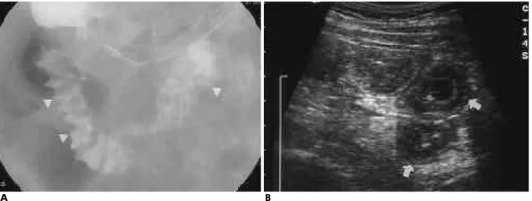
Fig. 2. Lupus enteritis.
A. UGI shows thickened duodenal and jejunal folds with tethered and spiculated pattern (arrowheads).
B. Transverse abdominal ultrasonography shows the typical target configuration of multiple inflamed bowel loops (arrows).
A B
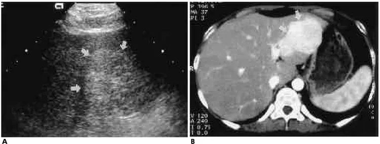
Fig. 5. Hepatocellular adenoma.
A. Abdominal ultrasonography shows well-defined hyperechoic mass (arrows) in the left lobe of the liver.
B. Enhanced abdominal CT scan shows relatively well-enhancing mass (arrows) in the left lobe of the liver.
A B
Fig. 3. Enhanced abdominal CT scan shows dilated duodenal loops with wall thickening and fluid retention as well as ring- like wall enhancement or target sign (arrows).
Fig. 6. Pancreas enlargement. Enhanced CT scan shows dif- fuse enlargement of pancreas without adjacent fat infiltration.
Fig. 7. Enhanced abdominal CT scan reveals minimally peri- toneal enhancement (arrows) with small paraaortic lym- phadenopathy (arrowhead).
Fig. 4. Isolated colonic wall thickening. Enhanced abdominal CT scan shows circumferential wall thickening of the rec- tosigmoid colon (arrows).
어 신장의 조영지연과 지연배설을 보인다(Fig. 14).
방광침범은 매우 드문 소견으로 활동성S L E와 동반되는 것
소적물리적압박 등이다. 그 밖에 하지의 심부정맥혈전과 폐혈 전색전증을 동반하는 경우도 있다( 9 ) .
Fig. 8. Enhanced abdominal CT scan reveals gallbladder with minimal wall thickening and enhancement (arrow), moderate enlargement of both kidneys with delayed enhancement, and ascites (arrowheads).
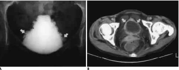
Fig. 9. Abdominal CT scan shows diffusely thickened urinary bladder wall with homogeneous enhancement (arrows). Also, enhancement of prevesical fascia, uterosacral ligament, and peritoneum is present.
Fig. 10. A. Paraaortic lymphadenopathies. The lymphadenopathies (arrows) are small and homogeneously enhanced without cen- tral low density.
B. Mesenteric lymphadenopathies have same findings to paraaortic lymphadenopathies. Bowel wall thickening and small amount of ascites are also seen.
A B
Fig. 11. Splenic infarction. Enhanced CT scan reveals multiple peripheral wedge shaped low attenuation area (arrows).
Hepatosplenomegaly and perisplenic ascites are also noted.
Fig. 13. Subcapsular hematoma. Enhanced CT scan shows hy- podense area to the posterior aspect of right kidney.
Fig. 12. Ileal lymphoma.
A. Double contrast colon study shows fungating mass (arrows) of the terminal ileum protruding in the ascending colon.
B. Abdominal CT scan shows well enhancing mass (arrows) and intussusception (arrowheads) of the ascending colon.
A B
Fig. 14. Lupus nephritis.
A. Sagittal ultrasonogram shows increased longitudinal and anteroposterior dimension of the kidney with increased cortical e- chogenicity (arrows) and prominent renal pyramids (arrowheads).
B. Portal phase CT scan reveals increased renal size and delayed renal enhancement.
A B
C
Fig. 15. Interstitial cystitis.
A. Voiding cystourethrogram reveals a small and contracted bladder with irregular wall and diverticula (arrow) of variable size as well as no evidence of vesicoureteral reflux on either side.
B. Abdominal CT scan shows well-enhancing urinary bladder with irregular wall thickening. Also, moderate amount of as- cites is present in pelvic cavity.
C. Abdominal CT scan, at the level of renal hilum, shows bi- lateral hydronephrosis.
Fig. 16. IVC thrombosis
A. Sagittal Abdominal ultrasonogram reveals small sized IVC (arrows) with echogenic thrombi (arrowheads).
B. Abdominal CT scan shows low-attenuating thrombi (arrowhead) in the IVC with multiple collateral vessels (arrows).
A B
B A
참 고 문 헌
1. Heiberg E, Wolverson MK, Sundaram M, Shields JB. Body com- puted tomography findings in systemic lupus erythematosus. J Comput Assist Tomogr 1 9 8 8 ; 1 2 : 6 8 - 7 4
2. Si-Hoe CK, Thng CH, Chee SG, Teo EK, Chng HH. Abdominal computed tomography in systemic lupus erythematosus. C l i n R a d i o l 1 9 9 7 ; 5 2 : 2 8 4 - 2 8 9
3. Kirshy DM, Gordon DH, Atweh NA. Abdominal computed to- mography in lupus mesenteric arteritis. Comput Med Imaging G r a p h 1 9 9 1 ; 1 5 : 3 6 9 - 3 7 2
4. Oddis C, McGlynn TJ. Abdominal computed tomography scan in acute lupus abdominal serositis. J Comput Assist Tomogr 1 9 8 4 ; 8 : 3 3 7 - 3 3 9
5. Hoffman BT, Katz WA. The gastrointestinal manifestation of sys- temic lupus erythematosus: a review of the literature. S e m i n Arthritis Rheum 1 9 8 0 ; 9 : 2 3 7 - 2 4 7
6. Shapira Y, Weinberger A, Wysenbeek AJ. Lymphadenopathy in systemic lupus erythematosus: prevalence and relation to disease manifestation. Clin Rheumatol 1 9 9 6 ; 1 5 : 3 3 5 - 3 3 8
7 . Abu-Shakra M, Gladman DD, Urowitz MB. Malignancy in sys- temic lupus erythematosus. Arthritis Rheum 1 9 9 6 ; 3 9 ( 6 ) : 1 0 5 0 - 1 0 5 4 8. Kim HJ, Park MH. Obstructive uropathy due to interstitial cystitis
in a patient with systemic lupus erythematosus. Clin Nephrol 1 9 9 6 ; 4 5 : 2 0 5 - 2 0 8
9. Mintz G, Acevedo-Vazquez E, Gutierrez-Espinosa G, Avelar- Garnica F. Renal vein thrombosis and inferior vena cava thrombo- sis in systemic lupus erythematosus. Arthritis Rheum 1 9 8 4 ; 2 7 : 5 3 9 - 5 4 4
J Korean Radiol Soc 1999;40:1 1 73- 1 1 7 9
Address reprint requests to : On-Koo Cho, M.D., Department of Radiology, Hanyang University Hospital,
#17, Haengdang-Dong, Seungdong-Gu, Seoul, 133-792, Korea.
Tel. 82-2-2290-9164 Fax. 82-2-2293-2111
Systemic Lupus Erythematosus : Abdominal Radiologic Findings1
Jae-Cheon Oh, M.D., On-Koo Cho, M.D., Yong-Joo Lee, M.D., Jae-Ik Bae, M.D., Yong-Soo Kim, M.D., Hyun-Chul Rhim, M.D., Byung-Hee Ko, M.D.
1Department of Diagnostic Radiology,College of Medicine, Hanyang University
Systemic lupus erythematosus(SLE) is a systemic disease of unknown etiology. Its main pathology is vasculi- tis and serositis, due to deposition of the immune complex or antibodies. Most findings are nonspecific ; ab- dominal manifestations include enteritis, hepatomegaly, pancreatic enlargement, serositis, lymphadenopathy, splenomegaly, nephritis, interstitial cystitis, and thrombophlebitis. We described radiologic findings of various organ involvement of SLE; digestive system, serosa, reticuloendothelial system, urinary system, and venous system. Diagnosis of SLE was done according to the criteria of American Rheumatism Association.
Understanding of the variable imaging findings in SLE may be helpful for the early detection of abdominal in- volvement and complications.
Index words :Abdomen, diseases Abdomen, CT Abdomen, US Lupus erythematosus