Endovascular Treatment for Arterioureteral Fistula of the Abdominal Aorta:
A Case Report and Literature Review
복부 대동맥에 발생한 동맥-요관 누공의 혈관 내 치료:
증례 보고와 문헌고찰
Hyoung Nam Lee, MD , Woong Hee Lee, MD*
Department of Radiology, Soonchunhyang University Cheonan Hospital, Cheonan, Korea
We present a rare case demonstrating successful endovascular management of an arterioure- teral fistula involving the abdominal aorta. Arterioureteral fistulas are rare but life-threatening, with mortality rates ranging from 7% to 23%. Early recognition and prompt management are essential for preventing catastrophic consequences, including hypovolemic shock. However, recognition of an arterioureteral fistula requires a high index of clinical suspicion due to its rari- ty and the lack of a sensitive diagnostic method. Arterioureteral fistulas could be induced by traumatic events in patients who have a history of pelvic surgery, radiation, and prolonged placement of a ureteral stent. Endovascular stent graft placement could be a valid treatment option for arterioureteral fistulas involving the abdominal aorta.
Index terms Urinary Fistula; Hematuria; Stents; Endovascular Procedures; Aneurysm, False
INTRODUCTION
The arterioureteral fistula (AUF) is a rare abnormal communication between the ar- tery and ureter. Since the first description of AUF in 1908 reported after bilateral ure- terolithotomies, the incidence of this condition is increasing (1). The mortality rate for patients with an undiagnosed AUF is reported up to 52% and early recognition and prompt management are essential for preventing catastrophic consequences (2, 3).
However, AUF remains a diagnostic challenge to most physicians due to its rarity and lack of sensitive diagnostic method (4). Herein, we present a case of AUF involving ab-
Received May 22, 2019 Revised September 22, 2019 Accepted September 27, 2019
*Corresponding author Woong Hee Lee, MD Department of Radiology, Soonchunhyang University Cheonan Hospital, 31 Suncheonhyang 6-gil, Dongnam-gu, Cheonan 31151, Korea.
Tel 82-41-570-3515 Fax 82-41-579-9026 E-mail c80128@schmc.ac.kr This is an Open Access article distributed under the terms of the Creative Commons Attribu- tion Non-Commercial License (https://creativecommons.org/
licenses/by-nc/4.0) which permits unrestricted non-commercial use, distribution, and reproduc- tion in any medium, provided the original work is properly cited.
ORCID iDs Hyoung Nam Lee https://
orcid.org/0000-0002-2135-9384 Woong Hee Lee
https://
orcid.org/0000-0002-9570-403X
dominal aorta successfully treated with endovascular stent graft placement.
CASE REPORT
A 62-year-old female was admitted for gross hematuria from both ileal conduit and left nephrostomy tube after slip-down. Twenty months before, the patient underwent pelvic ex- enterating with ileal conduit, radiation and ureteral stent placement for recurrent cervical cancer. Vital signs were as follows: blood pressure 100/60, heart rate 96/min, respiratory rate 18/min and temperature 36.4°C. Laboratory studies revealed a hemoglobin level of 7.0 g/dL and hematocrit level of 20.7%. Abdomen CT scan revealed small amount of hematoma in the left renal pelvis without evidence of active bleeding, suggesting possible renal injury. Two units of packed red blood cells were transfused. However, gross hematuria continued and significant drop of hemoglobin was noted on the following day; hemoglobin level of 6.3 g/dL and hematocrit level of 18.9%.
The patient was referred to the interventional radiology for diagnostic angiography and fur- ther therapeutic embolization if needed. Selective both renal angiography using a 5-F catheter (Yashiro; Cook Medical, Bloomington, IN, USA) showed no active bleeding focus. Subsequent aortography using a 5-F pigtail catheter (Cook Medical) revealed small pseudoaneurysm at distal abdominal aorta and fistula formation with adjacent left ureter, suggesting AUF (Fig.
1A, B). A retrospective review of abdomen CT scans identified corresponding minimal irregu- larity at the level of ureteroarterial crossing (Fig. 1C-E).
Superselective embolization of fistula tract using the glue-lipiodol mixture at a 1:2 ratio was attempted, but post-embolization angiography showed persistent flow into ureter. As a sec- ondary strategy, an endovascular stent graft treatment was adopted as a lifesaving procedure.
A 23 mm × 49 mm stent graft (Endurant IIs; Medtronic Vascular, Santa Rosa, CA, USA) was deployed at the distal abdominal aorta covering the opening of fistula. Following angiogra- phy revealed complete exclusion of pseudoaneurysm and immediate hemostasis can be achieved (Fig. 1B). After endovascular treatment, hematuria disappeared and hemoglobin level increased to normal levels. Broad spectrum antimicrobial prophylaxis with Piperacillin- tazobactam and oral antiplatelet therapy with aspirin were administered. The patient was eventually discharged in stable condition without significant complication after three weeks.
DISCUSSION
The pathophysiology of AUF is still not well understood, but inflammatory or ischemic in- juries to the ureters and adjacent vascular structures has been suggested. Previous surgery and radiation could induce fibrotic inflammatory process, which fixes the ureter to the ante-
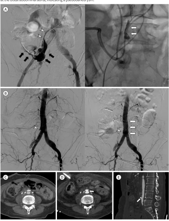
Fig. 1. A 62-year-old female with gross hematuria from both the ileal conduit and left nephrostomy tube af- ter a slip-down.
A. Aortography (left image) shows an arterioureteral fistula (arrowheads) and active contrast extravasation (arrows) along the left ureteral stent and ileal conduit. Superselective embolization of the fistula tract (right image) was performed using a glue-lipiodol mixture (arrows).
B. Aortography after glue embolization (left image) shows a small remnant fistula (arrowhead). A 23 mm × 49 mm self-expandable stent graft (right image, arrows) was deployed at the distal abdominal aorta with com- plete exclusion of the fistula (arrowhead).
C. Contrast-enhanced abdominal CT scan obtained three months before the slip-down demonstrates the intact anterior wall of the aorta (arrowhead) and an overlying ureteral stent.
D. Contrast-enhanced abdominal CT scan on admission shows subtle irregularity of the anterior wall of the aorta (arrow) at the level of the ureteroarterial crossing.
E. Sagittal reformatted contrast-enhanced CT scan reveals a corresponding small outpouching lesion (arrow) at the distal abdominal aorta, indicating a pseudoaneurysm.
A
B
C D E
rare condition, which reported in only 7 patients. In patients with urinary diversion, ureteroar- terial crossing takes place at a higher level than usual anatomy. Therefore, the arterial part of fistula could involve the proximal common iliac artery or even the distal abdominal aorta.
The AUF has been classified into primary and secondary types on the basis of etiology (4, 5).
The primary fistulas (15%) are related to the vascular abnormalities such as aneurysms, arte- riovenous malformations, or aberrant vessels. The secondary fistulas (85%) have been report- ed after prior pelvic interventions including pelvic surgery, combined with irradiation and with ureteral stent placement. Post-traumatic fistula is extremely rare subgroup with only one case report which describes a patient with a gunshot wound in the abdomen (7).
Gross hematuria is the dominating symptom of AUF, which can be intermittent or life- threatening massive with hypotension. Most patients experienced the first episode of hema- turia spontaneously and some patients during the change or insertion of ureteral stent (6).
However, hematuria provoked after blunt trauma has not been described before. There is no established explanation how blunt trauma affected AUF. The impact of trauma may disrupt already damaged ureter and arterial wall, promoting the formation of fistula. In the present report, recent trauma history and nonspecific CT finding raise a clinical suspicion of blunt renal injury and result in diagnostic delay. If noninvasive imaging workup failed to identify the cause of hematuria, diagnostic angiography can be helpful in patients with typical clini- cal triad of pelvic surgery, radiation and ureteral stenting (4).
Cross sectional imaging has a limited role in diagnosis of AUF, because of its low sensitivi- ty. Enhanced CT scan usually negative and the fistula tract is almost never identified. In a small subset of patients with pseudoaneurysm, enhancing lesion could be identified near the ureteroarterial crossing. In the present case, it was difficult to recognize the subtle irreg- ularity of aorta without retrospective careful review. The correct diagnosis could be made af- ter conventional angiography. Digital subtraction angiography remains the primary diagnos- tic tool, but its sensitivity is less than 50% (5, 8). Angiographic findings include direct extravasation into fistula, pseudoaneurysm and subtle irregularity or intimal defect (4). In cases of negative study, a provocative maneuver such as manipulation of ureteral stent could help visualization of extravasation (8). However, balloon tamponade should be prepared for subsequent massive hemorrhage.
A wide variety of treatment options have been suggested in the literature (6). Because the majority of fistulas are secondary to previous pelvic surgery and radiotherapy, open repair is often not feasible (9, 10). Currently, endovascular stent graft placement has become the treat- ment of choice for AUF with less morbidity and mortality (4, 6). Long-term follow-up data af- ter stent graft placement is limited due to its rare incidence. Previous study with mean fol- low-up of 15.5 months revealed possible complications including recurrent bleeding, lower extremity complications and stent graft complications, and recommended the use of antibi-
Author Contributions
Writing—original draft, all authors; and writing—review & editing, all authors.
Conflicts of Interest
The authors have no potential conflicts of interest to disclose.
REFERENCES
1. Moschcowitz AV. IX. Simultaneous ligation of both external iliac arteries for secondary hemorrhage. Ann Surg 1908;48:872-875
2. Dangle PP, Bahnson R, Patel A. Ureteral stent-related aortoureteric fistula: case report and literature re- view. Can Urol Assoc J 2009;3:E84-86
3. Krambeck AE, DiMarco DS, Gettman MT, Segura JW. Ureteroiliac artery fistula: diagnosis and treatment al- gorithm. Urology 2005;66:990-994
4. Pillai AK, Anderson ME, Reddick MA, Sutphin PD, Kalva SP. Ureteroarterial fistula: diagnosis and manage- ment. AJR Am J Roentgenol 2015;204:W592-W598
5. Madoff DC, Gupta S, Toombs BD, Skolkin MD, Charnsangavej C, Morello FA Jr, et al. Arterioureteral fistulas:
a clinical, diagnostic, and therapeutic dilemma. AJR Am J Roentgenol 2004;182:1241-1250
6. Van den Bergh RC, Moll FL, De Vries JP, Lock TM. Arterioureteral fistulas: unusual suspects-systematic re- view of 139 cases. Urology 2009;74:251-255
7. Dang C, Sullivan MJ. Traumatic arterio-ureteral fistula: “hematuria” without urine. J Trauma 1975;15:361- 362
8. Vandersteen DR, Saxon RR, Fuchs E, Keller FS, Taylor LM Jr, Barry JM. Diagnosis and management of ure- teroiliac artery fistula: value of provocative arteriography followed by common iliac artery embolization and extraanatomic arterial bypass grafting. J Urol 1997;158:754-758
9. Bergqvist D, Pärsson H, Sherif A. Arterio-ureteral fistula--a systematic review. Eur J Vasc Endovasc Surg 2001;22:191-196
10. Fox JA, Krambeck A, McPhail EF, Lightner D. Ureteroarterial fistula treatment with open surgery versus en- dovascular management: long-term outcomes. J Urol 2011;185:945-950
복부 대동맥에 발생한 동맥-요관 누공의 혈관 내 치료:
증례 보고와 문헌고찰
이형남 · 이웅희*
저자들은 복부 대동맥에 발생한 동맥-요관루를 혈관 내 접근을 통해 성공적으로 치료할 수 있었던 드문 증례를 보고하는 바이다. 동맥-요관루는 극히 드물지만, 사망률이 7~23%에 이 르는 치명적인 질환이다. 저혈량쇼크와 같은 치명적 합병증을 예방하기 위해서는 조기 진단 과 함께 즉각적인 치료가 필수적이다. 하지만 질환 자체가 희귀하고 민감도가 높은 검사 방 법이 없기 때문에, 진단을 위해서는 높은 수준의 임상적 의심이 반드시 필요하다. 복강 내 수 술, 방사선 치료 및 요관 스텐트의 장기 설치 등의 특징적인 과거력을 가진 환자에서 외상적 사건이 동맥-요관루 발생의 촉발 요인이 될 수 있다. 복부 대동맥에 발생한 동맥-요관루 환자 에서도 혈관 내 인조혈관 스텐트의 삽입은 효과적인 치료 방법이다.
순천향대학교 천안병원 영상의학과