Congenital Cytomegalovirus Infection of the Brain:
MR Imaging and Ultrasonographic Findings of Paraventricular Cysts
1Woo Mok Byun, M.D., Mi Soo Hwang, M.D.
Purpose: Although the neuroradiological findings of congenital cytomegalovirus (CMV) infection are well known, little has been reported concerning the imaging find- ings of paraventricular cysts occurring in patients with cytomegalovirus infection in- volving the brain. The purpose of this study is to describe the features of paraventricu- lar cysts observed at MRI and ultrasonography.
Materials and Methods: MR and ultrasonographic studies of ten patients with congeni- tal cytomegalovirus infections involving the brain were retrospectively reviewed.
Diagnosis was confirmed by positive culture of the virus in urine (n=4), the presence of CMV Ig G antibody (n=4), or positive CMV Ig M antibody (n=2), and on the basis of characteristic MR imaging findings. Initial MRI in all patients and initial ultrasonog- raphy in four of five with paraventricular cysts were performed. Three patients under- went follow-up MRI and ultrasonography for the evaluation of cystic change, and the size, location, bilaterality and morphology of the cysts were evaluated.
Results: Bilateral paraventricular cysts averaging 15 (range. 10-23) mm in size were found in five of the ten patients (50%). They were adjacent to the foramen of Monro in three cases, the occipital horn in one, and the temporal horn in one. MR imaging showed that the fluid content of all cysts was of similar signal intensity to cere- brospinal fluid (T1-WI, hypointense; T2-WI, hyperintense). The ultrasonographic find- ings varied: there was one pure cyst and one with a thick wall and septations, and two contained complex fluid. In three patients, follow up MRI and ultrasonography showed that the cysts disappeared after 4-23 months.
Conclusion: Although paraventricular cysts may appear at MRI to be purely cystic, ul- trasonography may indicate that their contents are more complex, or that septations are present.
Index words : Infant, newborn, central nervous system Brain, infection
Brain, MR Brain, US
1Department of Diagnostic Radiology, College of Medicine, Yeungnam University Received July 14, 2001 ; Accepted March 14, 2002
Address reprint requests to : Woo Mok Byun, M.D., Department of Diagnostic Radiology, College of Medicine, Yeungnam University, 317-1, Daemyung- dong, Nam-gu, Daegu 705-717, Korea.
Tel. 82-53-620-3046 Fax. 82-53-653-5484 E-mail: [email protected]
Cytomegalovirus (CMV), which causes transplacental infection, is a ubiquitous agent, and is the most common cause of congenital viral infections. In newborns, the re- ported incidence is 0.2-2.2%, and approximately 10%
of such cases are postnatally symptomatic (1-2). CMV infection has widespread manifestations, including those found in the central nervous system, namely mi- crocephaly, deafness, mental retardation, hypotonia, spastic quadriplegia, and varying degrees of perceptual, neurologic, psychomotor, and behavioral disturbances (3-4). Several reports have described the imaging find- ings of congenital CMV infection; these include periven- tricular calcification, ventriculomegaly, delayed myeli- nation, oligo/pachygyria, periventricular and subcortical signal intensity change, cerebellar atrophy, and par- aventricular cysts. The inflammatory process and necro- sis caused by congenital CMV infection involving the brain can lead to paraventricular cystic lesions.
Radiological studies have shown the presence of these in 36-60% of affected children (5-8).
Although a number of radiological findings have been reported in patients with congenital CMV infection, lit- tle has been written concerning the imaging findings of paraventricular cysts at MRI and ultrasonography. In this report, we describe the MRI and ultrasonographic findings of paraventricular cysts in patients with con- genital CMV infection.
Materials and Methods
MRI and ultrasonographic studies of ten patients with congenital CMV infections involving the brain were ret- rospectively reviewed. There were six females and four males, who at the time of initial cranial MR imaging studies were aged between 7 days and 23 months (mean, 5 months). Three of the infants were born pre- maturely and seven were born at term. All cases were diagnosed on the basis of characteristic clinical and imaging findings in conjunction with positive serologic
testing for CMV or culture of the virus from the urine of the child. Diagnosis was confirmed by positive culture of the virus (n=4), the presence of positive CMV Ig G antibody (n=4), or positive CMV Ig M antibody (n=2), and on the basis of typical MR findings. The diagnostic imaging findings were periventricular high signal inten- sity at T2-weigted imaging (n=8), cortical dysplasia (n=4), cerebellar atrophy (n=1) and noncommunicating hydrocephalus (n=6). The clinical signs for CMV infec- tion were microcephaly (n=6), thrombocytopenia (n=2), seizure (n=6), and sensory neural hearing loss (n=2).
For MR examinations, a 0.5T (Gyroscan T5-Ⅱ;
Philips, Netherlands) and a 1.5T unit (Vision; Siemens, Erlangen, Germany) were employed. Axial and coronal T1-weighted images (repetition time msec/ echo time msec: 400-600/25-30) were obtained before and after the administration of gadopentetate dimeglumine (Magnevist; Schering, Berlin, Germany; 0.1mmol per kilogram of body weight), and axial T2-weighted (2000- 2500/100-120) or turbo-T2-weighted images (3000- 4000/90-100) were obtained before the administration of gadopentetate dimeglumine. Four patients were scanned through the anterior fontanel using real-time ul- trasound (Ultramark 9; ATL, Bothell, Washington, U.S.A.) with a 7-MHz sector transducer. Initial MRI in all patients and initial ultrasonography in four with par- aventricular cysts were performed. Three patients un- derwent both second follow-up MR and ultrasonogra- phy for the evaluation of cystic change, and using these same modalities, one of the three was followed up a third time.
Diagnosis was established on the basis of MR imaging findings such as fluid signal intensity with a thin wall.
The size, location, bilaterality and morphology of the cysts were evaluated by MRI and ultrasonography, and the former was also used to determine whether wall en- hancement was present.
Table 1. Findings of Five Congenital CMV Patients with Paraventricular Cysts
Cases Ages Term (GA) Paraventricular cysts
Location Size (Cm) Disappearance
1 02 weeks Full (40w) Foramen of Monro 1.5 06 months
2 05 months Full (39w) Temporal horn 2.3 04 months
3 08 days Premature (36w) Occipital horn 1.5 23 months
4 10 days Premature (36w) Foramen of Monro 1.0 No follow up
5 07 days Full (39w) Foramen of Monro 1.3 No follow up
GA: Gestational age, w: weeks
Results
Bilateral paraventricular cysts averaging 15 (range, 10- 23) mm in size were found in five patients (50%). They were adjacent to the foramen of Monro in three cases, the occipital horn in one case, and the temporal horn in one case (see Table). MR imaging showed that the fluid content of all cysts was of similar signal intensity to cerebrospinal fluid (T1-WI, hypointense; T2-WI, hyper- intense) (Fig. 1A, B). For the four cysts examined ultra- sonographically, the findings varied: there was one pure cysts and one with a thick wall and septations, and two contained complex fluid (Fig. 1, 2, 3). The pure cyst was situated adjacent to the occipital horns, and the remain- ing three, located adjacent to the foramen of Monro, had a sonographic appearance which mimicked hematoma of the germinal matrix (Fig. 3A). Follow up MR imaging and ultrasonography indicated that the cysts of the three patients disappeared after 4, 6, and 23 months respec-
tively. Two cysts adjacent to the foramen of Monro dis- appeared after four and six months, respectively, while the one adjacent to the occipital horn became smaller af- ter four months and disappeared after 23 months (Fig.
1E). At follow-up MR imaging, we found no evidence of hematoma in cysts adjacent to the foramen of Monro.
Contrast T1-weighted images revealed that in all cases, the cystic wall was unenhanced. At the time a cyst dis- appeared, initial and follow-up MRI revealed no abnor- mal signal intensity caused by gliosis in surrounding brain parenchyma.
Discussion
In most infants with intrauterine CMV infection, clini- cal manifestations are not apparent at birth, while about 10% of neonates exhibit findings suggestive of congeni- tal infection (9-10). The diagnosis of CMV infection ul- timately rests on the recovery of the infectious virus from tissue or body fluid. Demonstration of seroconver-
D E
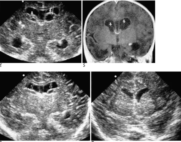
Fig. 1. Premature 8-day-old patient with delayed myelinaiton. Paraven- tricular cysts with clear content.
Bilateral cysts adjacent to occipital horns are seen. Cysts reveal low signal intensity (white arrows) at T1-weighted image (A) and high signal intensity (black arrows) at T2-weighted image (B). Ultrasonography (C) shows clear cysts without definite solid compo- nents (double white arrows). Follow up T1-weighted image (D) after four months shows significant reduction (white arrows) in the size of the par- aventricular cysts. Paraventricular cysts disappeared completely at T1- weighted image after 23 months (E).
A B C
sion or a fourfold change in CMV antibody titers strong- ly suggests recent CMV infection, while other tech- niques such as immunofluorescence, and light and elec- tron microscopic examination of tissue, may have diag- nostic utility in selected cases (11). The neuroradiologi- cal imaging of infants with congenital CMV infection is important, and several reports have described the find- ings. It is well known that intracerebral calcification is the most common abnormality revealed by neuroradio- logical imaging in infants with congenital CMV infec- tion: it has been found in up to 40% of affected children, either at plain radiography of the skull, or at CT (6, 12).
In our study, however, it was detected by neither ultra- sonography nor MRI. Radiological studies have demon-
strated the presence of paraventricular cysts, not un- commonly in the brain, in 36-60% of CMV-infected children. (5, 6, 8). Boesch et al. (6) reported that cranial MR imaging of ten children with congenital cy- tomegalovirus infection showed a dilated lateral ventri- cle (n=10) and subarachnoid space (n=8), oligo/pachy- gyria (n=8), delayed/ pathological myelination (n=7), paraventricular cysts, (n=6) and intracerebral calcifica- tion (n=1). They mentioned that six patients had charac- teristic paraventricular cysts; these were bilateral and adjacent to the occipital horns of the lateral ventricles.
Barkovich et al. (8) reported that in MRI and CT studies of 11 patients with congenital CMV infection involving the brain, four had paraventricular cysts adjacent to the
A B
C D
Fig. 2. Full term 2-week-old patient with delayed myelination. Paraventricular cysts with thick wall and septations.
Coronal (A) ultrasonographic scan shows cysts with thick wall (short arrows) and septations (long arrow) adjacent to foramen of Monro. Paraventricular cyst shows low signal intensity (arrows) similar to cerebrospinal fluid (CSF) signal intensity at postcontrast coronal T1-weighted image. No enhancement of the cyst wall is seen (B). Follow-up ultrasonography (C) after two months shows reduction (open arrow) in size of the paraventricular cysts. Noncommunicating hydrocephalus is seen on coronal ultrasonography (B, C). Bilateral cysts disappeared completely on follow-up ultrasonography after four months (D).
temporal horns. Butt et al. (5) stated that paraventricular cysts found in two of the four patients with congenital cytomegalovirus infection were observed at ultrasound examination. The prominent periventricular location of viral damage has been attributed to the predilection of CMV to infect rapidly-growing subependymal or germi- nal matrix cells (11). The necrotizing inflammatory process in CMV infection involving the brain reads to the growth of paraventricular or subependymal cysts (5). For the former, location and bilaterality may there- fore be characteristic but nonspecific imaging features of congenital CMV infection involving the brain.
Our results showed that although all paraventricular cysts were bilateral and had a pure cyst-like appearance at MR imaging (T1-WI, hypointense; T2-WI, hyperin- tense), ultrasonographic examination revealed different and various patterns. The ultrasonographic findings of paraventricular cysts included a pure cysts, one with a
thick wall and sepations, and two with complex fluid.
Three cysts with septation or complex fluid contents were situated adjacent to the foramen of Monro and their sonographic appearance mimicked hematoma of the germinal matrix. In our study, differentiation at ul- trasonography between germinal matrix hemorrhage and cysts adjacent to the foramen of Monro was diffi- cult, though at MRI, because a hematoma demonstrates varying signal intensities at different stages of resolu- tion, differentiation between a hematoma and a cyst was not. At MRI, cysts adjacent to the foramen of Monro show no signal intensities characteristic of a hematoma.
Sofer et al. (13) reported the occurrence of congenital CMV periventriculitis mimicking the classical sono- graphic picture of periventricular hemorrhage. They stated that sonographic examination of a CMV-infected brain revealed periventricular echogenic clumps, main-
A B
C D
Fig. 3. Premature 10-day-old patient with noncommunicating hydro- cephalus. Paraventricular cysts with complicated contents.
Coronal (A) and right sagittal (B) ultra- sonographic scan reveals echogenic complicated contents in cyst lumen (A:
black arrows, B: white arrow) adjacent to both foramen of Monro. Posterior enhancement below paraventricular cysts is noted. Hypointense cysts (open arrows) are noted on contrast enhanced coronal T1-weighted image No en- hancement of the cyst wall is seen (C).
Axial T2-weighted image (D) shows hy- perintense paraventricular cysts (ar- rows) similar to CSF intensity.
ly in the occipital region of the brain, that were isoe- choic with the choroid plexus. They also mentioned that at necropsy, examination of the brain revealed irregular- ity of the walls of the lateral ventricles accompanied by small irregular patches; at microscopic examination, these patches, containing CMV inclusion bodies, were shown to be foci of periventricular necrotizing en- cephalitis. Most reports have mentioned that the forma- tion of paraventricular cysts or echogenic materials con- current with congenital CMV infection involving the brain results from necrosis and an inflammatory process. Barkovich et al. (8) stated that the significance of paraventricular cysts is uncertain and that they are probably the result of localized tissue destruction. We suggest that necrotizing and inflammatory paraventricu- lar cysts may be either purely cystic or complex, mimic- king hematoma at ultrasonography. Complex contents in paraventricular cysts may be clumps or sludge caused by focal necrosis and inflammatory reaction.
Recognition of the various appearances of these cysts at ultrasonography may be important for proper diagnosis of congenital CMV infection in the brain.
Boesch et al (6) reported that in one of six patients with paraventricular cysts, a second MRI examination was performed seven months later, and at that time the cystic structure still persisted. In our study, three pa- tients underwent follow-up MR and ultrasonography, and it was found that the cysts disappeared after four to 23 months.
Paraventricular cysts occur in other congenital viral infections of the brain. In newborns, their most com- mon proven causes are rubella and CMV infections, and Beltinger et al (14) stated that in two newborns with con- genital rubella syndrome, cranial ultrasonography demonstrated bilateral cystic lesions in the subependy- mal germinal matrix. On the basis of our results and this report, we believe that congenital viral infections should be included in the differential diagnosis of neonates with various or characteristic paraventricular cysts. The sonographic detection of such cysts should prompt an intensive search for congenital viral infections.
In conclusion, we found that paraventricular cysts were most commonly located adjacent to the foramen of Monro. Although they may appear at MRI to be purely cystic in nature, ultrasonography may reveal that their
contents are complex, or that septations are present. The recognition of these characteristics at neuroradiological imaging may be important for the proper diagnosis of congenital CMV infection. Further studies of paraven- tricular cysts in more patients are needed.
Acknowledgements
We express our appreciation for support by neuro- science study groups at Yeungnam University College of Medicine
References
1. Stagno S, Pass RF, Dworsky ME, Alford CA. Maternal cy- tomegalovirus infection and perinatal transmission. Clin Obstet Gynecol1982;25:563-576
2. Stagno S, Pass RF, Dworsky ME, Britt WJ, Alford CA. Congenital and perinatal cytomegalovirus infections: Clinical characteristics and pathogenic factors. Birth Defects 1984;20:65-85
3. Birnbaum G, Lynch JI, Margileth AM, et al. Cytomegalovirus in- fection in newborn infants. J Pediatr 1969;75:789-795
4. Pass RF, Stagno S, Myers GJ, Alford CA. Outcome of symptomatic congenital cytomegalovirus infection: Result of long term longitu- dinal follow- up. Pediatrics 1980;66:758-762
5. Butt W, Mackay RJ, de Crespigny LC, Murton LJ, Roy RND.
Intracranial lesions of congenital cytomegalovirus infection detect- ed by ultrasound scanning. Pediatrics 1984;73:611-614
6. Boesch C, Issakainen J, Kewitz G, Kikinis R, Martin E, Boltshauser E. Magnetic resonance imaging of the brain in congenital cy- tomegalovirus infection. Pediatr Radiol 1989;19:91-93
7. Sugita K, Ando M, Makino M, Takanashi J, Fujimoto N, Niimi H.
Magnetic resonance imaging of the brain in congenital rubella virus and cytomegalovirus infections. Neuroradiology 1991;33: 239- 242
8. Barkovich AJ, Lindan CE. Congenital cytomegalovirus infection of the brain: Imaging analysis and embryologic considerations. AJNR Am J Neuroradiol 1994;15:703-715
9. Boppana SB, Pass RF, Britt WJ, Stagno S, Alford CA. Symptomatic congenital cytomegalovirus infection: Neonatal morbidity and mortality. Pediatr Infect Dis J 1992;11:93-99
10. Stagno S, Pass RF, Cloud G, et al. Primary cytomegalovirus infec- tion in pregnancy. Incidence, transmission to fetus, and clinical outcome. JAMA 1986;256:1904-1908
11. Bale JF. Human cytomegalovirus infection and disorders of the nervous system. Acta Neurol 1984;41:310-320
12. Bale JF, Bray PF, Bell WE. Neuroradiographic abnormalities in congenital cytomegalovirus infection. Pediatr Neurol 1985;1:42-47 13. Sofer S, Maor E, Barki Y. Cytomegalic virus periventriculitis: A
sonographic picture mimicking ventricular hemorrhage. J Clin Ultrasound 1985;13:574-576
14. Beltinger C, Saule H. Sonography of subependymal cysts in con- genital rubella syndrome. Eur J Pediatr 1988;148:206-207
대한방사선의학회지 2002;47:85-91
뇌의 선천성 Cytomegalovirus 감염:
뇌실주위낭종의 자기공명영상 및 초음파소견1
1영남대학교 의과대학 진단방사선과 변 우 목・황 미 수
목적:선천성 cytomegalovirus (CMV) 감염 의 신경방사선학적 소견은 잘 알려져 있지만 뇌를 침범하는 선천성 CMV 감염을 가진 환자에서 발생되는 뇌실주위 낭종의 영상소견에 대한 보고는 흔하지 않다. 이 연구의 목적은 MR 과 초음 파영상에서 나타난 뇌실주위 낭종의 형태에 대해 보고하고 자 한다.
대상과 방법:뇌를 침범한 선천성 CMV 감염을 가진 환자 10명의 MR과 초음파 소견을 후향적으로 분석하였다. 소변의
바이러스 배양에서 양성인 4명, CMV IgM 양성인 4명과 IgG 양성인 두 명에서 전형적인 자기공명영상소견으로 진단하 였다. 초기 자기공명영상은 10명에서 초기초음파는 뇌실주위낭종을 가진 5명중 4명에서 시행되었다. 뇌실주위낭종의 변화를 평가하기위해 추적 자기공명영상과 초음파소견은 3명의 환자에서 시행되었다. 뇌실주위낭종의 크기, 위치, 양 측성, 그리고 형태를 평가하였다.
결과:뇌실주위낭종은 10명의 환자 중 5명 (50%) 에서 발견되었다. 모든 뇌실주위낭종은 양측성이었다. 뇌실주위낭종 의 크기는 10-23 mm (평균 15 mm) 였다. 뇌실주위낭종의 위치는 Monro공 주위에 3예, 후두각주위 1예, 그리고 측 두각주위 1예였다. MR영상에서 뇌실주위낭종의 액체는 T1 강조영상에서는 저신호강도, T2강조영상에서는 고신호강 도로 뇌척수액과 비슷하였다. 초음파 검사를 시행하여 뇌실주위낭종의 소견은 다양하였는데 순수한 낭종은 1예, 두터 운 낭종벽과 격막을 가진 낭종은 1예, 그리고 복잡한 내용물을 가진 낭종들은 2예였다. 3증례에서 추적 MR과 초음파 검사를 시행하였고 뇌실주위낭종은 모두 4개월에서 23개월 후에 사라졌다.
결론:MR에서 뇌실주위낭종은 순수한 낭종으로 보이지만 초음파 소견에서는 복잡한 내용물(complicated contents) 혹 은 격막이 나타날 수 있다.