81
Copyright © 2019 The Korean Society of Fisheries and Aquatic Science pISSN:0374-8111, eISSN:2287-8815
서 론
알츠하이머병
(Alzheimer’s disease, AD),
프리온질환(prion diseases),
파킨슨병(Parkinson’s disease),
근위축성측색경화 증(amyotrophic lateral sclerosis)
과같은주요퇴행성신경질환(neurodegenerative disorders)
의공통적인병리학적특성은신 경세포의소실(neuronal loss)
및활성화된미세아교세포(acti- vated microglia)
가존재한다는것이다(Soto, 2003).
중추신경계에상주하는대식세포
(macrophages)
인 미세아교세포의활성화는신경염증
(neuroinflammation)
의주요특성이고,
그분 포가 퇴행성신경질환의 병리학적 병변부위에서 관찰됨으로써이들질환의발병에관여하는것으로알려져있다
(McGeer
et al., 2016; Molteni and Rossetti, 2017; Schain and Kreisl, 2017).
미세아교세포는내인성및외인성자극에의해활성화되면
interleukin (IL)-1β, IL-6, tumor necrosis factor-α (TNF-α)
와 같은염증촉진성cyokines
과nitric oxide
를포함하는유리래디 칼(free radical)
을생성함으로써염증반응에관여한다(Molteni and Rossetti, 2017; Schain and Kreisl, 2017).
이러한염증반응은손상및자극에대해숙주
(host)
를보호하기위한중요한방어기전이고일시적인반응이다
.
그러나그원인이완전히제 거되지않으면지속적인염증응답이생길수있고,
그러한만LPS로 유도한 마우스의 급성신경염증에 대한 톱니모자반(Sargassum serratifolium) 추출물의 효과
최민우·김형락·이형곤
1·김재일*
부경대학교 식품영양학과, 1산안토니오텍사스주립대학교 생물학과
Effect of a Sargassum serratifolium Extract on Neuroinflammation Induced by Lipopolysaccharides in Mice
Min-Woo Choi, Hyeung-Rak Kim, Hyoung-Gon Lee1 and Jae-Il Kim*
Department of Food Science and Nutrition, Pukyong National University, Busan 48513, Korea
1Department of Biology, University of Texas at San Antonio, Texas 78249, U.S.A.
The common hallmark of several neurodegenerative disorders, including Alzheimer’s disease (AD), is the presence of chronic neuroinflammation, which contributes to the loss of neuronal structure and function. This study investi- gated the effects of an ethanolic extract of Sargassum serratifolium (SSE) in a lipopolysaccharides (LPS)-induced murine neuroinflammation model. Mice were administered SSE (100 mg/kg body weight) or vehicle for 5 days by oral gavage, and then treated with LPS or saline by intraperitoneal injection. Thereafter, the brain tissues were col- lected, and the expression of pro-inflammatory cytokines was analyzed by quantitative real-time RT-PCR. There was a marked increase in the spleen weight index in the LPS-treated groups, which indicated the induction of acute systemic inflammation. Based on significant increases in the levels of IL-1 and IL-6 expression, the induction of neu- roinflammation was also evident in the cortex and hippocampus of the LPS-treated groups. The overall expression of IL-1 and IL-6 was decreased slightly by SSE administration, compared with the LPS group, and a marked change in IL-1 was observed in the cortex of the SSE-treated (SSE/LPS) group. These results suggest that SSE has potential as an anti-neuroinflammatory nutraceutical.
Key words: Neurodegenerative disorders, Neuroinflammation, Brown seaweeds, Sargassum serratifolium , Lipopoly- saccharides
*Corresponding author: Tel: +82. 51. 629. 5849 Fax: +82. 51. 629. 5842 E-mail address: [email protected]
This is an Open Access article distributed under the terms of the Creative Commons Attribution Non-Commercial Licens (http://creativecommons.org/licenses/by-nc/3.0/) which permits unrestricted non-commercial use, distribution, and reproduction in any medium, provided the original work is properly cited.
Received 15 February 2019; Revised 21 February 2019; Accepted 21 February 2019 저자 직위: 최민우(대학원생), 김형락(교수), 이형곤(교수), 김재일(교수) https://doi.org/10.5657/KFAS.2019.0081
Korean J Fish Aquat Sci 52(1), 81-86, February 2019
성적인염증이지속될경우숙주조직의손상이축적될수있 다
(Molteni and Rossetti, 2017; Schain and Kreisl, 2017).
상 기의주요 퇴행성신경질환들의경우 비정상적인단백질응집 물(protein aggregates)
이관찰되는것이그특징이고,
이러한 비정상적인단백질응집물들은뇌조직의만성적인염증반응을 유발함으로써신경세포소실을유발하는것으로알려져있다(Soto, 2003; Schain and Kreisl, 2017).
실험적으로in vitro
및in vivo
동물모델에서이들단백질응집물에의해신경염증이유발되는것이알려져있고
,
알츠하이머병및프리온질환과같 은환자뇌조직의병변부위에서이들단백질응집물과염증촉 진성매개체들이함께분포함으로써신경염증의병인적역할 이중요시되고있다(Soto, 2003; Molteni and Rossetti, 2017;
Schain and Kreisl, 2017).
따라서현재까지뚜렷한치료방법이 없는주요퇴행성신경질환의발병을예방또는지연시키기위 해서는과도한만성적인신경염증을억제할수있는방법의개 발이시급하다고할수있다.
모자반속
(Sargassum)
은 모자반과(Sargassace)
에 속하는갈 조류의하나로한국과 중국에서 식품및 전통약물로써 오랫 동안사용되어왔고, meroterpenoids, phlorotannins, fucoxan- thins, fucosterols
과같은다양한생리활성화합물을함유하고 있는것으로알려져있다(Liu et al., 2012).
톱니모자반(Sargas- sum serratifolium)
은한국과일본의연안에광범위하게서식하 고있고,
형태학적으로줄기가장자리에끝이가시형태를가지 고있다는것이특징이다(Oak and Lee, 2006).
최근톱니모자 반의추출물및함유화합물들을이용한in vivo
및in vitro
연 구에서다양한생리활성들이보고되었는데,
항암(anti-cancer) (Kang et al., 2015),
미백(hypopigmentation) (Azam et al., 2018),
항산화(anti-oxidant) (Lim et al., 2018; 2019),
항염증(anti-inflammatory) (Gwon et al., 2015; 2017; 2018; Joung et al., 2015; 2017),
간보호(hepatoprotective) (Lim et al., 2018),
신경보호(neuroprotective) (Oh et al., 2016; Choi et al., 2017;
Seong et al., 2017)
와같은활성들이확인되었다.
이러한톱니 모자반에는사가크로메놀(Sargachromenol, SCM),
사가퀴노 익산(Sargaquinoic acid, SQA),
사가하이드로퀴노익산(Sar- gahydroquinoic acid, SHQA)
과같은meroterpenoids
가고농 도로함유되어 있고(Gwon et al., 2015),
이들세화합물들의 공통적인특성은모두항산화(Lim et al., 2018; 2019)
및항염 증(Gwon et al., 2015; 2017; 2018; Joung et al., 2015; 2017)
활성을나타냄으로써톱니모자반의 주요활성성분으로 확인 되었다.
퇴행성신경질환과관련된연구로는베타-
아밀로이드(β-amyloid)
단백질생성억제에대한활성이보고되어있지만(Choi et al., 2017; Seong et al., 2017),
신경염증과관련된활 성은불분명하다.
따라서,
본연구에서는마우스에lipopolysac- charides (LPS)
를주입하여급성신경염증을유발하고,
이들동 물에톱니모자반주정추출물(ethanolic extract of S. serratifo- lium, SSE)
을투여하여그억제효과를구명하고자하였다.
재료 및 방법
실험재료
Poly (ethylene glycol)-400 (PEG-400), phosphate-buffered saline (PBS), 2,2,2-tribromoethanol (avertin), LPS
는Sigma- Aldrich (St Louis, MO, USA)
에서구매하였고, dimethyl sulf- oxide (DMSO)
는Life Sciences (Tewksbury, MA, USA)
에 서 구매하였다. RNA
추출을위한TRIzol
시약은Invitrogen (Carlsbad, CA, USA)
에서, tert-amyl alcohol (2-methyl-2-bu- tanol)
은Fisher Scientific (Pittsburgh, PA, USA)
에서각각구 매하였다, High Capacity cDNA Reverse Transcription Kits and RNase Inhibitor
와PowerUp™ SYBR™ Green Master Mix
는Applied Biosystems (Beverly, MA, USA)
에서구매하 였다.
톱니모자반 주정추출물(SSE) 시료의 제조
톱니모자반
(S. serratifolium)
은 이전의 보고(Gwon et al., 2015; Joung et al., 2015; Choi et al., 2017)
의방법에따라처 리하였다. 2016
년5
월에부산기장에서채취하였고,
종의확인 은부경대학교생태공학과최창근교수가수행하였다.
톱니모 자반은자연건조한후분쇄하였고,
분말50 g
에10
배양의주 정(95%
에탄올)
을넣고70℃
에서4
시간2
회반복하여환류냉 각추출하였다.
이후진공회전농축기로40℃
에서주정을제거 한후,
추출된잔사로추출물을얻었다.
이렇게얻은톱니모자 반주정추출물(SSE)
은사용전까지-20℃
냉동고에보관하였 다.
이전연구들에서SSE
에함유된주요성분은사가하이드로 퀴노익산(sargahydroquinoic acid, SHQA),
사가퀴노익산(sar- gaquinoic acid, SQA),
사가크로메놀(sargachromenol, SCM)
로 밝혀졌으며,
이들화합물은주정추출물의45-48%
를차지하고그중에서
SHQA
의함량이가장높은것으로확인되었다(Joung et al., 2017; Lim et al., 2018).
동물실험, 경구투여, LPS에 의한 급성신경염증의 유도
실험동물은12
주령의암컷C57BL/6J
마우스(9
마리)
를Jack- son Laboratory (Bar Harbor, ME, USA)
에서구입하였다.
일주 일의적응기간뒤3
마리씩무작위로세그룹으로나누고,
각각Control, LPS
군, SSE/LPS
군으로할당하였다.
SSE
는DMSO
에녹인후vehicle (25% DMSO/75% PEG-
400)
에최종농도를맞추어서경구투여시료를제조하였고(Ge-
renu et al., 2015), SSE/LPS
군을대상으로체중kg
당100 mg
의농도로5
일간경구투여(oral gavage)
하였다.
이기간동안Control
및LPS
군은동일한양의vehicle
을경구투여하였다.
LPS
에의한급성신경염증은Jo et al. (2017)
의방법에따라5
일째최종투여한이후2
시간뒤에LPS (5 mg/kg body weight)
를LPS
와SSE/LPS
군에복강투여하여유도하였다. Control
의 경우동일한양의PBS
를투여하였다. 4
시간이후avertin
용액(250-500 mg/kg body weight)
으로 안락사시키고 뇌조직을 적출하여대뇌피질(cerebral cortex)
과해마(hippocampus)
부위를분리해서
-80℃
에보관하였다.
또한이들마우스의비장(spleen)
을적출하여무게를측정하고,
체중대비비장의무게(spleen weight index)
를계산하였다.
이상의동물실험은샌안 토니오텍사스주립대학교(University of Texas at San Antonio)
의 동물실험윤리위원회(Institutional Animal Care and Use Committee, IACUC)
의규정에따라수행하였다.
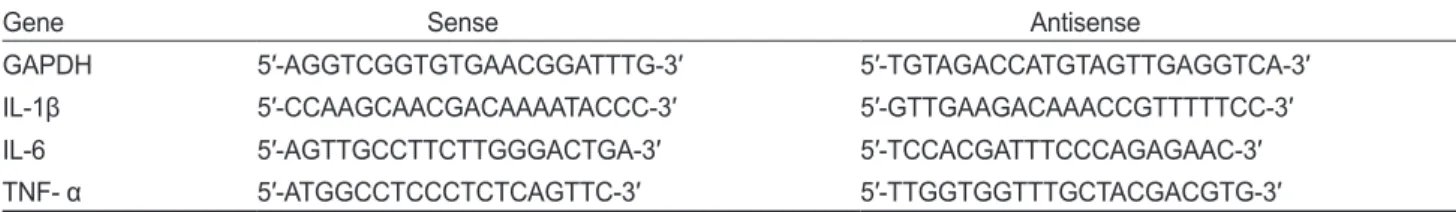
Quantitative Real-Time RT-PCR에 의한 염증성 cytokine 유전자 발현의 분석
TRIzol
용액(Invitrogen, Carlsbad, CA, USA)
을이용하여대 뇌피질및해마조직으로부터total RNA
를추출하였다. Total RNA (400 ng)
을사용하여high capacity cDNA reverse tran- scription kits (Applied Biosystems, Beverly, MA, USA)
로제 조사의방법에따라cDNA
를합성하였다.
합성된cDNA (10 ng)
를PowerUp™ SYBR™ green master mix (Applied Bio- systems, Beverly, MA, USA),
표적유전자특이적인primers
를 사용하여real-time PCR
을수행하였다.
유전자발현양의비교 는상대적인비교를위해ΔΔCt
방법을이용하였으며(Choi et al., 2017), real-time PCR
에이용된각primers
의염기서열은Table 1
에나타내었다.
통계 처리
분석결과는평균값및표준편차
(mean±SD)
를계산하여나 타내었고,
실험군간의유의성검증은Student’s t-test
로검증 하였다.
결과 및 고찰
본 연구에서는 신경염증에 대한 톱니모자반 주정추출물
(SSE)
의효과를알아보기위해서마우스에LPS
를투여하여급 성신경염증을유발한모델을이용하였다.
이러한LPS
투여에 의한신경염증모델은약물및천연물, nutraceutical
을포함하 여다양한화합물들의생리활성및신호전달기전구명을위한 모델로서널리이용되고있다(Catorce and Gevorkian, 2016).
LPS
투여에의해미세아교세포및 성상아교세포(astrocytes)
의활성화뿐만아니라cyclooxygenase-2 (COX-2), inducible nitric oxide synthase (iNOS)
와 같은염증매개효소및염증촉진성
cytokines
들의 발현이 뇌에서 유도되는 것으로 알려져있다
(Qin et al., 2007; Erickson and Banks, 2011; Jo et al.,
2017).
이외에LPS
를처리한알츠하이머병동물모델에서세포내
amyloid precursor protein (APP)
및β-amyloid,
과인산 화된(hyperphosphorylated) tau
단백질의축적이증가할뿐만 아니라기억력결핍이악화되는것이확인되었다(Sheng et al., 2003; Kitazawa et al., 2005). APP, β-amyloid
그리고hyper- phosphorylated tau
는알츠하이머병의환자의뇌조직에서관 찰되는병리학적인특징들이다(McGeer et al., 2016).
따라서LPS-
투여에의한신경염증모델은퇴행성신경질환예방물질의 활성을분석하기위한좋은모델이라고할수있다.
먼저
SSE
를5
일동안경구투여하였고마지막날LPS
를투 여하여신경염증을유도하였다.
각실험군의체중변화및비장 의무게를Table 2
에나타내었다.
세군모두투여첫날과비교 하여5
일째1 g
내외의체중증가는있었으나유의적인변화는Table 1. Primers used for quantitative real-time RT-PCR analysis
Gene Sense Antisense
GAPDH 5′-AGGTCGGTGTGAACGGATTTG-3′ 5′-TGTAGACCATGTAGTTGAGGTCA-3′
IL-1β 5′-CCAAGCAACGACAAAATACCC-3′ 5′-GTTGAAGACAAACCGTTTTTCC-3′
IL-6 5′-AGTTGCCTTCTTGGGACTGA-3′ 5′-TCCACGATTTCCCAGAGAAC-3′
TNF- α 5′-ATGGCCTCCCTCTCAGTTC-3′ 5′-TTGGTGGTTTGCTACGACGTG-3′
GAPDH, glyceraldehyde 3-phosphate dehydrogenase; IL-1β, interleukin-1β; IL-6, interleukin-6; TNF-α, tumor necrosis factors-α.
Table 2. Body weight change and spleen weight index in C57BL/6J mice treated with or without ethanolic extract of Sargassum serratifo- lium (SSE) followed by lipopolysaccharides (LPS)
Groups Body weight (g)
Spleen weight (g) Spleen weight index
Day 1 Day 5
Control 20.0±1.0 21.1±1. 2 0.067±0.006 3.17±0.17
LPS 19.8±1.6 20.6±1.1 0.084±0.009* 4.11±0.66*
SSE/LPS 19.3±1.4 19.5±1.4 0.089±0.006** 4.44±0.26**
Data are expressed as mean±SD (n=3). SSE or vehicle only was administered by oral gavage in mice for 5 days. On day 5, the mice were sacrificed after 4 h of intraperitoneal injection of LPS (5 mg/kg body weight) or PBS. The spleens from mice were isolated and weighed, and spleen weight index were calculated as organ weight (mg) per gram (g) of mouse body weight. *P<0.05 and **P<0.01 compared to control group.
없었고
,
또한vehicle
만을투여한대조군및LPS
군과비교하여SSE
투여군(SSE/LPS)
의체중도뚜렷한차이가없었다. LPS
를투여했을때 급성전신염증의유도는비장중량지수(spleen weight index)
를비교함으로써분석할수있다(Li et al., 2016;
Wang et al., 2017). Table 2
에나타낸것과같이비장(spleen)
의무게뿐만아니라비장중량지수가LPS
처리에의해현저하 게증가된것이확인되었다.
이는비장백혈구의급격한증가를나타내는것으로
, LPS
투여에의해급격한전신염증이유도됨을의미하는것이다
.
앞에서언급했듯이
LPS
투여에의한신경염증은이전여러 보고들에서확인되었다(Qin et al., 2007; Erickson and Banks, 2011, Jo et al., 2017).
본실험에서는퇴행성신경변화에취약 한뇌조직인대뇌피질(cortex)
과해마(hippocampuse)
영역에 서염증촉진성cytokines
의발현수준을real-time qunatitative RT-PCR
방법으로비교분석하였고그결과는Table 3
에나타 내었다.
두뇌조직모두에서LPS
투여에의해IL-1β
및IL-6
의발현이현저하게증가하였고(P<0.05-P<0.01),
그증가폭은IL-6
에서더큰것으로나타났다.
이들의cytokines
의발현수준 은SSE
투여(SSE/LPS
군)
에의해감소하는경향을보였고,
대뇌피질의경우
IL-1β
발현양이유의적으로감소를확인할수있었다
(P<0.05).
이러한결과에서SSE
에의한신경염증의억제 효과를확인할수있었다.
LPS
투여에의한신경염증유발모델은다양한형태로이루어 지고있다. LPS
투여농도및횟수,
마우스의종류, LPS
투여이 후동물을희생시킬때가지의시간등여러형태의조합이있고,
각각의실험조건에따라신경염증의결과는조금씩차이가있 는것으로알려져있다(Catorce and Gevorkian, 2016).
본연구 와동일한조건하에서수행된연구(Jo et al., 2017)
에서, LPS
투여이후의시간에따른염증성cytokines
의발현수준의변화 를분석하였을때, IL-1β
및IL-6
두가지모두2
시간째에가장 높게발현되고,
이후6
시간째에는점차감소하여12
시간이후부터는검출되지않는것으로확인되었다
.
본연구에서는LPS
투여이후4
시간째에뇌조직을분리하여분석하였다.
이시간대는뇌조직의급격한
cyokines
발현증가가계속높게유지되는시간이라고볼수있고
,
따라서SSE
의극적인효과를확인하 기는어려운조건일수도있었을것으로판단된다.
향후연구에 서는보다다양한시간대별발현의차이를비교분석하는연구 를수행하는것이필요할것으로생각된다.
주요퇴행성신경질환의공통적인특성은뇌조직에단백질응 집물이존재한다는것이다
(Soto C, 2003; Schain and Kreisl, 2017).
알츠하이머병의경우β-amyloid,
프리온질환의경우비 정상적인prion protein,
파킨슨병의경우α-synuclein
과같은 단백질로이루어진비정상적인응집물이관찰되고,
이러한특 성에서각질환들의발병의주요원인물질로알려져있다.
이 들단백질응집들은뇌조직에서신경염증을유발하는원인물질 로작용할수있고,
지속적으로축적되어만성적인염증을유 발함으로써신경세포소실에영향을미칠수있다(Soto, 2003;
Schain and Kreisl, 2017).
따라서본연구에서확인된SSE
의항 신경염증효과는주요퇴행성신경질환의예방혹은치료를위한 천연해조류성분의이용가능성을제시하는기초자료로이용될 수있을것이다.
사 사
이논문은부경대학교자율창의학술연구비
(2017
년)
에의하 여연구되었음.
References
Azam MS, Kwon M, Choi J and Kim HR. 2018. Sargaqui- noic acid ameliorates hyperpigmentation through cAMP and ERK-mediated downregulation of MITF in α-MSH- stimulated B16F10 cells. Biomed Pharmacother 104, 582- 589. https://doi.org/10.1016/j.biopha.2018.05.083.
Table 3. Gene expression profile of pro-inflammatory cytokines in the brains of C57BL/6J mice treated with or without ethanolic extract of Sargassum serratifolium (SSE) followed by lipopolysaccharides (LPS)
Brain regions Groups Genes
IL-1β IL-6 TNF-α
Cortex
Control 1.04±0.39 1.00±0.12 ND1
LPS 14.30±2.48** 76.88±26.26* ND
SSE/LPS 7.20±3.41*a 47.21±18.74* ND
Hippocampus
Control 1.02±0.30 1.05±0.39 ND
LPS 19.53±7.96* 66.19±9.03** ND
SSE/LPS 12.20±4.09* 45.97±25.59* ND
Gene expression levels were determined by reverse transcription followed by real-time PCR and were normalized with the housekeeping gene glyceraldehyde 3-phosphate dehydrogenase (GAPDH). The transcription levels were analyzed after 4 h of intraperitoneal injection of LPS (5 mg/kg body weight) or PBS. Data (mean±SD, n=3) are expressed relative to control group. IL-1β, interleukin-1β; IL-6, interleukin-6;
TNF-α, tumor necrosis factor-α. 1ND, not detected. *P<0.05 and **P<0.01 compared to control group; aP<0.05 compared
Catorce MN and Gevorkian G. 2016. LPS-induced mu- rine neuroinflammation model: Main features and suit- ability for pre-clinical assessment of nutraceuticals. Curr Neuropharmacol 14, 155-164. https://doi.org/10.2174/1570 159X14666151204122017.
Choi MW, Jung CG, Kim HR and Kim JI. 2017. Effect of Sar-
gassum serratifolium extracts on β-amyloid production. Ko-
rean J Fish Aquat Sci 50, 085-091. https://doi.org/10.5657/KFAS.2017.0085.
Erickson MA and Banks WA. 2011. Cytokine and chemokine responses in serum and brain after single and repeated in- jections of lipopolysaccharide: multiplex quantification with path analysis. Brain Behav Immun 25, 1637-1648. https://
doi.org/10.1016/j.bbi.2011.06.006.
Gerenu G, Liu K, Chojnacki JE, Saathoff JM, Martínez-Martín P, Perry G, Zhu X, Lee HG and Zhang S. 2015. Curcumin/
melatonin hybrid 5-(4-hydroxy-phenyl)-3-oxo-pentanoic acid [2-(5-methoxy-1H-indol-3-yl)-ethyl]-amide amelio- rates AD-like pathology in the APP/PS1 mouse model.
ACS Chem Neurosci 6, 1393-1399. https://doi.org/10.1021/
acschemneuro.5b00082.
Gwon WG, Joung EJ, Kwon MS, Lim SJ, Utsuki T and Kim HR. 2017. Sargachromenol protects against vascular inflam- mation by preventing TNF-α-induced monocyte adhesion to primary endothelial cells via inhibition of NF-κB activation.
Int Immunopharmacol 42, 81-89. https://doi.org/10.1016/j.
intimp.2016.11.014.
Gwon WG, Joung EJ, Shin T, Utsuki T, Wakamatsu N and Kim HR. 2018. Meroterpenoid-rich fraction of the ethanol extract from Sargassum serratifolium suppresses TNF-α- induced monocytes adhesion to vascular endothelium and vascular inflammation in high cholesterol-fed C57BL/6J mice. J Funct Foods 46, 384-393. https://doi.org/10.1016/j.
jff.2018.05.013.
Gwon WG, Lee B, Joung EJ, Choi MW, Yoon N, Shin T, Oh CW and Kim HR. 2015. Sargaquinoic acid inhibits TNF- α-induced NF-κB Signaling, thereby contributing to de- creased monocyte adhesion to human umbilical vein endo- thelial cells. J Agric Food Chem 63, 9053-9061. https://doi.
org/10.1021/acs.jafc.5b04050.
Jo M, Kim JH, Song GJ, Seo M, Hwang EM and Suk K. 2017.
Astrocytic orosomucoid-2 modulates microglial activation and neuroinflammation. J Neurosci 37, 2878-2894. https://
doi.org/10.1523/JNEUROSCI.2534-16.2017.
Joung EJ, Gwon YG, Shin T, Jung BM, Choi JS and Kim HR.
2017. Anti-inflammatory action of the ethanolic extract from Sargassum serratifolium on lipopolysaccharide-stim- ulated mouse peritoneal macrophages and identification of active components. J Appl Phycol 29, 563-573. https://doi.
org/10.1007/s10811-016-0954-9.
Joung EJ, Lee B, Gwon WG, Shin T, Jung BM, Yoon NY, Choi JS, Oh CW and Kim HR. 2015. Sargaquinoic acid attenuates
inflammatory responses by regulating NF-κB and Nrf2 path- ways in lipopolysaccharide-stimulated RAW264.7 cells. Int Immunopharmacol 29, 693-700. https://doi.org/10.1016/j.
intimp.2015.09.007.
Kang CW, Park MS, Kim NH, Lee JH, Oh CW, Kim HR and Kim GD. 2015. Hexane extrdact from Sargassum serratifo- lium inhibits the cell proliferation and metastatic ability of human glioblastoma U87MG cells. Oncol Rep 34, 2602- 2608. https://doi.org/10.3892/or.2015.4222.
Kitazawa M, Oddo S, Yamasaki TR, Green KN, LaFerla FM.
2005. Lipopolysaccharide-induced inflammation exacer- bates tau pathology by a cyclin-dependent kinase 5-medi- ated pathway in a transgenic model of Alzheimer's disease. J Neurosci 25, 8843-8853.
Li QH, Jin G, Wang JY, Li HN, Liu H, Chang XY, Wang FX and Liu SL. 2016. Live attenuated Salmonella display- ing HIV-1 10E8 epitope on fimbriae: systemic and mucosa- limmune responses in BALB/c mice by mucosal adminis- tration. Sci Rep 6, 29556. https://doi.org/10.1038/srep29556.
Lim S, Choi AH, Kwon M, Joung EJ, Shin T, Lee SG, Kim NG and Kim HR. 2019. Evaluation of antioxidant activities of various solvent extract from Sargassum serratifolium and its major antioxidant components. Food Chem 278, 178-184.
https://doi.org/10.1016/j.foodchem.2018.11.058.
Lim S, Kwon M, Joung EJ, Shin T, Oh CW, Choi JS and Kim HR. 2018. Meroterpenoid-rich fraction of the ethanolic extract from Sargassum serratifolium suppressed oxida- tive stress induced by tert-butyl hydroperoxide in HepG2 cells. Mar Drugs 16, pii: E374. https://doi.org/10.3390/
md16100374.
Liu L, Heinrich M, Myers S and Dworjanyn SA. 2012. Towards a better understanding of medicinal uses of the brown sea- weed Sargassum in traditional Chinese medicine: A phyto- chemical and pharmacological review. J Ethnopharmacol 142, 591-619. https://doi.org/10.1016/j.jep.2012.05.046.
McGeer PL, Rogers J and McGeer EG. 2016. Inflammation, antiinflammatory agents, and Alzheimer's disease: The last 22 years. J Alzheimers Dis 54, 853-857. https://doi.
org/10.3233/JAD-160488.
Molteni M and Rossetti C. 2017. Neurodegenerative diseases:
The immunological perspective. J Neuroimmunol 313, 109- 115. https://doi.org/10.1016/j.jneuroim.2017.11.002.
Oak JH and Lee IK. 2006. Taxonomy of the Genus Sargassum (Fucales, Phaeophyceae) from Korea II. Subgenus Bactro-
phycus section Halochloa and Repentia. Algae 21, 393-405.
Oh SJ, Joung EJ, Kwon MS, Lee B, Utsuki T, Oh CW and Kim HR. 2016. Anti-inflammatory effect of ethanolic extract of
Sargassum serratifolium in lipopolysaccharide-stimulated
BV2 microglial Cells. J Med Food 19, 1023-1031. https://doi.org/10.1089/jmf.2016.3732.
Qin L, Wu X, Block ML, Liu Y, Breese GR. Hong JS, Knapp DJ and Crews FT. 2007. Systemic LPS causes chronic neuro-
inflammation and progressive neurodegeneration. Glia 55, 453-462. https://doi.org/10.1002/glia.20467.
Schain M and Kreisl WC. 2017. Neuroinflammation in neuro- degenerative disorders-a Review. Curr Neurol Neurosci Rep 17, 25. https://doi.org/10.1007/s11910-017-0733-2.
Seong SH, Ali MY, Kim HR, Jung HA and Choi JS. 2017.
BACE1 inhibitory activity and molecular docking analysis of meroterpenoids from Sargassum serratifolium. Bioorg Med Chem 25, 3964-3970. https://doi.org/10.1016/j.
bmc.2017.05.033.
Sheng JG, Bora SH, Xu G, Borchelt DR, Price DL, Koliatsos VE. 2003. Lipopolysaccharide-induced-neuroinflammation increases intracellular accumulation of amyloid precursor protein and amyloid beta peptide in APPswe transgenic mice. Neurobiol Dis 14, 133-145.
Soto C. 2003. Unfolding the role of protein misfolding neurode- generative diseases. Nat Rev Neurosci 4, 49-60. https://doi.
org/10.1038/nm1007.
Wang Z, Xie J, Yang Y, Zhang F, Wang S, Wu T, Shen M and Xie M. 2017. Sulfated Cyclocarya paliurus polysac- charides markedly attenuates inflammation and oxidative damage in lipopolysaccharide-treated macrophage cells and mice. Sci Rep 7, 40402. https://doi.org/10.1038/srep40402.