서 론
젖소 유방염은 유선 내의 유방조직에 병원성 미생물 감염으로 인한 염증 반응을 일으키는 질병으로 원유 품질저하에 따른 생 산량 감소, 치료 비용, 다른 질병에 대한 감수성 증가, 감염우의 도태 등으로 인해 전 세계적으로 낙농산업에서 큰 경제적 손실 을 초래하고 있다(Halasa 등, 2007; Cheng 등, 2020).
유방염은 원인체의 발생 양상에 따라 전염성과 환경성 유방 염으로 분류된다. 전염성 유방염은 주로 착유 시 개체간 전염 으로 발생하며, 주요 원인균으로는 Staphylococcus aureus, Streptococcus agalactiae 등이 있다. 반면에 환경성 유방염 원인균은 대개 젖소의 유방이 아닌 우사 내 깔짚 등에 존재하며 주요 원인균으로는 Streptococcus uberis, coliform species, Pseudomonas spp. 등이 보고되어 있다(Bogni 등, 2011;
KJVS
Korean Journal of Veterinary Service ORIGINAL ARTICLE
젖소 유방염에서 분리한
Pseudomonasspp.의 분포 및 항생제 내성 비교
강혜정ㆍ김하영ㆍ홍세림ㆍ박다솜ㆍ윤순식ㆍ문진산*
농림축산검역본부 세균질병과
Comparison on prevalence and antimicrobial resistance of Pseudomonas spp. isolated from bovine mastitis milk in
South Korea
Hye Jeong Kang, Ha-Young Kim, Serim Hong, Dasom Park, Soon-Seek Yoon, Jin-San Moon*
Bacterial Disease Division, Animal and Plant Quarantine Agency, Gimcheon 39660, Korea
Received August 24, 2021 Revised September 3, 2021 Accepted September 10, 2021
Corresponding author:
Jin-San Moon
E-mail: [email protected] https://orcid.org/0000-0003-1057-9450
This study was aimed to investigate the prevalence and antimicrobial resistance of Pseudomonas spp.
isolated from bovine mastitis milk samples. A total of 50 (4.9%) Pseudomonas spp. was isolated from 1,023 samples, those collected between 2018 and 2021, derived from 110 dairy farms. The prevalence of the identified species of Pseudomonas isolates was as follows; P. aeruginosa (70.0%), P. fluorescens (14.0%), P. putida (10.0%), P. fragi (4.0%), and P. chlororaphis (2.0%). Most of somatic cell counts in the quarter milk carrying Pseudomonas spp. were less than 3,000,000 cell/ml (90.0%). The iso- lates of Pseudomonas spp. showed high susceptibility to cefepime (98.0%), ciprofloxacin (98.0%), ceftazidime (96.0%), and colistin (96.0%). The rate of antibiotic resistance in the isolates was highest to ceftiofur (92.0%), followed by the resistance rate to chloramphenicol (86.0%) and trimethoprim/
sulphamethoxazole (80.0%). In addition, there is a remarkable difference in antimicrobial resistance pattern among Pseudomonas species. P. aeruginosa and P. putida showed a similar resistance pattern, whereas P. fluorescens showed exceptionally lower resistance to trimethoprim/sulphamethoxazole and chloramphenicol than that of the other species. This study showed that prevalence of Pseudo- monas spp. other than P. aeruginosa were 30.0% in bovine mastitis milk, and the occurrence rate of antibiotic resistance were similar or higher level, compared with the previous reports on the mastitis- derived Pseudomonas spp. isolated in Korea.
Key Words: Antimicrobial resistance, Bovine mastitis, Pseudomonas spp., Somatic cell count
Copyright ⓒ The Korean Society of Veterinary Service.
This is an Open Access article distributed under the terms of the Creative Commons Attribution Non-Commercial License (http://creativecommons.org/licenses/
https://doi.org/10.7853/kjvs.2021.44.3.133 pISSN: 1225-6552 eISSN: 2287-7630 Korean J Vet Serv 2021;44(3):133-140
Cheng 등, 2020).
국내 낙농업의 전업화·규모화에 따른 농가별 평균 사육두수 가 지속적으로 증가하고 있지만 노동력 부족 등에 의하여 사육 환경이 열악해져 환경 유래 그람 음성균에 의한 유방염이 지속 적으로 문제되고 있다(Lee 등, 2007a; Nam 등, 2013). 주요 원인체로는 coliform 중에서는 E. coli, Enterobacter spp., Klebsiella spp. 등이, non-coliforms은 Pseudomonas spp., Acinetobacter spp., Serratia spp. 등이 보고되었다(Lee 등, 2007a; Nam 등, 2009). 그중에서도 Pseudomonas spp.는 목 장의 물탱크 벽, 호스, 착유기 라인 등 습한 장소에 존재하며 유 선 내 감염은 주로 P. aeruginosa에 의한 것으로 보고되고 있 다. 이들은 또한 생물막을 형성하여 다양한 항생제에 내성을 갖 고 재감염률이 높아 치료에 어려움이 있어 문제가 되고 있다 (Park 등, 2014; Kawai 등, 2017).
유방염 치료에 있어서 항생제 투여는 가장 주된 전략이다. 항 생제 감수성 양상은 사육환경 여건 및 사양관리 방법 등에 의 해 변할 수 있기 때문에 유방염 원인체에 대한 주기적인 항생제 감수성 양상 모니터링이 필요하다(Lee 등, 2007a; Gomes와 Henriques, 2016).
Pseudomonas spp.는 주요 환경성 유방염 원인균 임에도 불 구하고 국내외에서 발병률을 포함한 항생제 감수성 양상 등에 대한 연구가 P. aeruginosa에 집중되어 있으며 Pseudomonas spp.에 대한 정보가 부족한 실정이다. 따라서 본 연구에서는 지 난 4년간 전국 젖소 유방염 분방유에서 분리한 Pseudomonas spp.의 유방염 감염 빈도와 체세포수 분포, 항생제 감수성 양상 을 조사하였다.
재료 및 방법
대상목장 및 공시재료
2018년 1월에서 2021년 6월까지 경기도, 충청북도, 충청남 도, 경상북도 소재 110개 목장에서 유방염 감염 의심 분방유를 채취였다. 70% 알코올로 유두를 소독하고 2~3회 전착유를 실 시한 다음 분방유를 무균적으로 채취하여 냉장 상태로 운반한 후 체세포수 검사 및 유방염 원인균 분리에 사용하였다.
체세포수 검사
시료채취 중 오염 등에 의한 위양성 결과를 배제하기 위해 Fossmatic System 4000 (Foss Electric, HillerØd, Den-
mark)으로 분방유의 체세포수를 측정하여 유방염 감염여부를 판단하였다. 본 실험에서는 체세포수(cells/mL)가 20만개 이상 인 분방유를 유방염 감염 가능성이 높은 것으로 간주하고 유방 염 원인균 분리에 사용하였다.
Pseudomonas spp. 분리 및 동정
National mastitis council (NMC, 2017)의 기준에 따라 분 방유 시료에서 유방염 원인균 분리를 실시하였다. 즉, 분방유 시 료 10 μL을 혈액배지 (Komed, Gyeonggi, Korea)에서 37℃, 24~48시간 동안 배양한 후 Pseudomonas spp.로 의심되는 집 락을 순수 분리하여 MALDI-TOF MS (BioMérieux, Marcy L
‘Etoile, France)를 이용하여 동정하였다.
항생제 감수성 검사
분리된 Pseudomonas spp.를 대상으로 Clinical Labora- tory Standards Institute (CLSI M100, 2019)의 기준에 따 라 16종 항생제가 코팅된 KRNV5F Sensititre Panel (Trek Diagnostic Systems, Cleveland, OH, USA)을 이용하여 액 체배지희석법으로 최소억제농도(Minimum inhibitory con- centration, MIC)와 MIC50, MIC90을 측정하였다. 국내 가축에 서 사용 빈도가 높거나 사람에서 중요하게 사용되는 16종 항생 제를 다음과 같이 포함하였다(amoxicillin/clavulanic acid, ampicillin, cefepime, cefoxitin, ceftazidime, ceftiofur, chloramphenicol, ciprofloxacin, colistin, gentamicin, meropenem, nalidixic acid, streptomycin, sulfisoxazole, tetracycline, trimethoprim/sulphamethoxazole). 항생제 감수성 검사의 quality control 균주로는 Escherichia coli ATCC 25922를 사용하였다.
결 과
체세포수 검사 결과 체세포수가 20만개 cells/mL 이상인 110개 목장의 1,023개 분방유 중 20개 목장(18.2%)의 분방유 에서 Pseudomonas spp. 50균주(4.9%)가 분리되었다(Table 1). 전체 50개 분리주 중에서 P. aeruginosa가 70.0% (n=35) 로 가장 우세하였으며, P. fluorescens 14.0% (n=7), P. putida 10.0% (n=5), P. fragi 4.0% (n=2), P. chlororaphis 2.0%
(n=1) 순으로 동정되었다(Fig. 1).
Pseudomonas spp. 감염으로 확인된 50개 분방의 체세포 강혜정ㆍ김하영ㆍ홍세림ㆍ박다솜ㆍ윤순식ㆍ문진산
KJVS
수(cells/mL) 분포율은 20~50만, 50~100만, 100~300만, 300~500만, 500만 이상이 각각 40.0%, 20.0%, 30.0%, 4.0%, 6.0%로 조사되었다(Table 2). 균종별로 체세포수를 비교한 결 과 Non-P. aeruginosa는 모두 300만개 cells/mL 미만으로 나타났다.
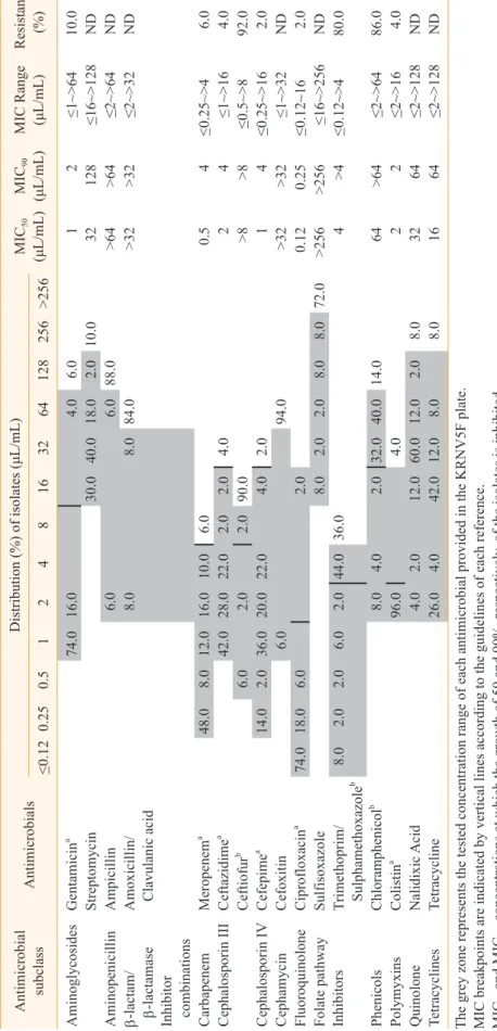
분리된 Pseudomonas spp.는 ceftiofur (92.0%), chlor- amphenicol (86.0%), trimethoprim/sulphamethoxazole (80.0%)에 높은 내성률을, ampicillin, amoxicillin/clavu- lanic acid, cefoxitin, sulfisoxazole에 높은 MIC50와 MIC90
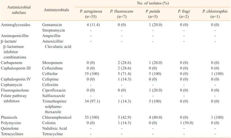
값을 나타내었다. 이에 반하여 cefepime (2.0%), ciprofloxa- cin (2.0%), ceftazidime (4.0%), colistin (4.0%), merope- nem (6.0%), gentamicin (10.0%)에는 낮은 내성률을 보였다 (Table 3). 균종별 항생제 내성률 비교에서는 P. aeruginosa 와 P. putida가 유사한 양상을 보였다. Trimethoprim/sul- phamethoxazole과 chloramphenicol에 대한 내성률은 P.
aeruginosa와 P. putida는 80.0~100%로 비교적 높게 나타난 반면, P. fluorescens는 각각 14.3%와 42.9%로 상대적으로 낮 게 나타나 일부 항생제의 경우 종별로 차이를 보였다(Table 4).
세 종류 이상의 antimicrobial subclass에 내성을 가지는 다 제내성균의 빈도는 P. aeruginosa, P. putida, P. fluorescens 에서 각각 97.1%, 80.0%, 28.6%로 나타났으며, P. fragi와 P.
chlororaphis에서는 확인되지 않았다(Table 5). Ceftiofur- chloramphenicol-trimethoprim/sulphamethoxazole 의 항생제 내성 유형이 P. aeruginosa와 P. putida에서 각각 85.7% (n=30), 60.0% (n=3)로 우세하게 나타났으며 P. fluore- scens는 6개의 다양한 유형을 나타내어 종간에 차이가 있는 것 으로 조사되었다.
고 찰
본 연구에서 유방염이 의심되는 분방유 시료에서 Pseudo- monas spp.의 분리율 4.9%를 보여 인도(6.5%)나 탄자니아 (9.0%)보다 낮았으며, 알제리(2.5%)보다는 높았다(Saidi 등, 2013; Banerjee 등, 2017; Suleiman 등, 2018). 또한 Kang 등(2001)이 1999년부터 2000년까지 제주에서 0.3%, Lee 등 (2007a)이 2001년부터 2004년까지 전국에서 2.3%, Kim 등 (2011)이 2009년 경남에서 2.8%, Nam 등(2013)이 2012년 전 국에서 2.0%로 조사한 국내의 결과 보다도 본 연구에서 다소 높은 분리율을 나타내었다. 본 연구에서 Pseudomonas spp.
의 균종별 분포는 P. aeruginosa, P. fluorescens, P. putida, P. fragi, P. chlororaphis 순으로 각각 70.0%, 14.0%, 10.0%, 4.0%, 2.0%로 조사되었다. 이러한 결과는 인도와 탄자니아뿐 아니라 일본에서도 Pseudomonas 유방염의 주된 원인균이 P.
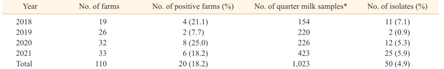
aeruginosa라는 보고와 유사한 결과를 나타내었다(Banerjee 등, 2017; Kawai 등, 2017; Suleiman 등, 2018). 하지만 국내 에서 Nam 등(2013)이 P. aeruginosa와 P. fluorescens의 분 리율이 동일했다는 보고와는 차이를 보였다. 이러한 분리율의 차이는 시료 채취 시기, 젖소의 사양관리 방법 및 환경여건의 변 화 등에 의한 것으로 생각된다(Smith와 Hogan, 1993; Nam 등, 2009). 또한 국내의 경우 목장 사육여건 및 환경의 변화와 더불어 유방염 5대 방제 프로그램의 적용에 의해 전염성 유방염 Table 1. Prevalence of Pseudomonas spp. isolated from bovine mastitis milk from 2018 to 2021
Year No. of farms No. of positive farms (%) No. of quarter milk samples* No. of isolates (%)
2018 19 4 (21.1) 154 11 (7.1)
2019 26 2 (7.7) 220 2 (0.9)
2020 32 8 (25.0) 226 12 (5.3)
2021 33 6 (18.2) 423 25 (5.9)
Total 110 20 (18.2) 1,023 50 (4.9)
*Bacterial examination of the quarter milk samples with somatic cell count ≥2×10⁶ cells/mL were conducted.
Fig. 1. Distribution of Pseudomonas spp. (n=50) isolated from bovine mastitis milk.
젖소 유방염에서 분리한 Pseudomonas spp.의 분포 및 항생제 내성 비교 KJVS
보다는 환경성 유방염이 더욱 문제되고 있음을 제시해 준다.
원유의 체세포수는 유방염을 진단하는 유용한 기준으로서 체 세포수의 증가는 젖소 유선내 감염과 밀접한 관계가 있다(Bog- ni 등, 2011). 본 연구에서 Pseudomonas spp.가 분리된 분방 의 체세포수는 20~50만 cells/mL이 40.0%였으며 일반적인 임 상형의 기준인 300만 cells/mL 이상은 10.0%로 조사되었다.
이러한 결과는 국내에서 Pseudomonas spp.를 포함한 그람 음 성균에 의한 유방염 시료의 체세포수가 300만 cells/mL 이상 이 10.8%인 것과 유사한 양상을 보였다(Lee 등, 2007a). 또한 Pseudomonas spp.로 인한 유방염은 임상형이 드물고 주로 준 임상형이라는 보고를 뒷받침한다(Banerjee 등, 2017; Schauer 등, 2021).
본 연구에서 검사한 16종 항생제 중 ceftiofur, chloram- phenicol, trimethoprim/sulphamethoxazole에 대한 Pseudomonas spp.의 감수성은 각각 8.0%, 14.0%, 20.0%로 비교적 낮게 나타났다. 또한, MIC50과 MIC90값을 비교했을 때 ampicillin (> 64 μL/mL), amoxicillin/clavulanic acid (>
32 μL/mL), cefoxitin (> 32 μL/mL), sulfisoxazole (> 256 μL/mL)에서 상대적으로 높게 나타나 이들 항생제에 대한 감수 성이 낮은 것으로 생각된다. Pseudomonas spp. 중에서도 가 장 높은 분포를 차지하는 P. aeruginosa에 대한 감수성 비율 은 미국의 경우 ampicillin, ceftiofur에 각각 0%, 일본의 경 우 ceftiofur와 chloramphenicol은 0%, trimethoprim/
sulphamethoxazole은 3.4%로서 본 연구와 유사한 양상을 나 타내었다(Erskine 등, 2002; Ohnish 등, 2011). Pseudomo- nas spp.에 대한 감수성 비율을 조사한 국내 연구 결과, Lee 등(2007a)이 2001년부터 2004년까지 ampicillin 16.7%, amoxicillin/clavulanic acid 35.7%, chloramphenicol 69.1%, Nam 등(2009)이 2003년부터 2008년까지 ampicillin 6.0%, amoxicillin/clavulanic acid 19.1%, chlorampheni- col 27.5%, Kim 등(2011)이 2009년 amoxicillin/clavulanic acid, trimethoprim/sulphamethoxazole에 각각 0%의 감
수성을 보이는 것으로 각각 보고하였다. 위와 같이 국내의 경우 Pseudomonas spp.에 대하여 chloramphenicol을 포함한 대 부분의 항생제에 대한 감수성이 시간이 지날수록 점차적으로 낮 아지고 있는 것으로 확인되었다. 이러한 결과는 국내에서 peni- cillins, cephalosporins, sulfonamides, aminoglycosides 등의 유방염 연고제 사용의 증가와 밀접한 관련이 있을 것으로 추정된다.
한편, 유방염 원인균은 균종별 항생제 감수성의 차이가 있음 이 보고된 바 있다(Lee 등, 2007b; Nam 등, 2010). 본 연구에 서 Pseudomonas spp.의 항생제 감수성을 균종별로 조사한 바, trimethoprim/sulphamethoxazole과 chloramphenicol에 대하여 P. aeruginosa와 P. putida는 높은 내성을 보인 반면 P.
fluorescens는 비교적 낮은 내성을 보였다. 이러한 결과는 젖소 유방염 치료제를 선택할 때 정확한 진단을 통한 원인균의 항생 제 감수성 검사를 우선적으로 실시하여 치료효과를 높이고 항생 제 오남용을 예방해야함을 뒷받침해준다.
위의 결과를 종합해 볼 때, 우리나라의 경우 2000년대 초반의 연구에 비하여 Pseudomonas spp.의 분리율이 높아졌으며, P.
aeruginosa 이외 다른 Pseudomonas 균종의 검출률이 30.0%
로 나타났다. Pseudomonas spp.에 감염된 분방의 90.0%가 체 세포수 300만 cells/mL 미만으로써 대부분이 준임상형 유방염 을 보였다. 또한 Pseudomonas spp.의 항생제 내성률이 과거 에 비해 높았으며 균종별로 내성률에 약간의 차이가 있는 것으 로 나타났다. 따라서 향후에 더 많은 시료를 대상으로 Pseudo- monas 유방염의 감염경로 규명 및 항생제 내성 획득 경로 등에 대한 체계적인 조사가 필요할 것으로 사료된다.
결 론
본 연구에서는 유방염 분방유 시료에서 Pseudomonas spp.
의 감염빈도 및 체세포수 분포와 균종별 항생제 감수성 양상을 조사하였다. 2018년 1월부터 2021년 6월까지 경기도, 충청북 Table 2. Distribution of somatic cell counts involved with Pseudomonas spp. isolates (n=50) from bovine mastitis milk
Species No. of isolates
Distribution of somatic cell count (×1,000 cells)
200~500 500~1,000 1,000~3,000 3,000~5,000 >5,000 P. aeruginosa 35 13 (37.1) 5 (14.3) 12 (34.3) 2 (5.7) 3 (8.6)
P. fluorescens 7 3 (42.9) 1 (14.3) 3 (42.9) 0 (0) 0 (0)
P. putida 5 2 (40.0) 3 (60.0) 0 (0) 0 (0) 0 (0)
P. fragi 2 2 (100) 0 (0) 0 (0) 0 (0) 0 (0)
P. chlororaphis 1 0 (0) 1 (100) 0 (0) 0 (0) 0 (0)
Total 50 20 (40.0) 10 (20.0) 15 (30.0) 2 (4.0) 3 (6.0)
강혜정ㆍ김하영ㆍ홍세림ㆍ박다솜ㆍ윤순식ㆍ문진산
KJVS
Table 3. Minimum inhibitory concentration of Pseudomonas spp. (n=50) isolated from bovine mastitis milk Antimicrobial subclassAntimicrobialsDistribution (%) of isolates (μL/mL)MIC50 (μL/mL)MIC90 (μL/mL)MIC Range (μL/mL)Resistant (%)≤0.120.250.51248163264128256>256 AminoglycosidesGentamicina 74.016.04.06.012≤1~>6410.0 Streptomycin30.040.018.02.010.032128≤16~>128ND AminopenicillinAmpicillin6.06.088.0>64>64≤2~>64ND β-lactam/ β-lactamase Inhibitor combinations
Amoxicillin/ Clavulanic acid8.08.084.0>32>32≤2~>32ND CarbapenemMeropenema 48.08.012.016.010.06.00.54≤0.25~>46.0 Cephalosporin IIICeftazidimea 42.028.022.02.02.04.024≤1~>164.0 Ceftiofurb 6.02.02.090.0>8>8≤0.5~>892.0 Cephalosporin IVCefepimea 14.02.036.020.022.04.02.014≤0.25~>162.0 CephamycinCefoxitin6.094.0>32>32≤1~>32ND FluoroquinoloneCiprofloxacina 74.018.06.02.00.120.25≤0.12~162.0
Folate pathway Inhibitors
Sulfisoxazole8.02.02.08.08.072.0>256>256≤16~>256ND
Trimethoprim/ Sulphamethoxazole
b8.02.02.06.02.044.036.04>4≤0.12~>480.0 PhenicolsChloramphenicolb 8.04.02.032.040.014.064>64≤2~>6486.0 PolymyxinsColistina 96.04.022≤2~>164.0 QuinoloneNalidixic Acid4.02.012.060.012.02.08.03264≤2~>128ND TetracyclinesTetracycline26.04.042.012.08.08.01664≤2~>128ND
The grey zone represents the tested concentration range of each antimicrobial provided in the KRNV5F plate. MIC breakpoints are indicated by vertical lines according to the guidelines of each reference. MIC
50 and MIC90, concentrations at which the growth of 50 and 90%, respectively, of the isolates is inhibited. ND, not determined. MIC breakpoints for P. aeruginosa in accordance with Clinical and Laboratory Standards Institute (CLSI). MIC breakpoint in accordance with Ohnishi et al., 2011.
젖소 유방염에서 분리한 Pseudomonas spp.의 분포 및 항생제 내성 비교 KJVS
도, 충청남도, 경상북도 소재 110개 목장에서 의뢰된 1,023개의 시료에서 Pseudomonas spp. 50균주(4.9%)를 분리하였다. 균 종별로는 P. aeruginosa가 70.0%로 가장 높은 비율을 차지하
였고 P. fluorescens가 14.0%로 그 뒤를 이었다. Pseudomo- nas spp.에 감염된 분방의 체세포수 분포율은 300만 cells/mL 미만이 90.0%로서 대부분이 준임상형 유방염인 것으로 나타 Table 4. Antimicrobial resistance profiles by species of Pseudomonas isolates from bovine mastitis milk
Antimicrobial
subclass Antimicrobials
No. of isolates (%) P. aeruginosa
(n=35) P. fluorescens
(n=7) P. putida
(n=5) P. fragi
(n=2) P. chlororaphis (n=1) Aminoglycosides Gentamicin 4 (11.4) 0 (0) 1 (20.0) 0 (0) 0 (0)
Streptomycin - - - - -
Aminopenicillin Ampicillin - - - - -
β-lactam/
β-lactamase inhibitor combinations
Amoxicillin/
Clavulanic acid - - - - -
Carbapenem Meropenem 0 (0) 2 (28.6) 1 (20.0) 0 (0) 0 (0)
Cephalosporin III Ceftazidime 0 (0) 2 (28.6) 0 (0) 0 (0) 0 (0)
Ceftiofur 35 (100) 5 (71.4) 5 (100) 0 (0) 1 (100)
Cephalosporin IV Cefepime 0 (0) 1 (14.3) 0 (0) 0 (0) 0 (0)
Cephamycin Cefoxitin - - - - -
Fluoroquinolone Ciprofloxacin 0 (0) 0 (0) 1 (20.0) 0 (0) 0 (0) Folate pathway
inhibitors Sulfisoxazole - - - - -
Trimethoprim/
sulphame- thoxazole
34 (97.1) 1 (14.3) 5 (100) 0 (0) 0 (0)
Phenicols Chloramphenicol 35 (100) 3 (42.9) 4 (80.0) 0 (0) 1 (100)
Polymyxins Colistin 0 (0) 1 (14.3) 0 (0) 1 (50.0) 0 (0)
Quinolone Nalidixic Acid - - - - -
Tetracyclines Tetracycline - - - - -
Table 5. Multidrug resistant and antimicrobial resistance patterns of Pseudomonas spp. isolated from bovine mastitis milk Species No. of MDR (%) Antimicrobial resistance pattern
Type No. of isolates (%)
P. aeruginosa (n=35) 34 (97.1) XNL-CHL 1 (2.9)
XNL-CHL-SXT 30 (85.7)
XNL-CHL-GEN-SXT 4 (11.4)
P. fluorescens (n=7) 2 (28.6) Pansusceptible 1 (14.3)
COL 1 (14.3)
XNL 1 (14.3)
XNL-CHL 2 (28.6)
FEP-TAZ-XNL-MERO 1 (14.3)
TAZ-XNL-CHL-MERO-SXT 1 (14.3)
P. putida (n=5) 4 (80.0) XNL-SXT 1 (20.0)
XNL-CHL-SXT 3 (60.0)
XNL-CHL-CIP-GEN-MERO-SXT 1 (20.0)
P. fragi (n=2) 0 (0) Pansusceptible 1 (50.0)
COL 1 (50.0)
P. chlororaphis (n=1) 0 (0) XNL-CHL 1 (100)
MDR, Multidrug resistant (≥3 CLSI subclass); FEP, Cefepime; TAZ, Ceftazidime; XNL, Ceftiofur; CHL, Chloramphenicol; CIP, Ciprofloxacin; COL, Colistin; GEN, Gentamicin; MERO, Meropenem; SXT, Trimethoprim/Sulphamethoxazole.
강혜정ㆍ김하영ㆍ홍세림ㆍ박다솜ㆍ윤순식ㆍ문진산
KJVS
났다. 전체 분리주에 대한 항생제 감수성 검사 결과 ceftiofur, chloramphenicol, trimethoprim/sulphamethoxazole에는 80.0~92.0%로 비교적 높은 내성을 보였고, cefepime, cipro- floxacin, ceftazidime, colistin, meropenem, gentamicin 에는 2.0~10.0%로 상대적으로 낮은 내성을 보였다. 항생제 내 성 양상은 균종별로 차이가 있는 것으로 나타났으며 그중에서도 P. fluorescens와 달리 P. aeruginosa와 P. putida는 유사한 양상을 보였다. 결론적으로 본 연구에서 P. aeruginosa 이외의 Pseudomonas spp.가 30.0% 분리되었으며, 이들의 항생제 내 성은 P. aeruginosa와 비슷하거나 낮게 나타났다.
감사의 글
본 연구는 농림축산검역본부의 농림축산검역검사기술개발 사업(과제명: 젖소 유방염 원인균 및 항생제 감수성 양상 조사, N-1543081-2017-36-01)의 연구비 지원에 의해 수행되었다.
CONFLICT OF INTEREST
No potential conflict of interest relevant to this article was reported.
ORCID
Hye Jeong Kang, https://orcid.org/0000-0002-4478-0147 Ha-Young Kim, https://orcid.org/0000-0002-1332-3832 Serim Hong, https://orcid.org/0000-0001-8005-8744 Dasom Park, https://orcid.org/0000-0001-8157-066X Soon-Seek Yoon, https://orcid.org/0000-0003-0908-8785 Jin-San Moon, https://orcid.org/0000-0003-1057-9450
REFERENCES
Banerjee S, Batabyal K, Joardar SN, Isore DP, Dey S, Samanta I, Samanta TK, Murmu S. 2017. Detection and characterization of pathogenic Pseudomonas aeruginosa from bovine subclinical mastitis in West Bengal, India. Vet World 10: 738.
Bogni C, Odierno L, Raspanti C, Giraudo J, Larriestra A, Reinoso E, Lasagno M, Ferrari M, Ducros E, Frigerio C, Bettera S, Pellegrino M, Frola I, Dieser S, Vissio
C. 2011. War against mastitis: Current concepts on controlling bovine mastitis pathogens. Science against microbial pathogens: Communicating cur- rent research and technological advances 483-494.
Cheng WN, Han SG. 2020. Bovine mastitis: risk factors, therapeutic strategies, and alternative treatments—
A review. Asian-Australas J of Anim Sci 33: 1699.
CLSI (Clinical and laboratory Standards Institute). 2019.
Performance standards for antimicrobial suscep- tibility testing; 29th edition. CLSI document M100.
29th ed. CLSI, Wayne, PA.
Erskine R, Walker R, Bolin C, Bartlett P, White D. 2002.
Trends in antibacterial susceptibility of mastitis pathogens during a seven-year period. J Dairy sci 85: 1111-1118.
Gomes F, Henriques M. 2016. Control of bovine mas- titis: old and recent therapeutic approaches. Curr Microbiol 72: 377-382.
Halasa T, Huijps K, Østeras O, Hogeveen H. 2007. Eco- nomic effects of bovine mastitis and mastitis man- agement: a review. Vet Q 29: 18-31.
Kang HJ, Kim IC, Kim JH, Son WG, Lee DS. 2001. Iden- tification and antimicrobial susceptibility of mi- croorganisms isolated from bovine mastitic milk.
Korean J Vet Res 41: 511-521.
Kawai K, Shinozuka Y, Uchida I, Hirose K, Mitamura T, Watanabe A, Kuruhara K, Yuasa R, Sato R, Onda K, Nagahata H. 2017. Control of Pseudomonas mas- titis on a large dairy farm by using slightly acidic electrolyzed water. Anim Sci J 88: 1601-1605.
Kim SE, Hah DY, Jang EH, Kwon HN, Jo SS, Kwon YT, Park DY, Lee KC, Kim JS. 2011. Survey of mastitis management and incidence of mastitis in high so- matic cell count of bulk milk at dairy farms in the Gyeongnam. Korean J Vet Serv 34: 349-388.
Lee ES, Kang HM, Chung CI, Moon JS. 2007a. Anti- microbial susceptibility and prevalence of gram- negative bacteria isolated from bovine mastitis.
Korean J Vet Res 47: 67-75.
Lee G, Kang HM, Chung CI, Moon JS. 2007b. Antimi- crobial susceptibility and genetic characteristics of
젖소 유방염에서 분리한 Pseudomonas spp.의 분포 및 항생제 내성 비교 KJVS
Streptococcus uberis isolated from bovine mastitis milk. Korean J Vet Res 47: 33-41.
Nam HM, Lim SK, Kang HM, Kim JM, Moon JS, Jang KC, Kim JM, Joo YS, Jung SC. 2009. Prevalence and an- timicrobial susceptibility of gram-negative bacteria isolated from bovine mastitis between 2003 and 2008 in Korea. J Dairy Sci 92: 2020-2026.
Nam HM, Lim SK, Jang GC, Joung DY, Kim HJ, Lee CS, Jung SC. 2013 Culture results from quarter milk samples submitted to veterinary diagnostic labo- ratories during january~November 2012 in Korea.
Prev Vet Med 37: 111-119.
Nam HM, Lim SK, Moon JS, Kang HM, Kim JM, Jang KC, Kim JM, Kang MI, Joo YS, Jung SC. 2010. An- timicrobial resistance of enterococci isolated from mastitic bovine milk samples in Korea. Zoonoses Public Health 57: e59-64.
NMC (National Mastitis Council). 2017. Laboratory Handbook on Bovine Mastitis, 3rd ed. New Prague:
NMC, USA.
Ohnishi M, Sawada T, Hirose K, Sato R, Hayashimoto M, Hata E, Yonezawa C, Kato H. 2011. Antimicrobial susceptibilities and bacteriological characteristics
of bovine Pseudomonas aeruginosa and Serratia marcescens isolates from mastitis. Vet Microbiol 154: 202-207.
Park HR, Hong MK, Hwang SY, Park YK, Kwon KH, Yoon JW, Shin S, Kim JH, Park YH. 2014. Charac- terisation of Pseudomonas aeruginosa related to bovine mastitis. Acta Vet Hung 62: 1-12.
Saidi R, Khelef D, Kaidi R. 2013. Subclinical mastitis in cattle in Algeria: frequency of occurrence and bac- teriological isolates. J S Afr Vet Assoc 23;84: E1-5.
Schauer B, Wald R, Urbantke V, Loncaric I, Baumgart- ner M. 2021. Tracing Mastitis Pathogens—Epidemi- ological Investigations of a Pseudomonas aerugi- nosa Mastitis Outbreak in an Austrian Dairy Herd.
Animals 11: 279.
Smith KL, Hogan JS. 1993. Environmental mastitis. Vet Clin North Am Food Anim Pract 9: 489-498.
Suleiman T, Karimuribo E, Mdegela R. 2018. Preva- lence of bovine subclinical mastitis and antibiotic susceptibility patterns of major mastitis pathogens isolated in Unguja island of Zanzibar, Tanzania.
Trop Anim Health Prod 50: 259-266.
강혜정ㆍ김하영ㆍ홍세림ㆍ박다솜ㆍ윤순식ㆍ문진산
KJVS