www.centauro.it Interventional Neuroradiology 20: 609-613, 2014 - doi: 10.15274/INR-2014-10051
Aneurysmal Bone Cyst in the Temporal Bone and Complete Resection
with Preoperative Embolization
A Case Report
BYOUNG JE KIM
1, EUN JU LEE
1, HYUK WON CHANG
1, HAE RA JUNG
2, EALMAAN KIM
3, SUNG IL SOHN
4, SANG PYO KIM
21
Department of Radiology,
2Department of Pathology,
3Department of Neurosurgery,
4Department of Neurology, Keimyung University Dongsan Medical Center; Daegu, Republic of Korea
Key words: bone cysts, aneurysmal, temporal bone, angiography, digital subtraction
Summary
We describe a rare case of aneurysmal bone cysts (ABCs) that occurred in the petrous por- tion of the temporal bone. The ABCs were treat- ed with preoperative embolization and complete removal of the mass from the adjacent tissue.
The technical details suggest that preoperative embolization is a good treatment option for ABCs.
Introduction
Aneurysmal bone cysts (ABCs), first de- scribed in 1942 by Jaffe and Lichtenstein, are a rare and benign disease showing rapid growth, with osteolysis and cystic lesions such as an ex- pansile arterial aneurysm
1. We describe a case of ABCs in the temporal bone, which were completely removed with the help of preopera- tive embolization, and were used to reduce in- traoperative bleeding. Forty cases of ABCs in the temporal bone have been reported in the literature, but only two cases described the use of preoperative embolization.
Case Report
A 17-year-old female visited our clinic due to headache, nausea, and vomiting. A history re- vealed a growing mass in the right temporal
area for three months. No history of trauma was noted. The general physical examination was unremarkable. However, a fundoscopic ex- amination showed bilateral papilledema and hemorrhage around the left optic disc.
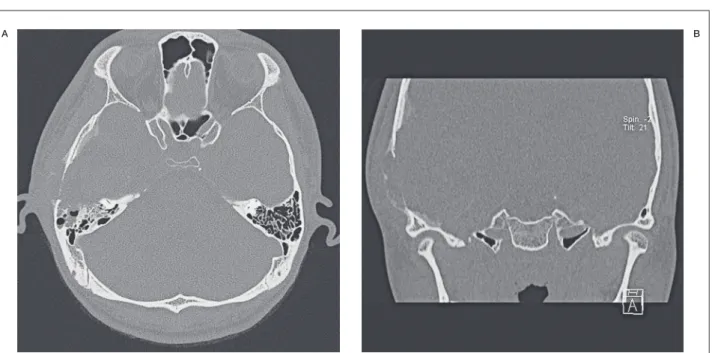
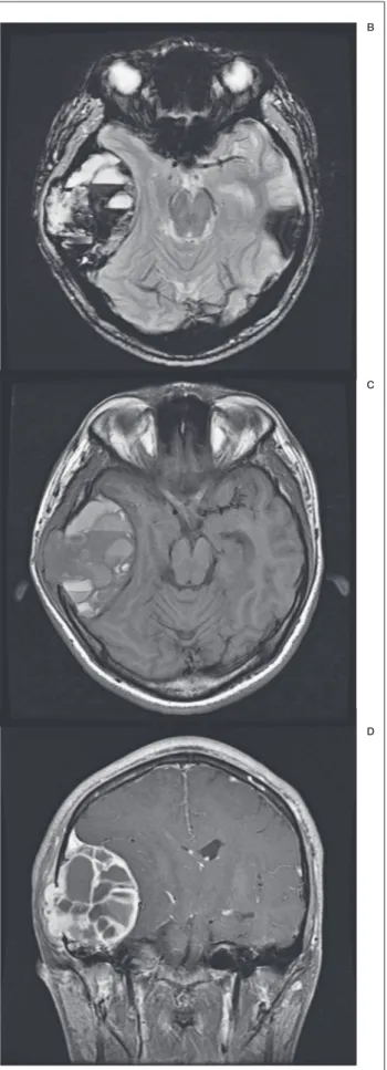
A computed tomography (CT) scan revealed an osteolytic and cystic lesion involving the in- ner and outer table of the temporal bone as well as the anterior portion of the mastoid air cells (Figure 1). A magnetic resonance image (MRI) revealed a 9 cm-sized multicystic mass with air fluid levels originating from the squamous por- tion of the temporal bone (Figure 2A). The axial gradient echo image showed that the lower lay- er of fluid-fluid level demonstrated low signal intensity suggestive of blood (Figure 2B). Low signal intensity was detected in the fibrous cap- sule and septa (Figure 2C) with homogeneous enhancement (Figure 2D). The mass did not in- volve the meninges or compress the brain pa- renchyma due to an indirect mass effect.
The surgeon decided to surgically excise the mass, considering the patient’s symptoms, the papilledema secondary to increased intracere- bral pressure, and the relatively rapid progres- sion of the mass; preoperative embolization was requested.
A large mass feeding from the middle me-
ningeal artery and superficial temporal artery
was detected on right external carotid angiog-
raphy. Particle embolization with polyvinyl al-
cohol 150-250 (Contour, Boston Scientific, Fre-
mont, CA, USA) was done with guidance from
years at the time of diagnosis have been re- ported
2, 3. ABCs shows a slight female predom- inance (male: female = 1:1.04-1.8)
2,3. About 52% of ABCs involve the metaphyseal region of long bones such as the femur, tibia, fibula, or humerus, 20% involve the spine, and only 3-6% involve the skull
2,4. About 70% of ABCs are primary lesions and the others are second- ary, representing other bone tumors such as gi- ant cell tumors, hemangiomas, and chondro- blastomas
5.
The pathogenesis of ABCs is controversial, and their exact cause remains unknown. Re- cent studies show a genetic aspect to ABCs, and it is accepted that they are a tumor rather than a reactive disease. Oliveira et al. revealed that 69% of primary ABCs are influenced by gene rearrangements localized to t (16,17) in which the ubiquitin-specific protease 6 onco- gene is located under the regulatory influence of the highly active cadherin-11 promoter.
However, such translocations are not detected in secondary ABCs
6. Leithner et al. showed that insulin-like growth factor-I (IGF-1) or mRNA coding for this growth factor is usually localized in multinucleate giant cells in all spec- imens. Normal human bone tissue does not show significant levels of IGF-1 expression
2.
The usual clinical presentation is tenderness and/or externally visible mass effects at the in- a 5 Fr Envoy catheter (Cordis Corp, Miami
Lakes, FL, USA) and superselection with an agility 10 soft (Cordis) and Prowler 14 (Cordis).
Surgical excision of the mass was uneventful.
Blood loss was about 200 ml, most of which oc- curred during the craniotomy. After the opera- tion, the patient’s symptoms and fundoscopic findings resolved, and no local recurrence has been detected after two years of follow-up.
The gross examination of the specimen re- vealed multiple honeycomb-like cystic lesions partially filled with non-coagulated blood. The microscopic examination showed that the septa of the mass comprised fibroblasts, multinuclear giant cells in the form of osteoclasts, granula- tion tissue, macrophages filled with hemosider- in, and extravasated red blood cells (Figure 4).
Thus, ABCs of the temporal bone were con- firmed pathologically.
Discussion
Aneurysmal bone cysts (ABCs) are benign but relatively rapidly growing vascular bone le- sions, representing about 1% of primary bone tumors
2.
ABCs occur mostly in patients ≤ 20-years- of-age, and the mean age of occurrence is 13 years, but cases of patients aged from one to 59
A B
Figure 1 A) Axial temporal bone computed tomography (CT) scan shows bony destruction of the anterior portion of the
mastoid air cells resulting in the meniscus sign. B) Coronal temporal bone CT scan shows severe bony thinning and destruc-
tion of the inner and outer tables of the temporal bone and a focal area of ground-glass attenuation along the inner table of
the temporal bone.
www.centauro.it Interventional Neuroradiology 20: 609-613, 2014 - doi: 10.15274/INR-2014-10051
volved site. Imaging studies are very important for the diagnosis and to make treatment deci- sions. Simple skull radiography shows the so- called “soap bubble”-like expansible osteolytic lesion. CT shows more details on the relation- ship between normal bone and ABC lesions and also demonstrates an expansive multilocu- lated osseous lesion invading the inner and outer tables of the skull. The CT image may show a fluid-fluid level of different attenua- tions. MRI shows a characteristic fluid-fluid level of different signal intensities. Contrast en- hancement in the peripheral capsule and inter- nal septation are detected on enhanced T1- weighted images. The osteolytic lesion is sur- rounded by inner and outer tables. The depend- ent portion of the fluid shows low signal inten- sity on a gradient echo image, due to non-coag- ulated hemorrhage, and this is an important finding of ABCs. However these findings are
A B
C
D
Figure 2 A) Axial fluid attenuated inversion recovery image
shows an expansile extra-axial mass with fluid-fluid levels
involving the temporal bone with a multicystic soap bubble
appearance. The upper layer of the fluid-fluid level is not
suppressed as cerebral spinal fluid. B) The axial gradient-
echo image shows that the lower layer of the fluid-fluid level
demonstrates low signal intensity suggestive of blood. C)
Axial T1-weighted non-contrasted image. D) Coronal T1-
weighted image with contrast enhancement. D) A well-en-
hanced internal septa and a soft tissue attenuated lesion
anchoring to the squamous portion of the temporal bone.
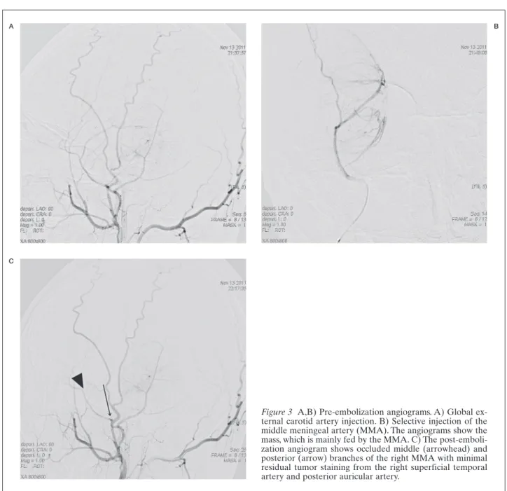
Figure 3 A,B) Pre-embolization angiograms. A) Global ex- ternal carotid artery injection. B) Selective injection of the middle meningeal artery (MMA). The angiograms show the mass, which is mainly fed by the MMA. C) The post-emboli- zation angiogram shows occluded middle (arrowhead) and posterior (arrow) branches of the right MMA with minimal residual tumor staining from the right superficial temporal artery and posterior auricular artery.
Figure 4 The microphotograph of the aneurysmal bone cyst shows cystic spaces including red blood cells which are sepa- rated by septa including spindle shaped cells and scattered multinucleate giant cells.
A B
C
www.centauro.it Interventional Neuroradiology 20: 609-613, 2014 - doi: 10.15274/INR-2014-10051
cent tissue by thrombosis of the feeding artery, and necrosis and shrinkage of the mass with reduced perioperative bleeding
9. Preoperative angiography reveals the vascularity and hemo- dynamic status of the tumor. Furthermore, oth- er researchers have shown the effectiveness of endovascular embolization with N-2-butyl cy- anoacrylate. They enrolled 55 cases of extremi- ty ABCs and demonstrated that 94% of cases were effectively treated with this method, with- out recurrence during 0.9–5 years of follow-up
10