http://dx.doi.org/10.14316/pmp.2014.25.4.199
Received 15 October 2014, Revised 24 November 2014, Accepted 28 November 2014
Correspondence: Suzy Kim ([email protected]) Tel: 82-2-870-1691, Fax: 82-2-870-1689 cc This is an Open-Access article distributed under the terms of the Creative Commons Attribution Non-Commercial License (http://creativecommons.org/licenses/by-nc/3.0) which permits unrestricted non-commercial use, distribution, and reproduction in any medium, provided the original work is properly cited.
Dosimetric Advantages of the Field-in-field Plan Compared with the Tangential Wedged Beams Plan for
Whole-breast Irradiation
Suzy Kim, Yunseok Choi
Department of Radiation Oncology, Seoul National University Boramae Medical Center, Seoul, Korea The purpose of this study is to evaluate the dosimetric outcome of the field-in-field (FIF) plans compared with tangential wedged beams (TWB) plans for whole breast irradiation of breast cancer patients. Twenty patients with right-sided breast cancer and 10 patients with left-sided breast cancer were retrospectively enrolled in this study. We generated a FIF plan and a TWB plan for each patient to compare dosimetric outcomes. The dose the homogeneity index (HI), the conformity index (CI) and the uniformity index (UI) were defined and used for comparison of the dosimetric outcome of the planning target volume (PTV). To compare the dosimetric outcome of the organs at risk, the mean dose (D
mean) and the percentage of volumes receiving more than 10, 20 and 30 Gy of the ipsilateral lung and heart were used. The FIF plans had significantly lower HI (p=0.002), higher UI (p=0.000) and CI (p=0.000) than those of the TWB plans, which means that the FIF plans were better than the TWB plans in the dosimetric comparisons of the PTV. The V10
lung(17.1±7.1 vs. 18.6±6.6%, p=0.020) and
V30
lung(10.3±5.1% vs. 10.7±5.2%, p=0.000) were lower with the FIF plans compared with those of the TWB
plans, with statistical significance. For the left-sided breast cancer patients, D
meanof the heart (2.6±1.3 vs.
3.2±1.4 Gy, p=0.000), V20
heart(3.4±2.6 vs. 3.6±2.8%, p=0.005) and V30
heart(2.6±2.3% vs. 2.9±2.4%, p=0.004) were significantly lower for the FIF plans in comparison with those of the TWB plans. The FIF plans increased the dose homogeneity, conformity and uniformity of the target volume for the whole-breast irradiation compared with the TWB plans. Moreover, FIF plans reduced the doses to the ipsilateral lung and heart.
Key Words: Breast cancer, Radiotherapy, Radiation dose, IMRT
Introduction
According to the nation-wide online registry data of the Korean Breast Cancer Society, a total of 16,967 patients were newly diagnosed with breast cancer in 2011. That number is 3 times the 5,401 new cases diagnosed in 2000.
1)The pattern of surgical treatment of breast cancer changed greatly during those 10 years. The proportion of patients who underwent breast-con- serving surgery (BCS) more than doubled, from 27.9% in 2000 to 65.7% in 2011 (135.5% rise).
The effects of radiation therapy (RT) after BCS are well proven by several clinical trials.
2-4)There is compelling evi- dence that RT after BCS not only improved local control but also long-term survival. Therefore, adjuvant whole-breast irra- diation after BCS is the standard of treatment for early-stage breast cancer.
For the conventional tangential wedged beams (TWB) tech- nique for whole breast irradiation, it is difficult to achieve homo- genous dose distribution because of breast contour irregularities.
Many studies reported the large dose inhomogeneity within the target volume.
5,6)Even with homogenous dose distribution in the central axis of the target volume, high-dose regions can be observed at the superior and inferior regions of the breast or at the medial or lateral aspects of the breast.
The development of 3-dimensional treatment planning with
computed tomography (CT) images and the intensity-modulated
radiation therapy (IMRT) planning technique enabled new treat-
ment techniques for whole breast irradiation. Several new breast irradiation techniques were used recently to achieve better dose distribution and lower the dose to surrounding normal tissues.
The field-in-field (FIF) technique, also called forward-planned IMRT technique, which uses the multileaf collimator, is rela- tively simple and a less time-consuming method than the in- verse-planned IMRT technique. In our institution, whole-breast irradiation has been carried out with the FIF technique.
This study was carried out to evaluate the dosimetric out- come of the FIF plans compared to TWB plans and to con- firm the advantages of the FIF plans.
Materials and Methods
1. Patients
The consecutive 20 patients with right-sided breast cancer and 10 patients with left-sided breast cancer, who were treated in our department between March 2012 and March 2013, were included in this analysis retrospectively. All patients had un- dergone BCS and were treated with postoperative RT, in which 50.4 Gy in 28 fractions were delivered to the whole breast using the FIF technique.
All patients underwent CT simulation. Patients were immo- bilized on the Breastboard (Civco, Orange City, IA, USA) and skin wires were placed on the medial and lateral borders. The medial border was set at the midline of the chest, and the lat- eral border was defined by physical examination. Superior and inferior borders were defined by the radiation oncologist, and they were usually set at the inferior edge of the medial head of the clavicle and 2 cm below the breast fold. CT scans were acquired with 5 mm thickness of slices using Siemens Somatom Definition AS Open (Siemens Medical Solutions, Malvern, PA, USA), and the acquired image sets were transferred to the Pinnacle radiation therapy treatment planning system (Phillips Medical Systems, Bothell, WA, USA).
2. Treatment planning
The clinical target volume (CTV) was defined as the whole breast tissue. The planning target volume (PTV) was the CTV with an extension of 0.5 to 1 cm margins and the build-up re- gion 5 mm beneath the skin surface was excluded from the PTV.
For each patient, the TWB plan and FIF plan were gen-
erated on a treatment planning system. Two plans for each pa- tient used the same isocenter, tangential beam angles, and col- limator sizes. Conventional TWB plans were made with a me- dial open beam and a lateral wedged beam with appropriate wedge angles for achieving proper dose distribution. For the FIF plans, two open tangential beams were generated and an initial calculation was performed without any beam modifiers. Then, hot dose areas were shielded by additional subfields. Hot dose areas were defined as areas receiving more than 103% of the prescribed dose. Two or three subfields using a multi-leaf col- limator were generated for each medial and lateral tangential field. This was done by sequentially blocking the hot dose areas with 3∼5% increments in the beam’s eye projections of the treatment field.
3. Indices used for dosimetric comparison
The homogeneity index (HI), the conformity index (CI) and the uniformity index (UI) were defined and used for compar- ison of the dosimetric outcome of the PTV.
7)The HI was used to evaluate the dose homogeneity within the PTV, and it was defined as the following formula:
D
maxis the maximum dose in the PTV and D
presciptionis the prescribed dose to the PTV.
The CI was used to evaluate the dose conformity and is de- fined as the ratio of volume enclosed by the prescription iso- dose to the PTV.
The UI was defined as the percentage of PTV volume in- cluded in the interval 97% to 103% of the prescribed dose and used to evaluate the PTV dose improvement.
For organs at risks (OARs), dose-volume histograms (DVHs) were generated from the treatment planning system. The V10, V20 or V30 represents the percentage of the volume of OAR receiving radiation doses of 10 Gy, 20 Gy or 30 Gy, respectively.
4. Statistical analysis
For comparison of mean values of the indices of the two
different treatment plans, the paired t-test was used. A p-value
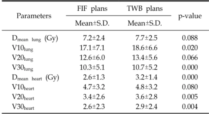
Table 1. Dosimetric comparison between FIF and TWB plans.
Parameters FIF plans TWB plans
p-value Mean±S.D. Mean±S.D.
Homogeneity index (HI) 1.038±0.001 1.053±0.022 0.002 Conformity index (CI) 0.362±0.073 0.244±0.086 0.000 Uniformity index (UI) 61.4±7.2 48.1±8.5 0.000 FIF: field-in-field, TWB: tangential wedged beams, S.D.: stan- dard deviation.
Table 2. Dosimetric parameters of the organs at risk for each treatment plan.
Parameters FIF plans TWB plans
p-value Mean±S.D. Mean±S.D.
D
mean lung(Gy) 7.2±2.4 7.7±2.5 0.088
V10
lung17.1±7.1 18.6±6.6 0.020
V20
lung12.6±6.0 13.4±5.6 0.066
V30
lung10.3±5.1 10.7±5.2 0.000
D
mean heart(Gy) 2.6±1.3 3.2±1.4 0.000
V10
heart4.7±3.2 4.8±3.2 0.080
V20
heart3.4±2.6 3.6±2.8 0.005
V30
heart2.6±2.3 2.9±2.4 0.004
FIF: field-in-field, TWB: tangential wedged beams, S.D.: stan- dard deviation.
of less than 0.05 indicates a statistically significant difference between the two data sets. All statistical analyses were per- formed using the SPSS version 20 software (IBM Corporation, Chicago, IL, USA).
Results
The dosimetric parameters of the PTV for each treatment plan are shown in Table 1.
The HI for FIF plans and TWB plans were 1.038±0.001 and 1.053±0.022, respectively. There was a statistically sig- nificant difference of HI between the two plans with a p-value of 0.002. The FIF plans had a lower HI than the TWB plans, which means that the FIF plans showed better dose homoge- neity within the PTV. Although the difference of HI was stat- istically significant, the absolute value of the difference seems to be small (≅0.015). Therefore, we selected 10 patients with HI of more than 1.060 in TWB plan, and compared the HI of the FIF plan with that of the TWB plan. For these patients, the HI of FIF plans and TWB plans were 1.039±0.002 and 1.080±0.023, respectively (p=0.000). Therefore, the FIF plans reduced the HI more significantly for patients with more in- homogeneous dose distribution in TWB plans than those with less inhomogeneous dose plans.
The CI of the FIF plans were higher than that of the TWB plans, and the differences were statistically significant (0.362±
0.073 vs. 0.244±0.086, p=0.000). If we compare the CI of the two plans for 15 patients whose CI is less than 0.20 in TWB plans, the difference was larger for these patients (0.314±0.041 vs. 0.150±0.033, p=0.000). Therefore, the FIF plans increased the dose conformity of PTV more significantly for the patients whose TWB plan shows lower CI than those with higher CI.
The UI of the FIF plans were also higher than that of the TWB plans, and the differences were also statistically significant (61.4±7.2 vs. 48.1±8.5, p=0.000).
The dosimetric parameters of the OARs are shown in Table 2.
The mean dose of the ipsilateral lung (D
mean lung) was not sig- nificantly different between the FIF plans and TWB plans (7.2±2.4 Gy vs. 7.7±2.5 Gy, p=0.088). However, the V10
lungof the FIF plans was 17.1±7.1, and that of the TWB plans was 18.6±6.6, showing that the FIF plan reduced the V10
lungwith statistical significance (p=0.02). The difference of the V20
lungbe- tween the two plans showed only marginal statistical sig- nificance (p=0.066), but the V30
lungwas lower with the FIF plans (10.3±5.1 vs. 10.7±5.2, p=0.000). For left-sided breast cancer patients, FIF plans reduced the mean dose to the heart (D
mean heart, 2.6±1.3 Gy vs. 3.2±1.4 Gy, p=0.000). The V20
heartand V30
heartwere lower with the FIF plan compared with TWB plans (p=0.005 and p=0.004, respectively). The V10
heartwas lower with the FIF plans compared with that of the TWB plans, but its difference showed only marginal significance (p=0.080).
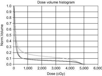
The DVH of a patient with left-sided breast cancer is shown in Fig. 1.
Discussion
The introduction of planning CT and the development of ra-
diation therapy planning systems enabled new techniques of
whole breast irradiation to improve the dose distributions in
the PTV and to reduce the doses to OARs. The FIF technique
Fig. 1. An example of dose-volume histograms of heart and ip-
silateral lung; comparison of TWB plan and FIF plan. Black solid line: heart, FIF plan, Black dashed line: heart, TWB plan, Gray solid line: lung, FIF plan, Gray dashed line: lung TWB plan.
is also called forward-planned IMRT technique, and it is less time-consuming than inverse-planned IMRT.
Several investigators compared the FIF plans and TWB plans for whole breast irradiation. In a RT planning study with 20 breast cancer patients, Sasaoka et al. showed that the FIF technique reduced the maximum dose and improved dose dis- tribution in the treated breast.
8)Their maximum dose was 111.2±3.4% for TWB plans and 105.8±1.4% for FIF plans (p=0.0051). They also found that dose homogeneity was better in the FIF plans compared with the TWB plans. Other inves- tigators also found that the dose homogeneity was improved with the FIF plans than with the TWB plans.
9)Most studies supported the idea that the FIF plans were bet- ter than the TWB plans, but a study by Taiwan investigators insisted that the FIF technique did not demonstrate superior dosimetric results.
7)They used the same indices used in our study-HI, CI and UI. They found that the FIF plan had a higher UI, but the HI and CI of the FIF plan were worse than those of the TWB plans, and the V20 of the lung did not ex- hibit a significant difference. They said that they used one or more subfields for FIF plan but we used at least two subfields.
Their technique for FIF plan might have not been as skillful as ours. In our study, the FIF plan had a significantly lower HI, higher CI and UI, that is, it demonstrated dosimetric ad- vantages over the TWB plans. The FIF plan was found to be
more advantageous for patients with less homogeneous or less conformal dose distribution in TWB plans. The difference of HI or CI between the two plans was larger for these patients.
There was a large randomized controlled trial of FIF techni- que for early breast cancer.
10)The study confirmed that breast dosimetry could be significantly improved with the FIF technique. Recently, 5-year follow-up results of the study proved that the improved dose homogeneity with FIF techni- que translated into superior overall cosmesis.
11)In the present study, the dosimetric comparisons of the OARs also showed that the FIF plan reduced the V10 and V30 of the ipsilateral lung. Clinically symptomatic radiation pneu- monitis occurs in 1∼10% of patients irradiated for breast cancer.
12)With the 3D-treatment planning system, many inves- tigators used lung-dose-volume histograms to predict the prob- ability of radiation pneumonitis after RT for lung cancer.
13,14)In a meta-analysis, mean lung dose, V5, V10 (≥34%), V20 (≥25%) and V30 (≥18%) of the lungs, were identified as significant risk factors for radiation pneumonitis.
15)Goldman et al. reported that they could lower the rate of radiation pneu- monitis with the dose volume constraint of V20 of the ipsi- lateral lung <30% in breast cancer irradiation.
16)Seventy five percent of their study population was patients who underwent total mastectomy as their surgical treatment, and they included internal mammary lymph node (IMN) area into the PTV in more than 80% of patients. They found that the V20 (35% vs.
26%) and V30 (24% vs. 16%) of ipsilateral lung were reduced with 3D-treatment planning compared with 2D planning using one anterior electron beam to cover the chest wall and the IMN.
In our study, the D
meanof the ipsilateral lung, V10
lung, V20
lungand V30
lungof TWB plans were much lower than the constraints for radiation pneumonitis, but FIF plans even lowered these values so that the risk of radiation pneumonitis could be minimized.
Several studies reported that left breast irradiation could be
a risk factor in the development of ischemic heart disease.
17,18)Recently, Darby et al. reported that the rates of ischemic heart
disease increased linearly with the mean dose to the heart by
7.4% per gray (Gy), with no apparent threshold.
19)If there is no
threshold, it is more important to lower the radiation dose to
the heart as low as possible in left breast irradiation. Although
the absolute value of difference was small, D
meanto the heart,
V20
heart, and V30
heartwere lower with FIF plans than with the TWB plans. Therefore, a lower risk of ischemic heart disease could be expected with FIF plans.
In the present study, the FIF plan improved dose homoge- neity, conformity and uniformity within the whole breast tissue in comparison with the TWB plan. The FIF plan also reduced the lung or heart volume receiving radiation doses that can in- duce radiation-related late toxicities. The FIF plan is a simple and clinically useful technique for whole breast irradiation.
References
1. Kim Z, Min SY, Yoon CS, et al: The basic facts of Korean breast cancer in 2011: results of a nationwide survey and breast cancer registry database. J Breast Cancer 17:99-106 (2014) 2. Fisher B, Anderson S, Bryant J, et al: Twenty-year fol-
low-up of a randomized trial comparing total mastectomy, lum- pectomy, and lumpectomy plus irradiation for the treatment of invasive breast cancer. N Engl J Med 347:1233-1241 (2002) 3. Clarke M, Collins R, Darby S, et al: Effects of radiotherapy
and of differences in the extent of surgery for early breast can- cer on local recurrence and 15-year survival: an overview of the randomized trials. Lancet 17:2087-2106 (2005)
4. Rapiti E, Fiorreta G, Vlastos G, et al: Breast-conserving surgery has equivalent effect as mastectomy on stage I breast cancer prognosis only when followed by radiotherapy. Radiother Oncol 69:277-284 (2003)
5. Buchholz TA, Gurqoze E, Bice WS, et al: Dosimetric analysis of intact breast irradiation in off-axis planes. Int J Radiat Oncol Biol Phys 39:261-267 (1997)
6. Solin LJ, Chu JC, Sontag MR, et al: Three-dimensional photon treatment planning of the intact breast. Int J Radiat Oncol Biol Phys 21:193-203 (1991)
7. Sun LM, Meng FY, Yang TH, et al: Field-in-field plan does not improve dosimetric outcome compared with the wedged beams plan for breast cancer radiotherapy. Med Dosim 39:
79-82 (2014)
8. Sasaoka M, Futami T: Dosimetric evaluation of whole breast radiotherapy using field-in-field technique in early-stage breast cancer. Int J Clin Oncol 16:250-256 (2011)
9. Onal C, Sonmez A, Arslan G: Dosimetric comparison of the field-in-field technique and tangential wedged beams for breast irradiation. Jpn J Radiol 30:218-226 (2012)
10. Barnett GC, Wilkinson J, Moody AM, et al: A randomized controlled trial of forward-planned radiotherapy (IMRT) for early breast cancer: Baseline characteristics and dosimetry results.
Radiother Oncol 92:34-41 (2009)
11. Mukesh MB, Barnett GC, Wilkinson JS, et al: Randomized controlled trial of intensity-modulated radiotherapy for early breast cancer: 5-year results confirm superior overall cosmesis.
J Clin Oncol 31;4488-4495 (2013)
12. Marks LB, Yu X, Vujaskovic Z, et al: Radiation-induced lung injury. Semin Radiat Oncol 13:333-345 (2003)
13. Graham MV, Purdy JA, Emami B, et al: Clinical dose-vol- ume histogram analysis for pneumonitis after 3D treatment for non-small cell lung cancer (NSCLC). Int J Radiat Oncol Biol Phys 45:323-329 (1999)
14. Claude L, Perol D, Ginestet C, et al: A prospective study on radiation pneumonitis following conformal radiation therapy in non-small cell lung cancer. Radiother Oncol 71:175-181 (2004) 15. Zhang XJ, Sun JG, Sun J, et al: Prediction of radiation pneumonitis in lung cancer patients: a systemic review. J Cancer Res Clin Oncol 138:2103-2116 (2012)
16. Blom Goldman U, Anderson M, Wennberg B, et al:
Radiation pneumonitis and pulmonary function with lung dose- volume constraints in breast cancer irradiation. J Radiother Pract 13:211-217 (2014)
17. Harris EE, Correa C, Hwang WT, et al: Late cardiac mor- tality and morbidity in early-stage breast cancer patients after breast conservation treatment. J Clin Oncol 24:4100-4106 (2006) 18. Borger JH, Hooning MJ, Boersma LJ, et al: Cardiotoxic effects of tangential breast irradiation in early breast cancer pa- tients: the role of irradiated heart volume. Int J Radiat Oncol Biol Phys 69:1131-1138 (2007)
19. Darby SC, Ewertz M, McGale P, et al: Risk of ischemic
heart disease in women after radiotherapy for breast cancer. N
Engl J Med 368:987-998 (2013)
유방암 환자의 방사선치료에 있어서 순치료계획 세기변조방사선치료법과 쐐기접선조사기법의 선량측정 비교