393
감마선을 조사한 마우스의 조혈 및 소장줄기세포에 대한 fucoidan의 방호효과
박은진·전성모·주홍구·황규계·지영흔*
제주대학교 수의과대학 (게재승인: 2008년 12월 5일)
Radioprotective effect of fucoidan against hematopoietic and small intestinal stem cells of
γ-ray irradiated mice
Eunjin Park, Seong Mo Jeon, Hong-Gu Joo, Kyu-Kye Hwang, Youngheun Jee*
College of Veterinary Medicine and Applied Radiological Science Research Institute, Cheju National University, Jeju 690-756, Korea
(Accepted: December 5, 2008)
Abstract :We investigated the potential of fucoidan for its ability to provide protection from gamma ray- induced damage. In our results, the fucoidan significantly improved the counts of endogenous colony forming unit to 9.5 ± 1.5, from 5.5 ± 2.5 compared with un-treated irradiated control group at 10 day after 7 Gy whole body irradiation. After 2 Gy irradiation, fucoidan treatment attenuated the percent of tail DNA of splenocytes, parameters of DNA damage, from 30.17 ± 1.7% to 13.67 ± 2.81% by comet assay and also accelerated the proliferation of splenocytes, compared with un-treated irradiated control group by 3H- thymidine incorporation assay. Furthermore, fucoidan decreased the number of apoptotic fragments per intestinal crypt by 31.8% at 1 days after 2 Gy irradiation. These results indicated that the fucoidan significantly improved the hematopoietic recovery, prevented the DNA damage in immune cells and enhanced their proliferation, which had been suppressed by ionizing radiation. in addition, fucoidan rescued intestinal cells from radiation-induced apoptosis. Thus, this study raises the possibility of using fucoidan as adjuvant therapeutic agent after radiotherapy.
Keywords :apoptosis, fucoidan, gamma-ray, hematopoiesis, mice
서 론
방사선및방사성동위원소의산업적및의학적이용 이현격히증가함에따라이와함께방사선피폭시일 어나는전신또는국소장기의장해에대한관심도가높 아지고있으며, 이런부작용을완화시켜줄수있는방사
선방어물질에대한연구가요구되고있다. 방사선은세 포에직접적으로영향을주어 DNA에손상을일으키거 나, 간접적으로세포내물분자의이온화로생긴산화적
라디칼에의해세포내 DNA, RNA, 단백질또는세포
막에손상을입힌다 [9]. 특히면역조혈계와소화기계등
의재생조직은방사선에의한생체손상에민감하여방 사선에의한급성장애는이들재생조직의손상에의해
나타난다 [10]. 따라서방사선에의한부작용을효과적
으로감소시키기위해서는방사선에의한재생조직의 손상을억제시킴과동시에손상된재생조직의회복을 증진시키는것이필수적이다 [4].
방사선방호제에대한연구는 Walter Reed 연구센터
에서개발한 thiol 복합체인 WR-2771(amifostine)을비롯
하여 interleukin-1과 같은 면역증강제, tumor necrosis
*Corresponding author: Youngheun Jee
College of Veterinary Medicine, Cheju National University, Jeju 690-756, Korea [Tel: +82-64-754-3374, Fax: +82-64-756-3354, E-mail: [email protected]]
factor, granulocyte colony-stimulating factor와같은조혈
증강제등의합성물질과생물제제의개발이많이이루 어졌음에도불구하고, 이런화합물은방어효능과함께 오심, 구토, 고혈압등의부작용이많을뿐만아니라고
가이기때문에실제임상적용이거의어려운실정이다
[6, 15, 17, 18, 21]. 이에더욱생체에안정적이고, 값이 싸며, 효과적인방사선방어제의개발필요성이제기되
고있다. 생약과같은천연물들은각종질병이나상해회 복에효과적이며, 독성이적어특별히부작용을나타내 지않는다. 따라서이들의약리학적작용기전또는성
분에대한연구및건강식품으로서의개발연구가활발 히진행되고있다.
미역, 다시마등의갈조류는우리나라와일본, 중국
등에서식품과약으로복용하여왔으며, 그중 fucoidan
은갈조류에다량함유되어있는다당류중의하나로최 근많은연구를통해항암작용, 면역조절작용, 항바이러
스작용그리고조혈줄기세포의말초혈액으로이동증 가등과같은생리활성이보고되었다 [5, 7, 13, 19]. 우 리는 최근 마우스 생체 실험에서 미역에서 추출한
fucoidan이항산화효과를가진다는보고를바탕으로 [3],
본연구에서 fucoidan이방사선에의한산화적세포손상
에방호효과및방사선장해경감효과를살펴보고자 방사선에의해유도된면역세포의 DNA 손상, 억제된 조혈기능과면역세포의증식능및소장움의 apoptosis에 대한방호효과를조사하였다.
재료 및 방법
실험 동물
실험동물은 6-10주령, 24-30 g의 C57BL/6 마우스(오
리엔트바이오, 한국)를사용하였다. 사육실온도를 23 ±
3oC, 습도를 50 ± 5%로유지시키고마우스전용고형사
료와 물을 자유롭게 섭취하도록 하였다. 실험군은
C57BL/6 마우스를정상대조군과방사선조사대조군,
추출물병행투여군으로각군마다 3마리로나누어실 험하였다. 본연구를위한동물실험계획서는제주대학
교동물관리및사용위원회의승인을받았으며, 모든실 험은제주대학교의동물실험규정에따라수행되었다.
시료와 처치
Fucus vesiculosus에서순수분리된화합물인 fucoidan
은 Sigma-Aldrich(USA)에서구입하였다. 정상군과방사
선조사대조군은 PBS(phosphate-buffered saline)를, 그리 고시료병행투여군은 fucoidan(10 mg/kg/마우스)을방사 선조사 17시간전과방사선조사후 1시간에복강내
에각각동량투여하였다.
방사선 조사
동물에대한방사선조사는마우스를 perspex box(3
× 3 × 11 cm)에 넣고 60Co γ 선 조사기(Theratron-780 teletherapy unit; 방사선응용과학연구소, 제주대학교, 한
국)를이용하여비장의회복과내재성비장집락형성시
험을위해 7 Gy를조사하였고, 비장세포의 DNA 손상정
도와증식능및소장움의 apoptosis 측정시험에서는 2
Gy를 1.5 Gy/min 선량률로 1회전신조사하였다.
비장의 회복과 내재성 비장집락 형성시험 방사선조사로억제된조혈기능에대한 fucoidan의효 능을알아보기위해준치사량인 7 Gy의방사선을조사 한후 10일에각실험군의마우스를희생시켜각각의
체중과비장의무게를재서체중에대한비장무게의비
율(SW/BW)을구하고, 비장표면에형성된조혈집락수
를세었다.
비장세포의 준비
마우스의비장을채취하여세포여과기를통해단일 세포 부유액을 얻었다. 이렇게 얻은 세포는 ACK (ammonium chloride)용액과함께 10분간실온에서배양 한 뒤 Dulbecco's phosphate-buffered saline(Gibco BRL, UK)로세정한다음, 10% 소태아혈청(Gibco BRL, UK)
과 1% 항생제(100 U/ml penicillin-streptomycin; Gibco BRL, UK)가포함된 RPMI-1640 배지(Gibco BRL, UK)
에부유시켰다.
비장세포에서의 comet assay를 이용한 DNA 손 상억제 측정
마우스의비장세포에서방사선에의해유도된 DNA
손상에대한 fucoidan의억제효과를확인하기위해 comet
assay를실시하였다. 방사선전신조사후 24시간에마우
스를희생시킨후비장을채취하여각마우스마다 5 ×
104의비장세포를얻었다. 이렇게얻은세포를 1 ml의 DPBS에세척을한다음, 0.7% low melting point agarose (LMA; Invitrogen, USA)와섞은후 1% normal melting
point agarose(Sigma, USA)가코팅되어 있는 슬라이드
위에 75 µl을도포하여 4oC 냉장고에넣어굳힌후그
위에다시 0.7% LMA 용액 75 µl를올려굳혔다. 슬라
이드를 lysing solution(2.5 M NaCl, 100 mM Na2-EDTA, 10 mM Tris-HCl pH 10, 1% DMSO, 1% Triton X-100, 1%
N-lauroulsarcosinate)에침전시키고, 4oC, 암실에서 1시간
동안침지시켜용해시켰다. 그후슬라이드를전기영동 장치에배열하고 4oC unwinding buffer(300 mM NaOH, 10 mM Na2-EDTA pH 8)를채워 20분동안 unwinding
시킨후, 25 V/300 mA의전압하에서 20분간전기영동
을실시하였다. 전기영동이끝난슬라이드는 neutrali- zation buffer(0.4 M Tris, pH 7)로 15분씩 세척한 뒤, ethidium bromide로 nucleotide를염색하여형광현미경에 서관찰하였다. DNA 손상정도는마우스마다 50개의
세포를관찰하여각각의세포핵이미지를 comet image
analyzing program인 Komet 5.5 프로그램(Kinetic Imaging, UK)을이용하여 tail DNA%, tail movement 및 tail length로나타내었다.
비장세포의 증식능 검사
말초면역세포의회복증진효과측정을위해비장세포 증식능력검사를3H-thymidine incorporation 방법으로측 정하였다. 방사선조사후 9일째에 비장으로부터분리
한비장세포(4 × 10⁵/well)를 96 well plate의각 well에
10% FBS(fetal bovine serum)와 1% streptomycin과
penicillin이포함되어있는 RPMI 배지와함께분주하고, 36.5oC, 5% CO2, 포화습도가있는 incubator에서 72시간 배양후, 3H-thymidine(42 Ci/mmol; Amersham, USA)를
1 µCi/well를첨가하여배양하였다. 18시간후에유리섬
유 여지에 포획하여 건조시킨 후 방사선 측정기
(MicroBeta TriLux; Perkin Elmer, Germany)를이용하여
3H-thymidine 양을측정하였다.
소장움에서의 apoptotic fragment 빈도 검사 방사선조사후 24시간에마우스를희생시켜소장을
채취하고 10% 중성포르말린에 1일이상고정시킨뒤각
마우스당 5-6개의소장편을통상적인 방법에따라파
라핀포매를하였다. 이후 5 µm 두께의절편을만들어
H&E 염색을한뒤, 광학현미경하에서 5-10개의가로절 편 조직을 검경하여 50개의 소장움에서 apoptotic
fragment 수를세어분석하였다. 방사선에의해유도된
apoptosis는핵의위축, 염색질의응축과변연화그리고
세포성분절등의형태학정특징을증거로판별하였다
[2].
통계처리
각 실험결과는 평균값 ± 표준편차로 나타내었고, Student t-test를이용하여통계처리한후 p< 0.05, p<
0.01 수준에서유의성을검정하였다.
결 과
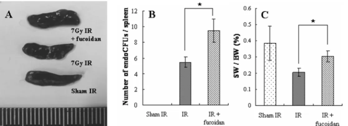
Fucoidan에 의한 비장의 회복과 조혈 촉진 정상대조군에서는비장집락을관찰할수없는반면
7 Gy 방사선조사대조군은 5.5 ± 2.5개로그수가증가
하였다. 그리고 fucoidan 병행투여군은 9.5 ± 1.5개로방
사선조사대조군에비해그수가유의성있게증가하 였다(Figs. 1A and B, p< 0.05). 이결과는방사선이조 사된마우스에서 fucoidan이비장집락수를방사선조사
대조군에비해약 1.7배증가시킨것으로, fucoidan이조
혈모세포의방호와재생성을촉진시키는효과가뛰어난 것을알수있다. 그리고정상대조군의체중에대한비
장무게의비율이 0.39 ± 0.11%인데 비해방사선조사
대조군은 0.21 ± 0.03%로확연히 작은체중에대한비
장의비율을나타내었다. 반면, fucoidan 병행투여군은
체중에대한비장무게의 비율이 0.31 ± 0.04%로방사
선조사대조군에비해 47.3%로뚜렷한증가를보였다
(Fig. 1C). 이렇게증가된체중에대한비장무게의비
율은방사선조사에의한손상으로부터비장의회복을 의미한다.
Fig. 1. The effect of fucoidan on endogenous spleen colonies in mice at 9 days after irradiation. Photograph of spleens excised from each group (A). The columns indicate the number of endogenous colony on spleen surface in each group (B). The columns indicate the percentage of spleen weight (SW) per body weight (BW) (C). Each data point represents the mean ± SEM (*p< 0.05). Although all spleens decreased in size after irradiation, the spleens of fucoidan recipients were bigger and had more endogenous splenocyte colonies.
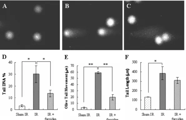
Fucoidan에 의한 비장세포에서의 DNA 손상억제
Comet assay는 in vivo와 in vitro system에서각각의 세포수준에서 DNA 손상을빠르고민감하게감지해낼
수있어, 많은화학적생물학적인자에의한 DNA 손상
을확인할수있는유용한기법이다 [8]. 따라서방사선
에의해유도된 DNA 손상에대한 fucoidan의효과를평
가하기위해 comet assay를수행하였다. 비장세포는감
마선조사를한군에서는 tail DNA%, tail movement 및
tail length가 각각 30.17 ± 1.7%, 60.27 ± 1.56 µm 및 384.36 ± 69.07 µm로크게증가한반면, fucoidan을병행 투여한 군에서는 13.67 ± 2.81%, 19.52 ± 4.34 µm 및
309.03 ± 31.97 µm로유의성있게감소하여각각 2.2, 3.6
및 1.2배억제되었다(Fig. 2, p< 0.05). 이는 fucoidan이 방사선조사에의해생기는비장세포의 DNA 손상을효 과적으로억제시키는효능을가지고있다는것을의미 한다.
Fucoidan에 의한 비장세포의 증식능 증가 내재성비장집락의형성결과를바탕으로방사선조
사후억제된면역세포의증식능에 fucoidan이미치는 영향을확인하기위해 3H-thymidine incorporation 실험 을수행하였다. 방사선조사후 9일째얻은비장세포에
서 fucoidan 병행투여군은 2,762 ± 231 cpm(counts per minute)으로방사선조사대조군이 2,003 ± 623 cpm에비 해약 1.4배증가를나타내었다(Fig. 3). Fucoidan 투여
는비장세포의손상을억제를시킬뿐만아니라비장세 포의증식을촉진시키는것으로사료된다.
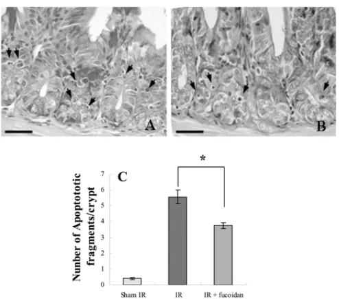
소장움의 apoptotic fragment 형성 억제
방사선조사후 24시간에 apoptotic cell은소장에서의 줄기세포에해당하는소장움의기저부에주로형성되었
으며, H&E 염색상에서핵염색질과세포질의농축및
세포성분절의특성을나타내었다. 반면 fucoidan 병행
투여군의소장에서각소장움당 apoptotic fragments의
수는 3.80 ± 0.31개(Fig. 4B)로 , 방사선조사대조군에서 의 5.57 ± 0.61개(Fig. 4A)에 비해 32%의 소장움의 apoptosis 억제효과가유의성있게나타났다(Fig. 4C, p
< 0.05). 이로써 fucoidan이방사선조사에의한비장세
Fig. 2. The effect of fucoidan on irradiation induced DNA damage in splenocytes using Comet assay. Mice were exposed to: sham irradiation (A), 2 Gy irradiation (B), 2 Gy irradiation plus fucoidan (10 mg/kg b.w., i.p.) treatment (C). The columns indicate tail DNA% (D), Olive tail movement (E), and tail length (F) in each group. The alkaline comet assay is an effective technique for monitoring the extent of DNA strand breaks. DNA damage is reflected in an increase of tail length and movement and a rise in percentage of DNA in the tail. When fucoidan was administrated with irradiation, there were a significant decrease in comet parameters. Each data point represents the mean ± SE of about 50 cells per mouse by Student's t-test (*p< 0.05).
포손상뿐만 아니라소장에서 재생력이있는 stem cell
에해당하는 소장움세포의 apoptosis를억제시켜방사
선장해에대한방호효과를나타냄을알게되었다.
고 찰
본연구에서 fucoidan이방사선에의해억제된조혈기
능을향상시키고비장세포의손상을억제시킬뿐만아 니라면역세포의증식능을증가시키며, 방사선에민감 한소장움세포의방사선에대한저항성을증가시킨다 는것을확인하였다.
Fucoidan은 1913년 Killing에의해처음보고된이후,
주로갈조류에함유되어있는것으로알려져있다. Li
등 [14]에따르면다시마(Laminaria japonica)에서추출 한 fucoidan(약 4,000 mg/kg)을쥐에게경구투여한단기
독성검사에서독성을관찰할 수없었으며, Cooper 등
은미역포자엽에서추출한 fucoidan 1~2.5 g을 1개월에 서 2년동안임상실험을진행한결과, 부작용을발견하
Fig. 3. The effect of fucoidan on proliferation ability of splenocytes. Splenocytes were separated from mice at 9 days after 2 Gy irradiation. Background proliferation is 177
± 28 cpm. Concanavalin A (Con A)-induced proliferation is 17,453 ± 1,828 cpm respectively. Each data point rep-
resents the mean ± SE.
Fig. 4. Morphological classification of typical apoptotic fragments in the small intestinal crypt cells using H&E staining.
(A) 2 Gy irradiation, (B) 2 Gy irradiation + fucoidan treatment, (C) the columns indicate the number of apoptotic fragments per small intestinal crypt in each group. Bars = 20.5µm. The mice were sacrificed at 24 h after 2 Gy gamma-ray irradiation and small intestines were separated. Apoptosis was assessed on the morphological evidence, such as cell shrinkage, chromatin condensation, margination and fragmentation. Values represent means ± SE of 50 crypts section per intestine and 5 small intestine sections per each mouse were recorded (*p< 0.05).
지못하였다고보고하였다 [12]. 이처럼 fucoidan은독성
이적고특별한부작용을나타내지않는다.
방사선조사는생체내말초혈액및말초림프기관의 면역세포수를급격히 감소시킨다. 이에대한보상작용
으로면역세포의재생성이일어나고, 골수내의조혈모
세포가말초림프기관으로이동하여증식하게된다. 이 때비장내에는조혈모세포의증식에의한집락이형성 되며, 방사선조사후비장에서형성된집락의수는조 혈모세포의방호및재생성능력의지표로사용되고있
다 [20]. 이전연구에서는 fucoidan을마우스에경구투여
하면조혈줄기세포가골수에서혈액으로의이동이증 가하고 [7], 사람에게매일 3 g씩 12일간경구투여시말 초혈구세포와줄기세포의수가증가한것을보고하였다
[11]. 본연구에서 fucoidan 투여에의해비장의무게와
집락의수가증가하였으며, 이는방사선조사로인해감 소한말초림프기관인비장내의면역세포수가조혈모세 포의증식으로인해증가하였기때문으로사료된다. 따
라서 fucoidan의투여는조혈계의보호및회복을촉진
시켜동물에서방사선조사후 10-15일에생기는급성
조혈계증후군을막을수있을것으로사료된다.
Apoptosis에의한세포사는 DNA손상에의한급성효
과의결과로서인식되고있으며특히분화증식능이높
은세포와기관에서쉽게나타난다 [1]. 분화증식능이높
은비장은면역체계에서매우중요한역할을하는기관 으로방사선손상에민감한장기로서방사선조사에의 한비장세포의 DNA 손상정도를 comet assay를이용하 여파악하는것은방사선조사에의한생물학적영향을 평가하는데중요한의의를갖는다. Comet assay에서관
찰된 tail length, tail movement 및 tail DNA% 등은 DNA
손상의지표로활용되고있는데, 이지표를기준으로하
였을때, fucoidan 병행투여군이정상대조군보다는높
은수치이지만, 방사선조사대조군과비교하였을때세
포손상즉 DNA 손상의현저한억제효과를나타내었
다. 이전연구에따르면다시마로부터추출한 fucoidan
을쥐에게투여하였을때항산화효소를조절함으로써 항산화효과를가진다고보고하였다 [3]. 따라서 fucoidan
이방사선조사에의해생기는산화적자유라디칼의발 생에대한항산화효과를가짐으로서비장세포의 DNA
손상을효과적으로억제시키는것으로사료된다.
방사선조사후에는면역세포수가급격히감소하며,
면역세포의재생성에의한면역세포수의회복은방사선
조사후회복에중요한역할을한다. 본연구에서fucoidan
은방사선조사로억제된비장세포의증식을촉진하였
다. 이는 fucoidan이면역세포의증가를촉진함으로써방
사선에의해손상받은면역계를보호할수있을것으 로보인다. 이로써말초혈액중면역세포의감소에의한
세균이나바이러스에의한감염을억제하는것에효과 적일것으로사료된다.
소장움은방사선에민감한재생조직으로서, 전신피 폭시위장관계상피세포들의손상에의해사망에이르 는위장관계증후군을일으키기도한다. 위장관계증구군 의주요요인은방사선조사에의해소장, 특히소장움 에서의세포수감소나세포가소장움로부터융모의끝 부분으로이행하는속도가저하되고, 소장점막상피세
포가탈락하기때문이다 [4, 16]. 따라서소장움은방사
선에의한산화적생체손상을측정하는대표적인지표 로이용되어지고있으며, 방사선에대한생체방호에있 어서소장움등의소화기관재생조직손상의억제및회 복이중요한요건의하나로받아들여지고있다. Fucoidan
은방사선조사에의한소장움세포의 apoptosis를억제
시켜방사선장해에대한방호효과를나타내었다. 이는
fucoidan이방사선에의한위장관계통장해에대한방어
효과를나타낼것으로생각되고 apoptosis와관련된질
병의예방과치료에적용할수있을것으로보인다. 하 지만방사선이조사된마우스에서이러한 fucoidan의방
사선방어효과와관련된기전은아직밝혀져있지않 아향후추가연구가필요할것으로사료된다.
결 론
본연구는 fucoidan의방사선장해경감효과를알아
보기위하여방사선을조사한마우스에서면역조혈계와 위장관의방호능력을관찰하였다. Fucoidan의투여는방 사선조사대조군에비해내재성비장조혈집락형성 을촉진시켰고, 비장세포의 DNA 손상을억제와함께증 식률을증대시켰다. 뿐만아니라 fucoidan은방사선에민
감한소장움세포의 apoptosis를억제시킴으로써방사선
에대한저항성을증가시킨다는것을확인하였다. 이상
의결과는 fucoidan이방사선조사로인한부작용인위
장관계통의장해나조혈기장해에대항하여방사선방 어제로서의임상적활용가능성을가질것으로사료된다.
감사의 글
이논문은 2006년도정부재원(교육인적자원부학술연
구조성사업비)으로학술진흥재단(KRF-2006-003-E00 366)과과학기술부의원자력연구개발사업(BAERI)의연 구비지원을받아연구되었음.
참고문헌
1. 김세라, 오헌, 이해준, 신동호, 김종춘, 박인철, 오기
석, 조성기, 김성호.고선량및저선량방사선조사
마우스에서누에동충하초 (Paecilomyces japonica)의 효과. 대한수의학회지 2003, 42, 181-188.
2.박상준, 정규식, 김태환, 임윤규, 박현정, Pham Duc Chuong, 지영흔.마우스소장 crypt cell에서방사선
조사에의해유도된 apoptosis의조절. 한국실험동물 학회지 2004, 20, 218-223.
3.정일윤.프로폴리스에서분리한플라보노이드화합물
의항산화활성및방사선방어효과. 한국식품영양과
학회지 2005, 34, 162-166.
4.조성기, 박혜란, 정우희, 오헌, 김성호, 이성태.방사
선에대한생약복합조성물(HemoHIM)의재생조직및
면역계방호 · 회복촉진효과. 한국식품영양과학회지 2005, 34, 805-813.
5.Aisa Y, Miyakawa Y, Nakazato T, Shibata H, Saito K, Ikeda Y, Kizaki M. Fucoidan induces apoptosis of human HS-sultan cells accompanied by activation of caspase-3 and down-regulation of ERK pathways. Am J Hematol 2005, 78, 7-14.
6.Bogo V, Jacobs AJ, Weiss JF. Behavioral Toxicity and efficacy of WR-2721 as a radioprotectant. Radiat Res 1985, 104, 182-190.
7.Frenette PS, Weiss L. Sulfated glycans induce rapid hematopoietic progenitor cell mobilization: evidence for selectin-dependent and independent mechanisms.
Blood 2000, 96, 2460-2468.
8.Gandhi NM, Gopalaswamy UV, Nair CK. Radiation protection by disulfiram: protection of membrane and DNA in vitro and in vivo against gamma-radiation. J Radiat Res (Tokyo) 2003, 44, 255-259.
9.Halliwell B, Gutteridge JM. Free Radicals in Biology and Medicine. 3rd ed. pp. 604-607, Oxford University Press, Oxford, 1999.
10. Hendry JH, Robert SA, Potten CS. The clonogen content of murine intestinal crypts: dependence on radiation dose used in its determination. Radiat Res 1992, 132, 115-119.
11. Irhimeh MR, Fitton JH, Lowenthal RM. Fucoidan ingestion increases the expression of CXCR4 on human CD34+ cells. Exp Hematol 2007, 35, 989-994.
12. Irhimeh MR, Fitton JH, Lowenthal RM, Kongtawelert P. A quantitative method to detect fucoidan in human plasma using a novel antibody.
Methods Find Exp Clin Pharmacol 2005, 27, 705-710.
13. Itoh H, Noda H, Amano H, Zhuaug C, Mizuno T, Ito H. Antitumor activity and immunological properties of marine algal polysaccharides, especially fucoidan, prepared from Sargassum thunbergii of Phaeophyceae.
Anticancer Res 1993, 13, 2045-2052.
14. Li N, Zhang Q, Song J. Toxicological evaluation of fucoidan extracted from Laminaria japonica in Wistar rats. Food Chem Toxicol 2005, 43, 421-426.
15. MacVittie TJ, Monroy RL, Patchen ML, Souza LM.
Therapeutic use of recombinant human G-CSF (rhG- CSF) in a canine model of sublethal and lethal whole- body irradiation. Int J Radiat Biol 1990, 57, 723-736.
16. Milas L, Hunter N, Ito H, Peters LJ. In vivo radioprotective activities of diethyldithiocarbamate (DDC). Int J Radiat Oncol Biol Phys 1984, 10, 2335- 2343.
17. Neta R. Role of cytokines in radioprotection.
Pharmacol Ther 1988, 39, 261-266.
18. Neta R, Douches S, Oppenheim JJ. Interleukin 1 is a radioprotector. J Immunol 1986, 136, 2483-2485.
19. Ponce NM, Pujol CA, Damonte EB, Flores ML, Stortz CA. Fucoidans from the brown seaweed Adenocystis utricularis: extraction methods, antiviral activity and structural studies. Carbohydr Res 2003, 338, 153-165.
20. Talmadge JE, Tribble H, Pennington R, Bowersox O, Schneider MA, Castelli P, Black PL, Abe F.
Protective, restorative, and therapeutic properties of recombinant colony-stimulating factors. Blood 1989, 73, 2093-2103.
21. Utley JF, Phillips TL, Kane LJ. Protection of normal tissues by WR2721 during fractionated irradiation. Int J Radiat Oncol Biol Phys 1976, 1, 699-703.