- 57 -
R eceived R e v i s e d
A ccepted
: June 27, 2018
: September 19, 2018(1차) / September 25, 2018(2차) September 27, 2018(3차)
: October 18, 2018 +Corresponding author: Minsu Kwon
Department of Otorhinolaryngology, Eulji University School of Medicine, Eulji Medical Center, 68 Hangeulbiseok-ro, Nowon-gu, Seoul 01830, Korea
Tel: +82-2-970-8276, Fax: +82-2-970-8275 E-mail: [email protected]
대한두경부종양학회지, 제34권 제2호, 2018. pp.57-60 Korean Journal of Head & Neck Oncology, Vol.34, No.2
https://doi.org/10.21593/kjhno/2018.34.2.57 ISSN 1229-5183(Print) / ISSN 2586-2553(Online)
이하선 Warthin 종양의 세침흡인검사 후 발생한 육아종성 괴사 1예
오현식⋅이은섭⋅조용태⋅권민수+
을지대학교 을지병원, 을지대학교 의과대학 이비인후과학 교실
A Case of Parotid Warthin Tumor with Granulomatous Necrosis After Fine-Needle Aspiration
Hyeon Sik Oh, MD, Eun Sub Lee, MD, Young Tae Jo, MD, Minsu Kwon, MD, PhD+ Department of Otorhinolaryngology, Eulji University School of Medicine, Eulji Medical Center, Seoul, Korea
= Abstract =
Fine needle aspiration is usually performed as a preoperative cytologic evaluation in salivary gland tumors, and complications of the lesion after fine needle aspiration are scarcely developed. The secondary change of tissue by fine needle aspiration can make the diagnostic and subsequent therapeutic difficulties for clinicians and require a careful approach. Fine needle aspiration can cause variety of changes in Warthin tumor, and it is presumed that those changes are mainly caused by the infarction of the mass due to vascular injury and following in- flammatory reactions. We would introduce our experience of granulomatous and necrotic change after fine needle aspiration on parotid Warthin tumor with consequent difficulties in diagnostic and surgical approach to the patient.
Key W ords : Fine needle aspiration, Granuloma, Parotid gland, Warthin tumor
서 론
Warthin 종양(Warthin tumor)은 이하선에 발생하는 타 액선 양성종양 중에서 두번째로 흔한 것으로 알려져 있으 며, 주로 고령의 남자에서 흡연과 관련되어 발생한다.1,2) Warthin 종양은 병리학적으로 papillary cystadenoma lym- phomatosum이라고 명명되는데, 명칭에서 유추할 수 있 듯이, 림프 조직 기원으로 다양한 림프구와 염증 관련 세포들이 내포되어 있고, 낭성 조직으로 혈관 발달이 잘 이뤄져 있지 않은 특성이 있다. 이로 인해 혈관손상에
따른 경색(infarction) 및 그에 따른 이차적 염증 변화가 나타날 수 있다.3)
세침흡인검사(fine needle aspiration)는 종양의 치료 전 조직학적 확인을 위해 사용되는 방법으로 두경부 병변에 대한 민감도 및 특이도가 각각 89.6%, 96.5%로 보고되고 있어 두경부 종양의 진단 및 치료계획 수립에 통상적으 로 이용되고 있다.4) Warthin 종양의 경우 세침흡인검사 의 진단 정확도가 53%로 상대적으로 떨어지는데, 이는 진단을 위해서 특징적인 두가지 성분인 호산성 종양세포 와 림프구들이 같이 나타나야 하는데 그렇지 못할 경우 진단하기 쉽지 않다는 점 때문이다.5)영상검사나 임상양 상으로 추정되는 종양과 세침흡인검사의 결과가 상이한 경우도 많고, 양성 종양과 악성 종양의 감별이 어려운 경우가 드물지 않아 진단에 어려움을 초래한다.
타액선 종양에서 세침흡인검사 후 출혈, 감염 및 기타 합병증 발생 빈도는 1.6-16.7% 정도로 알려져 있으며, 괴사, 낭성변화, 섬유화, 이형성(metaplasia) 등 검사 후 종양의 조 직학적 변화는 문헌보고 상 0.4% 정도로 매우 드물다.3,6)
- 58 - A
B
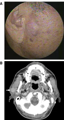
Fig. 1. Facial photography (A) and computed tomography (CT) scan (B) of the patient at initial visit. (A) A smooth and rounded mass was palpated in the right infraauricular area.
(B) Neck CT showed 2.7cm sized mass with a clear borderline and contrast enhancement in the right parotid gland (white arrow).
A
B
Fig. 2. Facial photography (A) and ultrasonography (B) of the patient after fine needle aspiration. (A) Overlying skin swelling and redness was observed after fine needle aspiration of right parotid gland. (B) Ultrasonography confirmed the mass occupying from the deep lobe to the superficial lobe of the parotid gland. The mass invades the superficial muscu- loaponeurotic system and is adjacent to skin (white arrow).
저자들은 Warthin 종양에서 세침흡인검사 이후 무혈관성 경색(avascular infarction)과 육아종성 괴사(granulomatous necrosis)를 보여 수술 전과 수술 중 악성종양과의 감별 진단과 수술 범위 결정에 있어 어려움을 겪은 증례를 보고하고, Warthin 종양의 세침흡인검사 후 발생할 수 있는 임상적 양상과 조직학적 변화에 관해 고찰하고자 한다.
증 례
73세 남자 환자가 3주 전부터 촉지된 우측 전이개 부위 의 종물을 주소로 내원하였다. 신체진찰에서 우측 전이개 부위에 2.5 cm 가량 직경의 부드럽고 둥근 종물이 촉진되 고 통증, 압통, 열감 등 동반 증상은 없었다. 경부 전산화 단층촬영에서는 우측 이하선 천엽에서 2.7cm 크기의 주 변과 경계가 분명하고, 조영증강되는 종괴가 관찰되었 으며, 영상 판독 상 다형선종 또는 Warthin 종양으로 진 단되었다(Fig. 1). 외래에서 세침흡인검사를 시행하였으 며 의미 있는 세포는 발견되지 않았다.
상기 소견을 바탕으로 이하선 양성종양으로 진단 후 수술적 절제를 계획하고 있던 중 세침흡인검사 후 40일째 검사 부위에서 노란색 장액성 분비물과 함께 종물을 덮고 있는 피부에 어두운 발적을 보이는 변화가 관찰되어 예정 일보다 일찍 환자가 내원하였다(Fig. 2A). 세침흡인검사 부작용으로 인한 감염 발생 가능성을 고려하였으나 검사 후 40일이나 지난 시점이며, 통증이나 열감, 압통 등을 동 반하지 않았고 혈액검사 상 염증을 시사하는 소견이 보 이지 않아 감염보다는 세침흡인검사 후 종물에 이차적 변화가 발생한 것으로 판단하였고, 영상의학과에 의뢰하 여 초음파 유도 하 세침흡인검사를 재시행하였다. 초음 파 검사상 종괴는 이하선의 심엽부터 천엽까지 차지하고 있었으며, 표층근건막체계(superficial musculoaponeurotic system)를 뚫고 피부에 인접해 있었다(Fig. 2B). 세침흡인 검사 결과는 Warthin 종양이었지만 임상 양상 및 초음파 소견을 바탕으로 악성 종양의 가능성도 염두에 두고 수술 을 진행하였다.
Modified Blair 절개 후 피부피판을 거상하자 이하선 피막을 뚫고 피하조직에 접해 있는 종양이 관찰되었고 (Fig. 3A), 수술 중 종양이 안면 신경에 심하게 유착되어 있었으며, 심엽까지 침범하고 있었다(Fig. 3B). 수술 중
- 59 -
A B C
Fig. 3. Intraoperative findings. (A) Tumor was adjacent to skin and invasion of parotid gland capsule. (B) The tumor and facial nerve were severely adherent. (C) All branches of the facial nerve were identified and tumor occupying the deep lobe of the superficial lobes of the parotid gland was removed.
A B C
Fig. 4. Pathologic findings. (A) A pathologic examination revealed typical features of Warthin's tumor, including dense lymphatic matrix, variable germinal center, and cystic space at the center of the mass. (B) Necrosis and inflammatory tissue were observed in the outer part of the tumor. (C) Multinucleated giant cells were also observed. (white arrow) (H&E stain).
동결절편검사에서는 만성 염증 및 괴사 조직만 관찰되었 으며 악성세포는 확인되지 않았다. 안면신경의 모든 분 지를 확인하면서 이하선 심엽까지 차지하고 있던 종물을 제거하고 수술을 종료하였다(Fig. 3C).
병리 검사 결과 종괴 중심부에서는 조밀한 림프성 기 질, 가변 배중심(variable germinal center), 낭포성 공간 등 Warthin 종양의 전형적인 모습이 관찰되었고(Fig. 4A), 종 양과 연접해 있던 피하조직과 안면신경 주변부위의 검체 에서는 심한 괴사 및 염증 조직이 관찰되었다(Fig. 4B).
또한, 다핵거대세포(multinucleated giant cell)도 관찰되었 으며(Fig. 4C) 결핵 등 육아종성 병변을 보이는 기타 질환 과 감별을 위해 항산균 도말염색 등을 추가적으로 시행 하였으나 음성으로 확인되었다. 병리 결과는 Warthin 종 양으로 확진 되었다.
수술 직후 House-Brackmann 4등급의 안면마비가 관찰 되었으며, 수술 후 7일째 퇴원하였다. 외래 경과관찰 중 수술 19일째부터 배액관을 삽입했던 부위에서 타액으로 추정되는 장액성 분비물이 나왔고, 압박드레싱으로 보 존적 치료하였으며 수술 33일째에 분비물은 더 이상 나 오지 않았다. 안면신경 마비는 조금씩 호전되어 수술 후 99일째 이마 부위 주름의 비대칭만 약간 남기고 모두 호전되었다.
고 찰
Warthin 종양은 유두상 성장, 낭종의 형성, 림프조직의 침윤을 보이는 특징을 지닌 양성종양이지만 드물게 악성 으로 전환되는 사례들이 보고되고 있다.6-9)두경부 종괴의 진단적 접근을 위해 흔히 세침흡인검사를 시행하는데, 타 액선 악성 종양 양성 예측도는 91%로, 검사상 양성 소견 을 보인 경우라도 악성 종양 가능성을 배제할 수 없다.10) 본 증례의 이하선 종물에 대해 저자들은 초기에는 양성 종양으로 진단하였다가, 세침흡인검사 이후 종양이 피부 누공을 형성하는 점, 표층근건막체계를 침범하는 초음파 소견, 종물 주변의 섬유화, 안면 신경과의 심한 유착을 보이는 점을 이유로 악성 종물 가능성을 염두하였으나 최종 병리 결과는 양성인 Warthin 종양으로 확인되었다.
세침흡인검사가 이하선 검사 부위의 부작용 및 종괴의 성질 변화를 일으킨 사례에 대해 보고된 문헌을 살펴보면, Bahar 등11)은 세침흡인검사를 시행한 35예의 Warthin 종양 환자 중에 5예 에서 급성 이하선염이 발생하였고, Suzuki 등12)은 35예의 Warthin 종양 환자 중에 3예에서 이하선염 이 발생하였으며, 동반 증상이 없었던 본 증례와는 다르게 통증, 부종, 농양 형성 등의 증상을 동반하였다. 또한 기 존 보고에서는 세침흡인검사 후 1일에서 7일 이내 단기 간에 합병증이 일어난 반면, 본 증례의 경우는 검사 후 1개월이 넘은 시점에서 종물의 변성에 따른 문제가 확인
- 60 - 되었다는 것에서 보고 가치가 있을 것이다. Jung 등13)은 Warthin 종양의 1% 정도에서 육아종성 변화를 관찰하였 으며, Ryska 등3)도 이하선 세침흡인검사 후 1059예 중 4예에서 육아종성 변화를 일으킨 것을 보고하였다. 감염, 염증 및 종양 등 다양한 원인으로 인해 발생한 조직 손상 후 활성화된 조직구(histiocyte)가 효율적으로 죽은 세포의 섭식(phagocytosis)을 못하게 되면, 이차적으로 복합적인 면역반응이 지속되어 각종 면역세포의 군집과 특징적인 육아종성 변화가 나타나게 된다.13)본 증례의 경우에도 세침흡인검사 후 손상된 세포의 제거와 급성기 염증 반 응의 조절이 효율적으로 이뤄지지 못하면서 육아종성 염증반응이 점진적으로 진행되었고, 그에 따라 검사 후 1개월이 넘게 지난 뒤늦은 시점에서 육아종성 염증반응 의 파급으로 인한 피부 누공과 이하선 심엽의 조직변화 를 초래한 것으로 설명할 수 있을 것이다.
Warthin 종양에서 세침흡인검사 후 검사 부위의 병리 학적 변화를 설명할 수 있는 합리적인 가설은 검사로 인해 혈관 손상이 유발되어 이차적인 변화가 일어난다는 것이다.14)즉, 혈관 손상으로 인해 종물의 허혈이 발생하 여 주변부 세포의 출혈, 괴사, 섬유화, 이형성 변화 등의 이차성 변화가 일어나며, 경색된 Warthin 종양은 다양한 병리적 변성을 보이게 된다. 괴사, 유두 소실, 편평세포화 생, 포말대식세포, 다핵거대세포, 괴사성 육아종, 섬유화 등의 같은 형태학적 변성이 관찰될 수 있으며, 이는 양성 종양을 악성 종양으로 오인하게 할 수 있다.15)
따라서 본 증례를 통해 Warthin 종양에서 세침흡인검 사 후 일어날 수 있는 병리학적 변화 가능성을 숙지해 두는 것이 향후 유사한 경험을 하게 될 수 있는 두경부외 과의들에게 올바른 치료계획을 세우는데 도움이 될 것으 로 생각한다.
References
1) Spiro RH. Salivary neoplasms: Overview of a 35-year experience with 2,807 patients. Head Neck Surg. 1986;8:177-184.
2) Woo JS, Lee HM, Kim HY, Choi JH, Jung JY, Jung KY, et al.
Effect of smoking to Warthin's tumor of parotid gland. Korean J
Otolaryngol-Head Neck Surg. 2002;45:380-382.
3) Ryska A, Seifert G. Adenolymphoma (Warthin’s tumor) with multiple sarcoid-like granulomas. Pathol Res Pract. 1999;195:
835–839.
4) Tandon S, Shahab R, Benton JI, Ghosh SK, Sheard J, Jones TM.
Fine-needle aspiration cytology in a regional head and neck can- cer center: Comparison with a systematic review and meta- analysis. Head Neck. 2008;30:1246-1252.
5) Choi G, Kim HY, Park HJ, Woo JS, Jung KY, Choi JO. Clinical Study of Warthin's Tumors. Korean J Otorhinolaryngol- Head Neck Surg. 1996;39:1820-1827.
6) Mukunyadzi P, Bardales RH, Palmer HE, Stanley MW. Tissue ef- fects of salivary gland fine needle aspiration. Does this proce- dure preclude accurate histologic diagnosis? Am J Clin Pathol.
2000;114:741–745.
7) Yoo GH, Eisele DW, Askin FB, Driben JS, Johns ME. Warthin’s tumor: A 40-year experience at the Johns Hopkins Hospital.
Laryngoscope. 1994;104:799-803.
8) Yaranal PJ, Umashankar T. Squamous Cell Carcinoma Arising in Warthin’s Tumour: A Case Report. J Clin Diagn Res. 2013;7:
163–165.
9) Yamada S, Matsuo T, Fujita S, Suyama K, Yamaguchi A, Mizuno A. Mucoepidermoid carcinoma arising in Warthin's tu- mor of the parotid gland. Pathol Int. 2002;52:653-656.
10) Kim JY, Pae KH, Choi SH, Kim SY, Nam SY. Clinical Values of Fine Needle Aspiration Biopsy in Salivary Gland Diseases.
Korean J Otolaryngol-Head Neck Surg. 2006;49:639-643.
11) Bahar G, Dudkiewicz M, Feinmesser R, Joshua BZ, Braslavsky D, Popovtzer A, et al. Acute parotitis as a complication of fine- needle aspiration in Warthin's tumor. A unique finding of a 3-year experience with parotid tumor aspiration. Otolaryngol Head Neck Surg. 2006;134:646-649.
12) Suzuki K, Iwai H, Kaneko T, Sakaguchi M, Hoshino S, Inaba M.
Induction of parotitis by fine-needle aspiration in parotid Warthin’s tumor. Otolaryngol Head Neck Surg. 2009;141:282-284.
13) Shah KK, Pritt BS, Alexander MP. Histopathologic review of granulomatous inflammation. J Clin Tuberc Other Mycobact Dis. 2017;7:1-12.
14) Jung SM, Hao SP. Warthin's tumor with multiple granulomas: A clinicopathologic study of six cases. Diagn Cytopathol. 2006;
34:564-567.
15) Tan Y, Kryvenko ON, Kerr DA, Chapman JR, Kovacs C, Arnold DJ, et al. Diagnostic pitfalls of infarcted Warthin tumor in frozen section evaluation. Ann Diagn Pathol. 2016;25:26-30.