LED 파장이 Saccharomyces cerevisiae의 생장에 미치는 영향
쉬시아오통1․이지은1․정소미2․강우신1․류시형1․김한호1․김수룡1․이가혜2․안동현1
1부경대학교 식품공학과/식품연구소
2부경대학교 수산과학연구소
Effect of Light-Emitting Diode (LED) Wavelengths on Growth of Saccharomyces cerevisiae
Xiaotong Xu1, Ji-Eun Lee1, So-Mi Jeong2, Woo-Sin Kang1, Si-Hyeong Ryu1, Han-Ho Kim1, Su-Ryong Kim1, Ga-Hye Lee2, and Dong-Hyun Ahn1
1Department of Food Science and Technology/Institute of Food Science and
2Institute of Fisheries Sciences, Pukyong National University
ABSTRACT Light-emitting diodes (LEDs) are divided into ultraviolet LEDs (UV-LEDs), visible LEDs (Vis-LEDs), and infrared LEDs (IR-LEDs). LEDs of different wavelengths have different functions. This study examined the effects of different wavelengths on the growth of Saccharomyces cerevisiae. S. cerevisiae cells were incubated for 48 h at 30°C. Subsequently, S. cerevisiae suspensions were diluted to 1×10³∼1×10⁴ CFU/mL and 1×10⁷ CFU/mL. The S. cerevisiae suspensions were then exposed to UV-LED (270 nm and 365 nm), Vis-LED (465∼475 nm and 620∼630 nm), and IR-LED (850 nm and 5,000∼7,000 nm), with a lamp-to-suspension distance of 4.5 cm. The microorganisms were irradiated by 270 nm UV-LED for 10 and 30 min and 365 nm UV-LED, Vis-LED, and IR-LED for 60 and 180 min, respectively. Among the LEDs used, the 465∼475 nm Vis-LED had the strongest inhibitory ability on S.
cerevisiae. In contrast, the 620∼630 nm Vis-LED and infrared LED showed either low antibacterial activity or even no bacteriostatic ability.
Key words: light-emitting diode (LED), Saccharomyces cerevisiae, inhibitory ability, different wavelengths
Received 7 Jan 2021; Revised 19 Feb 2021; Accepted 22 Feb 2021 Corresponding author: Dong-Hyun Ahn, Department of Food Sci- ence & Technology, Pukyong National University, 45, Yongso-ro, Nam-gu, Busan 48513, Korea, E-mail: [email protected] Author information: Xiaotong Xu (Graduate student), Ji-Eun Lee (Graduate student), Woo-Sin Kang (Graduate student), Si-Hyeong Ryu (Graduate student), Han-Ho Kim (Graduate student), Su-Ryong Kim (Graduate student), Dong-Hyun Ahn (Professor)
서 론
지난 수십 년 동안 식품 보존에 있어 미생물 오염과 식품 부패를 방지하는 것이 식품 안전의 가장 중요한 문제가 되어 왔다. 식품이 제조업체에서 소비자에게 전달되기 전에 식품 공급 체인의 모든 단계에서 식품 부패가 발생한다. 따라서 식품 부패 방지는 식품 엔지니어가 해결해야 하는 중요한 문제이다.
최근 식품의 품질을 개선하고 유지하며 유통기한을 연장 하기 위한 방법으로 비가열 가공기술인 발광다이오드(light- emitting diode, LED) 조사 방법이 주목 받고 있다(Do 등, 2015). LED는 전류가 통과할 때 가시광선을 방출하는 반도 체 소자로서 매우 좁은 스펙트럼 파장에서 발광할 수 있으며 단색 파장을 갖고 있다(Held, 2016). 또한 가시광원에 비해
낮은 에너지 소비 및 높은 내구성 등의 장점이 있다(Ghate 등, 2013).
최근에는 LED 기술을 살균 용도로 활용하고 있을 뿐만 아니라 특정 파장의 LED는 프로바이오틱스의 성장을 촉진 하고, 발효과정에서 생리 활성 물질을 증가시키는 것으로 밝 혀진 바 있다(Jeong 등, 2018). 또한 식물의 재배, 의료 영역 및 피부 미용 등 특수 응용 분야에서도 LED의 사용이 점차 확대되고 있다(Bang과 Kim, 2012). 이와 같이 다양한 용도 에 사용되고 있는 LED는 방출하는 빛의 종류에 따라 자외선 LED(UV-LED), 가시광선 LED(Vis-LED), 적외선 LED(IR- LED)로 구분되고(Lee 등, 2011), 다양한 파장에 따라 신제 품 및 용도 개발이 가능하다. UV 스펙트럼은 다양한 파장과 용도에 따라 세분화될 수 있는데, 파장에 따라 UV-A(315~
400 nm), UV-B(280~315 nm) 및 UV-C(<280 nm)로 나 눌 수 있다(Soni 등, 2016). 살균파장으로 알려진 UV-C는 DNA에 잘 흡수되기 때문에 병원성 및 부패 미생물의 DNA 를 손상시킬 수 있다(Bolton과 Cotton, 2008). UV-A는 주 로 활성산소의 생성에 의해 단백질과 지질을 포함한 다른 생체분자에 산화 저해를 일으켜 미생물을 비활성화시킨다 (Brem 등, 2017). 가시광선 LED는 LED 시장에서 가장 큰 비중을 차지하고 있고, 주로 실내・외 조명, 전자제품, 간판
Table 1. Inactivation of S. cerevisiae by ultraviolet C irradiation (270 nm) (Unit: log CFU/mL)
Irradiation time (min) Control 107 1) 0
10 30
6.80±0.28b2) 7.10±0.21a 7.49±0.09a
<33)
<3
1)Initial strain concentration.
2)Means in the same column bearing different letters (a,b) in samples are significantly different (P<0.05).
3)No growth.
및 자동차 등에 이용되고 있다(BIR Research Group, 2010). 최근에는 가시광선 LED의 살균작용과 손상된 피부 에 대한 재생 및 재활에 대한 연구가 활발하게 이루어지고 있다(Kim과 Lee, 2019; Do와 Bang, 2013). 적외선 LED는 주로 광통신, 리모컨 및 계측기 등 다양한 분야에 활용되고 있는데, Sadick의 연구(2009)에서는 피부에 가시광선 LED 와 적외선 LED를 조합하여 처리했을 때 여드름을 감소시킬 수 있다고 보고하였다.
효모의 일종인 Saccharomyces cerevisiae는 작은 단일 세포로서 쉽게 배양할 수 있고 유전자 조작이 가능하다는 장점이 있어, 식품산업에서 주요 발효제로 사용되고 있을 뿐만 아니라(Legras 등, 2007; Park 등, 2003) 여러 다양한 분야에서 활용되고 있다(Stewart, 2014). S. cerevisiae는 인체에 무해한 GRAS(generally recognized as safe) 미생 물로서 오래전부터 빵, 와인 및 맥주의 발효를 포함하여 식 품산업에서 널리 사용되어 왔으며, 단백질, nucleic acids, 비타민, 지질 등 다양한 유용물질을 생산하기 위한 원료로도 사용되고 있다(Park 등, 2003). 또한, 항생제 투약에 의한 설사치료제와 영양보충제로도 사용되고 있는데(Liu 등, 2017) S. cerevisiae를 식품산업에 효율적으로 활용하기 위해서는 S. cerevisiae의 생육 또는 발효 과정을 적절히 조절할 수 있는 다양한 방법에 대한 연구가 필요할 것으로 여겨진다.
따라서 본 연구에서는 식품산업에서 S. cerevisiae를 효 율적으로 활용하기 위해 다양한 파장의 빛을 방출하는 LED 를 이용하여 S. cerevisiae의 생육에 미치는 영향을 조사하 였고, 식품산업에 있어 파장별 LED 조사의 효율적 이용 가 치를 제시하고자 하였다.
재료 및 방법
시험 균주 및 배지
본 실험에서 사용한 S. cerevisiae 균주는 한국생명공학 연구원 생명자원센터(Korea Culture Type Collection;
KCTC, Daejeon, Korea)에서 분양받아 사용하였다. S. cere- visiae는 YM배지(0.3% yeast extract, 0.3% malt extract, 1% dextrose, 0.5% peptone)에 접종하여 30°C에서 48시 간 배양하였다. YMA는 YM배지에 1.2% agar를 첨가하여 사용하였다.
LED 광원
파장의 크기에 따른 LED가 S. cerevisiae의 생장에 미치 는 영향을 알아보기 위해 총 6종(270 nm, 365 nm, 465~
475 nm, 620~630 nm, 850 nm 및 5,000~7,000 nm)의 LED 파장을 실험에 사용하였다. 실험장비는 가로×세로의 크기가 26×13.5 cm가 되도록 판을 제작하였고, 270 nm 및 365 nm 빛 장비는 판 하부에 LED 소자 40개를 가로, 세로 2×2 cm 간격으로, 465~475 nm 빛 장비는 판 하부에 LED 소자 168개를 가로, 세로 0.8×1.6 cm 간격으로, 620~
630 nm 빛 장비는 판 하부에 LED 소자 168개를 가로, 세로 0.8×1.2 cm 간격으로, 850 nm 빛 장비는 판 하부에 LED 소자 160개를 가로, 세로 0.9×1.3 cm 간격으로, 5,000~
7,000 nm 빛 장비는 판 하부에 LED 소자 204개를 가로, 세로 0.6×1.5 cm 간격으로 부착하여 제작하였다. 전류 전 원은 AC adapter(0~8 A, 0~24 V)를 이용하여 공급하였다.
LED 조사
파장별 LED 조사가 S. cerevisiae에 미치는 영향에 대해 알아보기 위해 1×10³~1×10⁴ CFU/mL 또는 1×10⁷ CFU/
mL의 S. cerevisiae를 60×15 mm Petri dish에 1 mL씩 접 종하고 파장별 LED를 조사하였다. 270 nm UV-LED 램프 의 조사 시간은 10분과 30분으로 하였고, 365 nm UV-LED 램프, 465~475 nm, 620~630 nm visible-LED 램프, 850 nm visible-LED 램프 및 5,000~7,000 nm infrared-LED 램프의 조사 시간은 60분과 180분으로 하였다. 조사 시 배 지의 온도는 30~35°C로 일정하게 유지하였으며 램프와 배 지와의 거리는 4.5 cm이고, 465~475 nm, 620~630 nm, 850 nm 및 5,000~7,000 nm의 조사 강도는 150 W/cm2, 270 nm의 조사 강도는 0.5 W/cm2, 360 nm의 조사 강도는 35 W/cm2로 하였다. 파장별 LED를 조사한 후 30°C에서 1시간 정치하였고, 균수가 약 10~102 CFU/mL가 되도록 10배 희석법으로 희석하였다. 희석한 각 균주는 주입평판법 을 이용하여 배지에 접종하고 30°C에서 48시간 동안 배양 하여 최종적으로 생성된 콜로니를 계수하였다.
통계처리
실험 결과의 통계처리는 SAS program(Statistical ana- lytical system V8.2, SAS Institute Inc., Cary, NC, USA) 을 이용하여 분산분석을 실시하였다. Duncan의 다중검정법 으로 P<0.05 수준에서 항목 간의 유의성을 검정하였다.
결과 및 고찰
270 nm UV-LED 조사 시 S. cerevisiae의 생장억제 효과 270 nm UV-LED를 1×10⁷ CFU/mL의 S. cerevisiae에 조사한 결과는 Table 1과 같다. 초기 S. cerevisiae의 균수 는 6.80 log CFU/mL였고, 10분과 30분 비조사구의 S. cerevisiae 균수는 각각 7.10 log CFU/mL와 7.49 log
Table 2. Inactivation of S. cerevisiae by ultraviolet a light emit- ting diode irradiation (365 nm) (Unit: log CFU/mL)
Irradiation time (min) Control 107 1) 0
60 180
7.83±0.10c2) 8.85±0.01b 9.19±0.01a
7.00±0.00
<33)
1)Initial strain concentration.
2)Means in the same column bearing different letters (a-c) in samples are significantly different (P<0.05).
3)No growth.
Table 3. Inactivation of S. cerevisiae by visible light emitting diode irradiation (465∼475 nm) (Unit: log CFU/mL)
Irradiation time (min) Control 107 1) 0
60 180
7.30±0.17b2) 8.57±0.07aA3)
7.29±0.36bA
7.63±0.07aB 7.47±0.03aA
1)Initial strain concentration.
2)Means in the same column bearing different letters (a-c) in samples are significantly different (P<0.05).
3)Means in the same row bearing different letters (A,B) in sam- ples are significantly different (P<0.05).
Table 4. Activation of S. cerevisiae by visible light emitting diode irradiation (620∼630 nm) (Unit: log CFU/mL)
Irradiation time (min) Control 103 1) 0
60 180
3.41±0.01a2) 3.59±0.16aB3)
3.65±0.07aB
4.24±0.09bA 4.63±0.03aA
1)Initial strain concentration.
2)Means in the same column bearing different letters (a,b) in samples are significantly different (P<0.05).
3)Means in the same row bearing different letters (A,B) in sam- ples are significantly different (P<0.05).
CFU/mL로 확인되었다. 270 nm UV-LED로 10분과 30분 동안 조사 시 대조구에 비해 모두 100% 생장억제 효과를 보였다.
UV-C는 식품 표면의 미생물을 효과적으로 줄일 수 있는 비가열 기술이다. 자외선 광자의 흡수는 DNA와 RNA의 구 조에 변화를 일으켜 병원체가 복제할 수 없게 하여 세포사멸 을 유도한다(Beukers와 Berends, 1960; Elmnasser 등, 2007). Green 등(2018)의 연구에서는 259~275 nm의 UV- LED가 미생물의 성장을 효과적으로 억제할 수 있다고 보고 하였고, Kim 등(2016)도 UV-C를 이용하여 슬라이스 치즈 의 병원균을 억제한다고 보고하였다. 또한 Moazzami 등 (2021)은 265 nm UV-C LED가 오염된 닭 운송 장치에 대한 항균효과를 나타낸다고 보고하였다. 따라서 270 nm UV-LED 조사는 S. cerevisiae를 비활성화하기 위한 효과 적인 살균기술로 활용될 수 있을 것으로 판단된다.
365 nm UV-LED 조사 시 S. cerevisiae의 생장억제 효과 365 nm UV-LED를 S. cerevisiae에 60, 180분 동안 조 사한 결과는 Table 2에 나타냈다. 초기 S. cerevisiae의 균 수는 7.83 log CFU/mL, 60분과 180분 비조사구 S. cer- evisiae의 균수는 각각 8.85 log CFU/mL와 9.19 log CFU/
mL로 확인되었다. 365 nm UV-LED를 60분 동안 조사 시 대조구에 비해 1.85 log CFU/mL만큼 생장이 감소하였다.
또한 180분 조사 시에는 100% 생장억제율을 보였다.
Lee 등(2019)은 365 nm UV-LED의 살균 실험에서 365 nm UV-LED가 포장재를 투과하여 Bacillus subtilis의 생 육을 억제한다고 보고하였고, Mori 등(2007)은 365 nm UV-LED를 이용한 물 소독에 관한 연구에서 Escherichia coli DH5α, enteropathogenic E. coli, Vibrio parahae- molyticus 및 Staphylococcus aureus 등의 균주가 365 nm UV-LED 조사에 의해 비활성화될 수 있을 것이라고 밝 혔다. 또한 Lee 등(2012)은 400 nm의 UV-A LED를 이용 한 Pseudomonas aeruginosa와 S. aureus의 살균효과를 입증한 바 있다. 따라서 본 연구를 통해 365 nm UV-LED를 S. cerevisiae에 대한 살균광원으로 사용할 수 있으며, 조사 시간이 길어질수록 우수한 살균효과가 있는 것을 확인하였 다.
465~475 nm Vis-LED 조사 시 S. cerevisiae의 생장억제 효과
가시광선 영역인 465~475 nm Vis-LED를 S. cerevisiae 에 60, 180분 동안 조사한 결과는 Table 3과 같다. 초기 S. cerevisiae의 균수는 7.30 log CFU/mL, 60분 비조사구 의 S. cerevisiae 균수는 8.57 log CFU/mL로 확인되었다.
180분 비조사구의 S. cerevisiae 균수는 7.29 log CFU/mL 로 60분 비조사구에 비해 약간 감소하지만 초기 균수와 비 교하여 유의한 차이는 없었다. 465~475 nm Vis-LED를 60분 동안 조사 시 대조구에 비해 약 0.94 log CFU/mL의 감소를 보였으나, 180분 동안 조사한 경우에는 대조구에 비 해 유의한 차이가 없는 것으로 확인되었다.
465~475 nm의 파장은 청색 빛을 방출하며, 청색 빛은 세균과 곰팡이에 대해 광범위한 항균효과를 나타내는 것으 로 밝혀졌다(Maclean 등, 2009; Murdoch 등, 2013). Do와 Bang(2013)은 461 nm 청색 LED를 E. coli O157:H7, S. aureus 및 V. parahaemolyticus에 조사한 결과 살균효과가 있음을 보고하였으며, Ghate 등(2016)의 연구에서는 460 nm의 파장이 오렌지 주스에서 Salmonella를 비활성화시킬 수 있어 주스의 유통기한을 연장시킬 수 있다고 보고하였다.
또한 Kim과 Han(2015)은 453 nm 청색 LED가 발효 초기 단계에서 효모의 성장을 자극한다고 보고하였다. 따라서 가 시광선 영역인 465~475 nm Vis-LED 조사에 의해 S. cer- evisiae의 생장을 억제할 수 있을 것으로 판단하였다.
620~630 nm Vis-LED 조사 시 S. cerevisiae의 생장증식 효과
Table 5. Activation of S. cerevisiae by infrared light emitting diode irradiation (850 nm) (Unit: log CFU/mL)
Irradiation time (min) Control 103 1) 0
60 180
3.39±0.12b2) 4.26±0.17aA3)
4.34±0.08aA
4.56±0.02aA 4.57±0.01aA
1)Initial strain concentration.
2)Means in the same column bearing different letters (a,b) in samples are significantly different (P<0.05).
3)Means in the same row bearing different letter in samples are significantly different (P<0.05).
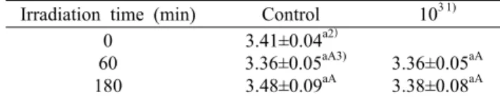
Table 6. Effect of infrared light emitting diode irradiation (5,000
∼7,000 nm) on S. cerevisiae growth (Unit: log CFU/mL) Irradiation time (min) Control 103 1)
0 60 180
3.41±0.04a2)
3.36±0.05aA3) 3.48±0.09aA
3.36±0.05aA 3.38±0.08aA
1)Initial strain concentration.
2)Means in the same column bearing different letter in samples are significantly different (P<0.05).
3)Means in the same row bearing different letter in samples are significantly different (P<0.05).
620~630 nm Vis-LED가 S. cerevisiae의 생육에 미치 는 영향을 확인하기 위해 620~630 nm Vis-LED를 S. ce- revisiae에 60, 180분 동안 조사하였다(Table 4). 초기 S. cerevisiae의 균수는 3.41 log CFU/mL였고, 620~630 nm Vis-LED를 60분 동안 조사한 경우 대조구에 비해 0.83 log CFU/mL 만큼 증가하였다. 조사 시간이 길어질수록 S. cer- evisiae 집락수가 대조군에 비해 유의하게 증가하여 180분 조사 시 S. cerevisiae의 균수는 4.63 log CFU/mL로 나타 났다.
620~630 nm Vis-LED는 적색 빛을 방출하는데 Jo 등 (2014)의 LED 광을 이용한 눈꽃동충하초 생산 연구에서는 적색 LED가 눈꽃동충하초의 생육 활성을 높여 생산성을 향 상한다고 보고하였다. 또한 Kim 등(2013)은 625 nm Vis- LED의 조사에 의한 S. aureus, E. coli 및 Porphyro- monas gingivalis의 살균효과는 없으며, 8시간 동안 조사 시 P. gingivalis의 성장은 대조군 값보다 2.5배 더 증가한다고 보고하였다. 이러한 결과에 의하여 620~630 nm Vis-LED 조사가 S. cerevisiae의 생장을 촉진할 수 있을 것으로 판단 되었다.
850 nm IR-LED 조사 시 S. cerevisiae의 생장증식 효과 850 nm IR-LED 조사가 S. cerevisiae의 생장에 미치는 효과를 확인한 결과는 Table 5와 같다. S. cerevisiae의 초 기 균수는 3.39 log CFU/mL로 확인되었고, 60, 180분 비조 사구의 균수는 각각 4.26 log CFU/mL와 4.34 log CFU/mL 로 나타났다. S. cerevisiae에 850 nm IR-LED를 60분 동 안 조사 시 S. cerevisiae의 균수는 4.56 log CFU/mL로 확인되었고, 180분 동안 조사 시 4.57 log CFU/mL로 확인 되었다. 조사구와 비조사구 사이에 미미한 증가를 보였지만 유의적인 차이는 없었다.
가시광선이나 자외선에 비해 적외선은 열작용이 강하여 공업용이나 의료용으로 널리 사용된다(Ahn과 Kwon, 2018).
Nussbaum 등(2002)의 연구에서는 810 nm의 레이저를 조 사했을 때 E. coli의 성장을 증가시킬 수 있다고 보고하였고, Qi 등(2017)의 연구에서는 적색 LED가 무산소성 광합성 세균의 성장을 효과적으로 촉진할 수 있음을 확인하였다.
그러나 Guffey와 Wilborn(2006)의 연구에서는 405 nm와 880 nm의 빛을 동시에 S. aureus와 P. aeruginosa에 조사
했을 때 항균효과를 나타낸다고 보고하였다.
5,000~7,000 nm IR-LED 조사 시 S. cerevisiae의 생육 에 미치는 영향
5,000~7,000 nm IR-LED가 S. cerevisiae 생육에 미치 는 영향을 확인하기 위해 5,000~7,000 nm IR-LED를 S. cerevisiae에 60, 180분 동안 조사하였고 그 결과는 Table 6과 같다. 초기 S. cerevisiae의 균수는 3.41 log CFU/mL 로 확인되었으며 60, 180분 비조사구는 각각 3.36 log CFU/mL, 3.48 log CFU/mL로 나타났다. 60분 동안 5,000
~7,000 nm IR-LED를 조사한 경우 S. cerevisiae의 균수는 3.36 log CFU/mL로 나타났고 180분 동안 조사한 경우 3.38 log CFU/mL로 확인되었으며, 대조구에 비해 유의적 인 변화는 확인할 수 없었다.
5,000~7,000 nm의 파장은 원적외선에 속하며 Bang과 Kim(2012)은 1,000 nm 이상의 파장은 식물에 특별한 영향 을 미치지 않고 식물체에 흡수되어 열로 변한다고 보고하였 다. 따라서 5,000~7,000 nm IR-LED는 S. cerevisiae 생장 에 특별한 작용이 없음을 확인할 수 있었다.
요 약
본 연구에서는 다양한 LED 파장이 S. cerevisiae의 생육에 미치는 영향을 확인하였다. 270 nm UV-LED를 S. cerevi- siae에 10분 동안 조사한 경우 100% 생장억제로 가장 뛰어 난 억제 효과를 보였고, 360 nm UV-LED를 S. cerevisiae 에 180분 동안 조사한 경우 유의적인 생장억제 효과를 보였 다. 465~675 nm Vis-LED 조사의 경우 UV-LED보다 낮 은 생장억제 효과를 보였고, 60분 조사 시 대조구에 비해 약 0.94 log CFU/mL의 감소를 확인하였다. 반면, 620~
630 nm Vis-LED를 S. cerevisiae에 180분 동안 조사한 경우, S. cerevisiae의 생장이 유의적으로 촉진됨을 확인할 수 있었다. 850 nm IR-LED의 경우 S. cerevisiae의 생육이 미미하게 촉진되는 것으로 나타났고, 5,000~7,000 nm IR- LED를 조사한 경우에는 S. cerevisiae 생장에 특별한 작용 이 없음을 확인할 수 있었다. 따라서 S. cerevisiae의 생장 특성에 따른 다양한 LED 파장을 이용한다면 식품산업에서 S. cerevisiae의 효율적인 활용이 가능할 것으로 판단되며,
더불어 다양한 LED 파장의 활용이 식품산업에 있어 잠재적 이용가치가 있는 것으로 사료된다.
감사의 글
이 논문은 부경대학교 자율창의학술연구비(2019년)에 의하 여 연구되었다.
REFERENCES
Ahn SS, Kwon KJ. Design of 850 nm near infrared and galvanic current based eyeglass-type device for periorbital wrinkle treatment and verification of treatment performance through image analysis. Journal of Korea Multimedia Society. 2018.
21:1379-1386.
Bang GW, Kim YH. LED for plant growth regulators for the study of light on the device. J Digit Converg. 2012. 10:267- 272.
Beukers R, Berends W. Isolation and identification of the irradi- ation product of thymine. Biochim Biophys Acta. 1960. 41:
550-551.
BIR Research Group. Eco-friendly, high-efficient LED technol- ogy development trends and market outlook. BIR Inc., Seoul, Korea. 2010. p 27-188.
Bolton JR, Cotton CA. The ultraviolet disinfection handbook.
American Water Works Association, New York, NY, USA.
2008. p 1-70.
Brem R, Guven M, Karran P. Oxidatively-generated damage to DNA and proteins mediated by photosensitized UVA. Free Radic Biol Med. 2017. 107:101-109.
Do JS, Bang WS. Bactericidal effect of 461 nm blue light emit- ting diode on pathogenic bacteria. Korean J Food Preserv.
2013. 20:419-423.
Do JS, Chung HJ, Bang WS. Bactericidal effect of pathogenic bacteria on acid treatment combined with red, green, and blue LED light at a low temperature environment. J Korean Soc Food Sci Nutr. 2015. 44:1725-1732.
Elmnasser N, Guillou S, Leroi F, Orange N, Bakhrouf A, Federighi M. Pulsed-light system as a novel food decontami- nation technology: a review. Can J Microbiol. 2007. 53:813- 821.
Ghate V, Kumar A, Zhou W, Yuk HG. Irradiance and temper- ature influence the bactericidal effect of 460-nanometer light- emitting diodes on Salmonella in orange juice. J Food Prot.
2016. 79:553-560.
Ghate VS, Ng KS, Zhou W, Yang H, Khoo GH, Yoon WB, et al. Antibacterial effect of light emitting diodes of visible wavelengths on selected foodborne pathogens at different il- lumination temperatures. Int J Food Microbiol. 2013. 166:
399-406.
Green A, Popović V, Pierscianowski J, Biancaniello M, Warri- ner K, Koutchma T. Inactivation of Escherichia coli, Listeria and Salmonella by single and multiple wavelength ultraviolet- light emitting diodes. Innov Food Sci Emerg Technol. 2018.
47:353-361.
Guffey JS, Wilborn J. Effects of combined 405-nm and 880-nm light on Staphylococcus aureus and Pseudomonas aeruginosa in vitro. Photomed Laser Surg. 2006. 24:680-683.
Held G. Introduction to light emitting diode technology and ap- plications. CRC Press, Boca Raton, FL, USA. 2016. p 1-170.
Jeong SY, Velmurugan P, Lim JM, Oh BT, Jeong DY. Photobio-
logical (LED light)-mediated fermentation of blueberry (Vac- cinium corymbosum L.) fruit with probiotic bacteria to yield bioactive compounds. LWT-Food Sci Technol. 2018. 93:
158-166.
Jo YY, Kweon HY, Lee KG, Lee HS, Yeo JH. Effects of LED on the growth of P. tenuipes. J Seric Entomol Sci. 2014.
52:59-63.
Kim B, Lee J. A study on design development of acne treatment device applying LED-wave. J Indust Des. 2019. 13:23-33.
Kim S, Kim J, Lim W, Jeon S, Kim O, Koh JT, et al. In vitro bactericidal effects of 625, 525, and 425 nm wavelength (red, green, and blue) light-emitting diode irradiation. Photomed Laser Surg. 2013. 31:554-562.
Kim SJ, Kim DK, Kang DH. Using UVC light-emitting diodes at wavelengths of 266 to 279 nanometers to inactivate food- borne pathogens and pasteurize sliced cheese. Appl Environ Microbiol. 2016. 82:11-17.
Kim SW, Han GD. Changes in fermentation properties and phe- nolic contents of Muscat bailey A wine by LED irradiation treatment. Microbiol Biotechnol Lett. 2015. 43:350-356.
Lee CW, Jeong KI, Hwang KH, Lee SJ, Yoo GC. A study of sterilization effect of long-wavelength UVA-LED irradiation on bacteria causing eye diseases. J Korean Oph Opt Soc.
2012. 17:99-105.
Lee DH, Jeong SM, Xu X, Kim KBWR, Ahn DH. Inhibition effect of Bacillus subtilis on 365 nm UV-LED irradiation ac- cording to packaging materials. Microbiol Biotechnol Lett.
2019. 47:332-336.
Lee JY, Kim PJ, Kang DS, Kim HJ, Yu SY, Lee WH. A biblio- metric analysis on LED research. J Inf Manage. 2011. 42:1- 26.
Legras JL, Merdinoglu D, Cornuet JM, Karst F. Bread, beer and wine: Saccharomyces cerevisiae diversity reflects human his- tory. Mol Ecol. 2007. 16:2091-2102.
Liu J, Lin L, Zhou L, Li B, Xu Z. Effect of ultrasound treatment conditions on Saccharomyces cerevisiae by response surface methodology. Microb Pathog. 2017. 111:497-502.
Maclean M, MacGregor SJ, Anderson JG, Woolsey G. Inactiva- tion of bacterial pathogens following exposure to light from a 405-nanometer light-emitting diode array. Appl Environ Microbiol. 2009. 75:1932-1937.
Moazzami M, Fernström LL, Hansson I. Reducing Campylobac- ter jejuni, Enterobacteriaceae and total aerobic bacteria on transport crates for chickens by irradiation with 265-nm ultra- violet light (UV-C LED). Food Control. 2021. 119:107424.
https://doi.org/10.1016/j.foodcont.2020.107424
Mori M, Hamamoto A, Takahashi A, Nakano M, Wakikawa N, Tachibana S, et al. Development of a new water sterilization device with a 365 nm UV-LED. Med Biol Eng Comput. 2007.
45:1237-1241.
Murdoch LE, McKenzie K, Maclean M, MacGregor SJ, Ander- son JG. Lethal effects of high-intensity violet 405-nm light on Saccharomyces cerevisiae, Candida albicans, and on dor- mant and germinating spores of Aspergillus niger. Fungal Biol. 2013. 117:519-527.
Nussbaum EL, Lilge L, Mazzulli T. Effects of 630-, 660-, 810-, and 905-nm laser irradiation delivering radiant exposure of 1-50 J/cm2 on three species of bacteria in vitro. J Clin Laser Med Surg. 2002. 20:325-333.
Park JH, Kang MS, Kim HI, Chung BH, Lee KH, Moon WK.
Study on immuno-stimulating activity of β-glucan isolated from the cell wall of yeast mutant Saccharomyces cerevisiae IS2. Korean J Food Sci Technol. 2003. 35:488-492.
Qi X, Ren Y, Tian E, Wang X. The exploration of monochro-
matic near-infrared LED improved anoxygenic photosynthetic bacteria Rhodopseudomonas sp. for wastewater treatment.
Bioresour Technol. 2017. 241:620-626.
Sadick N. A study to determine the effect of combination blue (415 nm) and near-infrared (830 nm) light- emitting diode (LED) therapy for moderate acne vulgaris. J Cosmet Laser Ther. 2009. 11:125-128.
Soni A, Oey I, Silcock P, Bremer P. Bacillus spores in the food industry: a review on resistance and response to novel in- activation technologies. Compr Rev Food Sci Food Saf. 2016.
15:1139-1148.
Stewart GG. Saccharomyces cerevisiae. 2nd ed. In: Batt CA, editor. Encyclopedia of Food Microbiology. Academic Press, London, UK. 2014. p 309-3015.