Biological Activities of Pharbitis nil and Partial Purification of Anticancer Agent from Its Extract
Hyeun Deok Choi1,4, Sun Nyoung Yu1, Sul-Gi Park1, Young Wook Kim1,2, Hyo Won Nam1,2, Hyun Hee An1,2, Sang Hun Kim1, Kwang-Youn Kim3 and Soon Cheol Ahn1,2*
1Department of Microbiology & Immunology, Busan National University School of Medicine, Yangsan 50612, Korea
2Immunoregulatory Therapeutics Group in Brain Busan 21 Project, Pusan National University, Yangsan 50612, Korea
3Department of Herbal Formula, Medical Research Center (MRCGHF), College of Oriental Medicine, Daegu Haany University, Gyeongsan 38610, Korea
4Institute of Fisheries Sciences, Pukyong National University, Busan 46041, Korea
Received December 24, 2016 /Revised February 3, 2017 /Accepted February 3, 2017
This study aimed to evaluate several biological activities of Pharbitis nil and to isolate an anticancer agent from its methanol extract. Pharbitis nil seeds were extracted with methanol (PNM). Then, PNM was fractionated into solvent layers such as ethyl acetate fraction (PNE), butanol fraction (PNB), and water fraction (PNW). The biological activities of the fractions were analyzed for tyrosinase inhibition, lipase inhibition, DPPH-free radical scavenging, and cell growth inhibition. PNM showed strong growth inhibition of prostate cancer PC-3 cells. PNM was subjected to Diaion HP-20 and eluted step- wise with 50%, 80%, and 100% methanol. Then, for activity-guided fraction, each fraction was ana- lyzed for growth inhibition of prostate cancer PC-3 cells by using an MTT assay. Because the 100%
fraction showed significantly strong inhibitory activity, the fraction was further separated in the re- verse phase C18, which was eluted with 80% and 90% methanol. The 90% fraction was further sub- jected to Sephadex LH-20 using a mobile solvent of 100% methanol. Finally, the compound PN was partially purified for HPLC analysis. PN showed cell growth inhibitory activity and induced the apop- tosis and cell cycle arrest of prostate cancer PC-3 cells, as measured by flow cytometry. The results together suggest that Pharbitis nil possesses various biological activities, especially the inhibitory activ- ity for the proliferation of prostate cancer PC-3 cells, suggesting the possibility of its use as an anti- cancer agent.
Key words : Biological activity, compound PN, Pharbitis nil, proliferation, purification
*Corresponding author
*Tel : +82-51-510-8092, Fax : +82-55-382-8090
*E-mail : [email protected]
This is an Open-Access article distributed under the terms of the Creative Commons Attribution Non-Commercial License (http://creativecommons.org/licenses/by-nc/3.0) which permits unrestricted non-commercial use, distribution, and reproduction in any medium, provided the original work is properly cited.
Journal of Life Science 2017 Vol. 27. No. 2. 225~232 DOI : https://doi.org/10.5352/JLS.2017.27.2.225
서 론
서구화된 식습관과 불규칙한 생활로 인해 현대 사회에서 각종 성인병 환자들이 증가하고 있는 추세이며, 현대인들은 식품으로 건강을 유지하고, 질병을 치료하거나 예방할 수 있 는 웰빙(Well-being)형 기능성 식품에 많은 관심을 가지고 있 다[1, 2]. 예로부터, 동양의학에서는 한약재와 같은 천연물을 이용하여 질병을 치료하거나 예방을 해왔으며, 현대에서도 이 를 바탕으로 다양한 물질을 탐색하려는 연구가 활발히 진행되 고 있다[4, 21]. 천연물은 식품, 동물, 미생물 등과 같은 자연계 에 존재하는 다양한 물질을 말하며, 이를 통하여 의약품, 식품 등으로 이용하고 있다. 특히, 최근에는 천연물을 이용하여 의
약품을 만드는 고부가가치 산업이 각광받고 있으며, 합성 의 약품이 아닌 천연 의약품으로 부작용을 최소화 할 수 있는 신약 개발에 전세계 제약회사가 뛰어들고 있는 상황이다[12].
Apoptosis는 programmed cell death 중의 하나이며, 외 인성 경로와 내인성 경로로 진행되고, 내외부의 다양한 신호 에 의해 여러 단계를 거쳐 일어나게 되며 세포의 수축, 핵의 응축 및 세포막의 기포 형성 등과 같은 현상이 일어난다[6, 8]. 이러한 외인성 경로와 내인성 경로에 의한 apoptosis는 미 토콘드리아에서 세포질로 방출되는 cytochrome c에 의해 유 도 되며, Bcl-2 family 및 caspase가 활성화되고, 이로 인해 poly (ADP-ribose) polymerase (PARP)가 조절된다[5, 16].
전래로부터 사용되어 온 천연물의 하나로서 나팔꽃 씨앗인 견우자는 메꽃과에 속하는 식물로서, 동의보감에서는 사하 작 용 및 이뇨 작용 등에 사용되었다고 전해진다[7]. 최근 몇몇 연구자를 중심으로 견우자에 대한 연구가 진행되어 여러가지 생리활성[9, 10] 및 암세포주에 대한 항암 효과[14, 15] 등의 연구 결과가 보고되고 있으나, 견우자에 대한 연구가 미진한 상태이므로 본 연구에서는 견우자의 다양한 생리활성을 조사 하고 그 중 생리활성이 가장 우수한 세포증식 저해활성을 대 상으로 인간 전립선암 세포주인 PC-3 세포에 항암효과를 나타
Table 1. HPLC condition for compound PN from Pharbitis nil
Parameters Condition
Instrument Column Detector Flow rate Mobile phase Injection volume
Sample
YL9100 HPLC
YMC PAK C8 (Ø 4.6x250 mm, 5 μm) sofTA ELSD
0.8 mL/min 100% MeOH
10 μl Compound PN 내는 물질을 분리하고 그 특성을 조사하였다.
재료 및 방법 실험 재료 및 시약
본 실험에서 사용된 견우자는 부산 한약 시장의 삼세당에서 주문하여 사용하였다. 3-(4,5-Dimethylthiazol-2-yl)-2,5-diphe- nyltetrazolium bromide (MTT), 1,1-diphenyl-2-picryl hydra- zyl (DPPH), pancreatic lipase, p-nitrophenyl palmitate (pNPP), mushroom tyrosinase, ascorbic acid, orlistat, kojic acid 등은 Sigma Chemical Co. (St. Louis, MO, USA)에서 구 입하였다. Annexin V/PI staining kit는 BD Bioscience (San Jose, CA, USA)에서 구입하여 사용하였다.
견우자 추출물의 제조
견우자 1 kg을 분쇄하고 2 l methanol (MeOH)을 가하여 실온에서 3회 추출하였다. MeOH 추출물을 5 μm filter paper 로 여과한 후, rotary evaporator에서 감압 농축하여 조 추출물 108 g을 얻었다. 그 중 10 g의 MeOH 추출물을 증류수에 현탁 시킨 뒤, ethyl acetate (EtOAc), n-butanol (BuOH)로 3회 분획 하여, EtOAc 분획물(0.82 g), BuOH 분획물(6.9 g), water 분획 물(2.2 g)을 얻었다. 이들 용매 분획물을 정량하여 각각의 생리 활성 분석에 사용하였다.
견우자 추출물의 생리활성 측정 DPPH free radical 소거활성
견우자 추출물의 항산화 활성은 DPPH radical scavenging 활성을 이용하여 측정하였다[17]. 1,1-Diphenyl-2-picryl hy- drazyl (DPPH)을 0.15 mM로 ethanol에 녹여 DPPH 용액을 제조하였으며, 4℃ 냉장고에 빛이 닿지 않도록 하여 보관하였 다. DPPH 시약 180 μl에 견우자 추출물을 농도별로 20 μl를 넣어 암실에서 30분간 실온에서 반응시킨 후, VERSAMAX mi- croplate reader (Molecular Devices, Toronto, Canada)를 이용 하여 510 nm에서 흡광도를 측정하였다. 시료 무첨가구는 neg- ative control로 methanol과 증류수를 사용하였고, 효소 무첨 가구는 blank로 하였다. Positive control로는 ascorbic acid를 사용하였으며 DPPH radical scavenging 활성(%) 은 다음과 같이 계산하였다.
DPPH radical scavenging 활성(%) = [1-(B-C) / A)] ×100 [A: 시료 무첨가구의 흡광도, B: 시료 첨가구의 흡광도, C:
효소 무첨가구의 흡광도]
Pancreatic lipase 저해활성
Pancreatic lipase 효소활성은 기질로 사용된 p-nitrophenyl palmitate (pNPP)가 lipase 효소에 의하여 p-nitrophenol로 가 수분해되어 나타나는 흡광도의 차이를 이용하였다[3]. 1.5 ml tube에 견우자 추출물 10 μl와 100 mM Tris-HCl (pH 8.5) buf-
fer를 100 μl, 2 mM pNPP를 80 μl를 첨가 하였다. 여기에 pan- creatic lipase 10 μl (10 mg/ml)를 첨가하고 37℃ water bath에 30분간 반응한 후, ice에서 10분간 반응을 정지하고 13,000 rpm으로 1분간 원심분리하여 그 상층액을 96-well plate에 분 주하여 VERSAMAX microplate reader로 405 nm에서 흡광도를 측정하였다. 시료 무첨가구는 negative control로 methanol과 증류수를 시료 대신 사용하였고, 효소 무첨가구는 blank로 하 였다. Positive control로는 orlistat을 사용하였다. Lipase 저해 활성(%) 은 다음과 같이 나타내었다.
Lipase 저해활성(%) = [1-(B-C) / A)] ×100
[A: 시료 무첨가구의 흡광도, B: 시료 첨가구의 흡광도, C:
효소 무첨가구의 흡광도]
Mushroom tyrosinase 저해활성
Mushroom tyrosinase 활성은 L-tyrosine이 tyrosinase에 의 해 DOPA quinone으로 전환되는 것을 측정하였다[13]. 1 mM L-Tyrosine과 50 mM sodium phosphate buffer (pH 6.5) 와 증류수를 6:6:4 (v/v/v)의 비율로 넣은 tyrosine buffer 160 μl, 견우자 추출물 10 μl, mushroom tyrosinase (200 U/ml) 30 μl를 96 well plate에 넣어 37°C 에서 30분간 반응 시켰다. 반응 후 VERSAMAX microplate reader를 이용하여 475 nm에서 흡 광도를 측정하였다. 시료 무첨가구는 negative control로 methanol과 증류수를 시료 대신 사용하였고, 효소 무첨가구는 blank로 하였다. Positive control로는 kojic acid을 사용하였 다. Tyrosinase 저해활성(%)은 다음과 같이 나타내었다.
Tyrosinase 저해활성(%) = [1-(B-C) / A)] ×100 [A: 시료 무첨가구의 흡광도, B: 시료 첨가구의 흡광도, C:
효소 무첨가구의 흡광도]
세포 생존율 측정 및 세포형태 변화 관찰
PC-3 세포는 1×10⁴ cells/well의 농도로 세포 배양용 48- well plate에 분주하여 24시간 동안 안정화시킨 후 2, 4, 6, 8 μg/ml 농도의 견우자 추출물을 처리하였다[17]. 24시간 반응 후, MTT 시약을 0.5 mg/ml 농도로 각각 250 μl 첨가하여 37℃
에서 2시간 동안 반응시켰다. 반응이 끝난 후, 상등액을 제거 하고 생성된 formazan에 DMSO 250 μl를 각각 분주하여 96-well plate로 100 μl씩 옮긴 후, VERSAMAX microplate read- er를 이용하여 570 nm에서 흡광도를 측정하였다. 세포의 형태
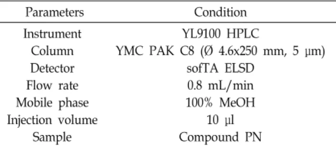
Fig. 1. DPPH free radical scavenging activity of solvent extracts from methanol ex- tract of Pharbitis nil seeds. PNM: meth- anol extract of Pharbitis nil seeds, PNE:
ethyl acetate layer of Pharbitis nil seeds, PNB: butanol layer of Pharbitis nil seeds, PNW: water layer of Pharbitis nil seeds. Ascorbic acid was used as a positive control. Data represent the mean ± SD (n=3 in each group) from three separate experiments. **p<0.01,
***p<0.001 compared with negative control. N.S = non-significantly.
변화를 관찰하기 위하여 Olympus CKX41/31 도립 현미경 (Olympus, Tokyo, Japan)을 이용하여 각 농도에 따른 세포의 형태 변화를 400배 배율로 관찰하였다.
Flow cytometry 분석에 의한 cell cycle 측정
PC-3 세포를 1×10⁵ cells/well의 농도로 세포 배양용 6-well plate에 분주하여 24시간 동안 안정화 시킨 후, compound PN 을 다양한 농도로 처리하였다[20]. 24시간 후, PC-3 세포를 회 수하여 phosphate-buffered saline (PBS)로 세척하고 70%
ethanol을 넣어 4℃에서 12시간 고정하고 RNase A를 처리하 여 37℃에서 1시간 반응 시킨 후, DNA staining dye인 propi- dium iodide (PI)로 염색하여 flow cytometry (Becton Dickin- son Co., Franklin Lakes, NJ, USA)를 이용하여 분석하였다.
Annexin V/PI double staining assay에 의한 apop- tosis 측정
견우자에 의한 apoptosis의 유도를 확인하기 위하여 annex- in V/PI detection kit을 이용하여 PC-3 세포를 flow cy- tometry로 분석하였다[19]. PC-3 세포를 1×10⁵ cells/well의 농 도로 6-well plate에 분주하고 24시간 동안 안정화시킨 후, compound PN을 다양한 농도로 PC-3 세포에 처리하여 24시 간 배양하였다. 24시간 후, 세포를 회수한 다음 PBS로 세척하 고, cell pellet을 annexin V/PI 시약을 처리하여 20분간 염색 시킨 후, 염색된 세포는 flow cytometry를 이용하여 분석하였다.
통계처리
모든 실험의 결과는 3번 반복 수행하여 얻어진 것으로, 통계 분석은 ANOVA에 의해 분석하여 mean±S.D로 표시하였고, 통계적 유의성은 p≤0.05로 판정하였다.
결과 및 고찰 DPPH free radical 소거활성
용매 분획을 통해 얻은 견우자 분획물을 이용하여 DPPH 소거능을 측정하였다. 견우자의 methanol 추출물(PNM), eth- yl acetate 분획물(PNE), butanol 분획물(PNB), water 분획물 (PNW)의 최종 농도를 100, 300, 1,000 μg/ml로 처리하여 측정 한 결과, 1,000 μg/ml 농도의 PNM, PNE, PNB, PNW에서 각 각 64.2, 80.9, 80.3, 25.8%의 free radical 소거능을 보였으며 PNM, PNE, PNB의 IC50 값은 271, 221.9, 149.3 μg/ml로 측정 되었다(Fig. 1). 이를 통해 PNE, PNB에서 가장 높은 DPPH free radical 소거활성을 보였으며, 견우자의 분획물에 따라 비 슷한 항산화 활성이 보고된 기존의 결과와 유사하였다[11]. 따 라서 이러한 DPPH free radical 소거활성을 통해 견우자가 천연 항산화제로서 이용될 수 있는 가능성을 확인하였다.
Pancreatic lipase 저해활성
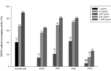
췌장의 지방분해 효소인 lipase는 지방질 가수분해 효소로 triglyceride를 분해하여 지방의 흡수 및 소화에 중요한 역할을 한다. 게다가 lipase로 인한 지방의 과도한 흡수는 비만을 유발 하게 되고 여러가지 질병을 야기한다. 따라서 lipase의 활성을 저해하여 triglyceride 및 cholesterol의 흡수를 감소시킬 수 있 음이 보고되었다[3]. 이러한 기존의 보고를 근거로 하여 견우 자 추출물의 pancreatic lipase 저해활성을 평가한 결과, 견우 자의 PNM, PNE, PNB, PNW을 500 μg/ml의 최종 농도로 처리하였을 때 각각 64.6, 56.9, 52.8, 15.1%의 저해활성을 보였 으며, 그 중, PNM, PNE, PNB의 저해활성이 매우 높은 것을 확인할 수 있었다(Fig. 2).
Mushroom tyrosinase 저해활성
견우자 추출물의 tyrosinase 저해활성을 측정하기 위해 mushroom tyrosinase를 이용하였다. 각 추출물의 농도에 따 른 tyrosinase의 저해 활성은 PNM, PNE, PNB의 농도에 따라 tyrosinase의 저해율이 증가하는 것을 확인할 수 있었다(Fig.
3). 또한 PNM, PNE, PNB를 최종농도 500 μg/ml로 처리하였 을 경우, 각각 27, 41, 25%의 저해율을 나타냈으며, 그 중 PNE
Fig. 2. Inhibitory activity of solvent extracts from methanol extract of Pharbitis nil seeds on pancreatic lipase activity.
PNM: methanol extract of Pharbitis nil seeds, PNE: ethyl acetate layer of Pharbitis nil seeds, PNB: butanol layer of Pharbitis nil seeds, PNW: water lay- er of Pharbitis nil seeds. Orlistat was used as a positive control. Data repre- sent the mean ± SD (n=3 in each group) from three separate experiments. *p<
0.05, ***p<0.001 compared with neg- ative control. N.S = non-significantly.
Fig. 3. Inhibitory activity of solvent extracts from methanol extract of Pharbitis nil seeds on mushroom tyrosinase activity.
PNM: methanol extract of Pharbitis nil seeds, PNE: ethyl acetate layer of Pharbitis nil seeds, PNB: butanol layer of Pharbitis nil seeds, PNW: water lay- er of Pharbitis nil seeds. Kojic acid was used as a positive control. Data repre- sent the mean ± SD (n=3 in each group) from three separate experiments. *p<
0.05, **p<0.01, ***p<0.001 compared with negative control. N.S = non-sig- nificantly.
의 저해활성이 가장 높은 것을 확인할 수 있었다. 이러한 결과 들은 견우자의 methanol 추출물이 mushroom tyrosinase 및 B16 melanoma tyrosinase 활성을 농도의존적으로 저해한다 는 보고들과 일치하였다[4, 22]. 따라서 이런 결과들을 통해 견 우자가 천연 미백제로서 이용될 수 있는 가능성을 확인하였다.
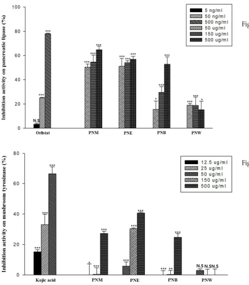
암세포주의 증식 저해활성
견우자 추출물의 암세포주의 증식에 대한 저해활성을 확인 하기 위하여, 인간 전립선암 세포주인 PC-3 세포를 이용하여 세포의 성장을 측정하였다. 견우자 추출물을 전립선암 세포주 인 PC-3 세포에 농도별로 처리하여 실험한 결과, PNM, PNE, PNB에서 농도 의존적으로 세포 성장에 대한 저해효과가 나타 나는 것을 확인할 수 있었다(Fig. 4). 특히, 최종 농도로 50, 100 μg/ml를 처리하였을 때, PNM과 PNB에서 각각 92%와 94%의 저해율을 보였다. 이를 통해 세포성장 저해 활성 물질 이 PNM에서 PNB로 이동하여 강한 저해활성을 나타내는 것 을 확인 할 수 있었다. 이러한 결과들은 견우자의 methanol 추출물이 AGS 위암 세포의 증식을 저해한 보고와 유사하였다 [15]. 따라서, 전립선암 세포주인 PC-3 세포에 대한 성장을 저
해하는 물질을 분리하여 그 특성을 조사하고자 하였다.
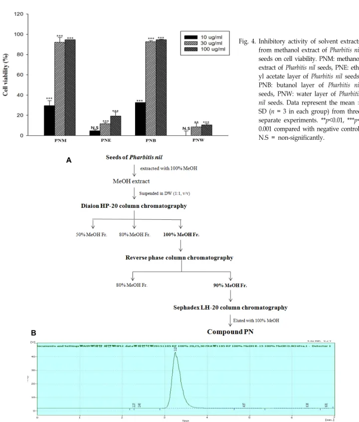
암세포주의 증식 저해물질의 정제
암세포주 증식을 저해하는 물질을 정제하기 위하여 견우자 의 methanol 추출물 5 g을 50% methanol에 현탁한 뒤, Diaion HP-20 column chromatography를 이용하여 정제를 수행하였 다. 이동상 용매를 50, 80, 100% methanol로 단계적으로 증가 시켜 용출하였다. 그 중, 100% methanol 분획물(800 mg)에서 가장 높은 암세포의 증식 저해 활성을 보였고, 이 분획물을 이용하여 reverse phase column chromatography (RP-18)(Ø 1.5 10 cm, 230-400 mesh)를 실시하였다. 이동상 용매를 80, 90% methanol로 단계적으로 증가시키면서 용출하였다.
RP-18로부터 용출된 분획물 중에서 90% methanol 분획물(640 mg)에서 암세포 증식 저해활성이 가장 높았으며, 이 분획물을 이용하여 Sephadex LH-20 column chromatography (Ø 4×80 cm)를 실시하였다. 이동상으로 100% methanol을 이용하여 용 출하였으며, 그 중 저해활성이 가장 높은 분획물을 모아서 compound PN (250 mg)이라 명명하고, high performance liq- uid chromatography (HPLC)를 이용하여 그 순도를 확인하였
Fig. 4. Inhibitory activity of solvent extracts from methanol extract of Pharbitis nil seeds on cell viability. PNM: methanol extract of Pharbitis nil seeds, PNE: eth- yl acetate layer of Pharbitis nil seeds, PNB: butanol layer of Pharbitis nil seeds, PNW: water layer of Pharbitis nil seeds. Data represent the mean ± SD (n = 3 in each group) from three separate experiments. **p<0.01, ***p<
0.001 compared with negative control.
N.S = non-significantly.
A
B
Fig. 5. Purification procedure of compound PN from methanol extract of Pharbitis nil seeds. (A) Schematic procedure for purification.
(B) HPLC analysis of compound PN. High performance liquid chromatography (HPLC) was performed by using C8 re- verse-phase column (Ø 4.6x250 mm, 5 μm) eluted with 100% MeOH. Flow rate was 0.8 ml/min. Detector was evaporative light-scattering detector (ELSD).
다(Fig. 5A, Fig. 5B). 그 결과, 여러가지의 HPLC 분석 조건에 서 단일 피크를 보였으나, NMR과 같은 기기분석을 통해 유사 한 구조의 물질이 혼재된 부분 정제된 상태로 분석되어 저해
물질의 정확한 구조 동정을 위해 순수분리를 위한 추가적인 정제 과정이 필요한 것으로 사료되었다(data not shown).
A B
Fig. 6. Compound PN inhibits cell viability and changes the morphology in prostate cancer PC-3 cells. (A) Cell viability. PC-3 cells were treated with compound PN in a dose dependent manner for 24 and 48 hr. It was determined by MTT assay.
(B) Cell morphology. PC-3 cells were treated with compound PN in a dose dependent manner for 24 hr. Its morphology was observed by inverted microscopy at 400x magnification ratio. Data are presented as mean ± SD (n=3 in each group).
*p<0.05, **p<0.01, ***p<0.001 compared with untreated control.
A
B
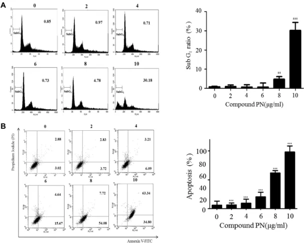
Fig. 7. Compound PN induces cell cycle arrest and apoptosis in prostate cancer PC-3 cells. (A) Cell cycle arrest. (B) Apoptosis.
PC-3 cells were treated with compound PN in a dose dependent manner for 24 hr. Cells were stained with propidium iodide (PI) and analyzed by flow cytometry. Apoptosis was detected by annexin V/PI double staining analysis. Data are presented as mean ± SD (n=3 in each group). *p<0.05, ** p<0.01, *** p<0.001 compared with untreated control.
Compound PN의 암세포 증식 저해 및 형태 변화 견우자의 부분 정제물질인 compound PN을 전립선암 세포 주인 PC-3 세포에 농도별로 처리하고, 각각 24, 48시간 배양한
후 세포의 증식에 대한 저해활성을 확인하였다(Fig. 6A). 24시 간 동안 배양한 세포주에서는 7.1 μg/ml, 48시간 동안 배양한 세포주에서는 5.6 μg/ml에서 IC50 값을 나타냈다. 또한, com-
pound PN을 PC-3 세포에 농도별로 처리하여 24시간 동안 배양 시킨 후 광학 현미경으로 세포의 형태를 관찰한 결과, 무처리군에서는 세포막을 유지하고 기벽에 부착되어 세포의 정상적인 형태를 나타냈지만, compound PN 처리군에서는 농도가 증가할수록, 세포의 수가 줄어들고 점차 팽창되면서 기벽에서 떨어져 배지에 부유하고 있는 형태를 관찰할 수 있 었다(Fig. 6B).
Compound PN의 cell cycle arrest 및 apoptosis 유도 Compound PN에 의한 전립선 암세포주인 PC-3의 세포증 식 저해 효과가 cell cycle에 영향을 끼치는지 확인하기 위해, compound PN을 농도별로 처리하여 24시간 동안 배양한 후, propidium iodide (PI)로 염색하여 flow cytometry로 cell cy- cle을 분석하였다(Fig. 7A). 그 결과, compound PN에 의해 sub-G1에서 arrest된 세포의 수가 농도 의존적으로 증가하는 것을 확인 할 수 있었다. 또한 annexin V/PI staining을 이용하 여 apoptosis를 분석한 결과(Fig. 7B), compound PN의 농도에 따라 apoptosis가 증가하는 것을 확인 할 수 있었다. 이러한 결과를 통하여 compound PN이 cell cycle arrest와 apoptosis 의 유도를 통하여 전립선암 세포주인 PC-3에서 세포사멸을 유도한다는 것을 확인 할 수 있었다[18, 19].
감사의 글
이 논문은 부산대학교 기본연구지원사업(2년)에 의하여 연 구되었음.
References
1. Ahn, I. S., Park, K. Y. and Do, M. S. 2007. Weight control mechanisms and antiobesity functional agents. J. Kor. Soc.
Food Sci. Nutr. 36, 503-513.
2. Bahn, K. N., Lee, C. H., Cho, T. Y., Lee, J. Y., Lee, Y. J.
and Chae, G. Y. 2005. Determination of phosphatidylcholine in Korea functional foods contatining lecitins using HPLC with evaporative light-scattering detector (ELSD). J. Fd. Hyg.
Safety 20, 267-271.
3. Bitou, N., Nimomiya M., Tsjita, T. and Okuda, H. 1999.
Screening of lipase inhibitors from marine algae. Lipids 34, 441-445.
4. Cha, E. J. and Kim, A. K. 2003. The effects of two plant extracts on tyrosinase activity. Yakhak Hoeji 47, 20-24.
5. de Oliveira, J. R., de Aguiar Almeida, R. B., das Graças Figueiredo Vilela, P., de Oliveira, F. E., da Rocha, R. F., Jorge, A. O. and de Oliveira, L. D. 2014. Control of micro- organisms of oral health interest with Arctium lappa L.
(burdock) extract non-cytotoxic to cell culture of macro- phages (RAW 264.7). Arch. Oral. Biol. 59, 808-814.
6. Huerta, S., Goulet, E. J. and Livingston, E. H. 2006. Colon cancer and apoptosis. Am. J. Surg. 191, 517-526.
7. Ji, H. Y., Liu, K. H., Jeong, J. H., Lee, D. Y., Shim, H. J., Son, M. W. and Lee, H. S. 2012. Effect of a new prokinetic agent DA-9701 formulated with Corydalis Tuber and Pharbiti- dis Semen on cytochrome P450 and UDP-glucuronosyl- transferase enzyme activities in human liver microsomes.
Evid. Based Complement. Alternat. Med. 2012, 1-8.
8. Kamata, H. and Hirata, H. 1999. Redox regulation of cellular signalling. Cell. Signal. 11, 1-14.
9. Kim, A. K. and Cha, E. J. 2007. Effect of Pharbitidis seed extract on the antioxidant enzyme activity in B16F10 murine melanoma cells by oxidative Stress. Yakhak Hoeji 48, 93-98.
10. Kim, K. H., Choi, S. U., Son, M. W. and Lee, K. R. 2010.
Two new phenolic amides from the seeds of Pharbitis nil.
Chem. Pharm. Bull. 58, 1532-1535.
11. Kim, J. H. and Ko, Y. S. 2011. The biological activities of extracts and fractions of herbal plants. J. Ori. Rehab. Med.
21, 47-56.
12. Kim, J. W., Kim, J. K., Song, I. S., Kwon, E. S. and Youn, K. S. 2013. Comparison of antioxidant and physiological properties of Jerusalem artichoke leaves with different ex- traction processes. J. Kor. Soc. Food Sci. Nutr. 42, 68-75.
13. Kim, Y. J. and Uyama, H. 2005. Tyrosinase inhibitors from natural and synthetic sources: structure, inhibition mecha- nism and perspective for the future. Cell. Mol. Life Sci. 62, 707-1723.
14. Ko, S. G., Jun, C. Y., Park, C. H. and Bae, H. S. 2003. Anti -cancer effects and apoptosis by Korean medicinal herbs.
J. Physiol. Pathol. Kor. Med. 17, 819-825.
15. Ko, S. G., Koh, S. H., Jun, C. Y., Nam, C. G., Bae, H. S. and Shin, M. K. 2004. Induction of apoptosis by Saussurea lappa and Pharbitis nil on AGS gastric cancer cells. Biol. Pharm.
Bull. 27, 1604-1610.
16. Kong, E. H., Kim, Y. J., Kim, Y. J., Cho, H. J., Yu, S. N., Kim, K. Y., Chang, J. H. and Ahn, S. C. 2008. Piplartine induces caspase-mediated apoptosis in PC-3 human pros- tate cancer cells. Oncol. Rep. 20, 785-792.
17. Lee, S. Y., Choi, H. D., Yu, S. N., Kim, S. H., Park, S. K.
and Ahn, S. C. 2015. Biological activities of Mesembryanthemum crystallinum (ice plant) extract. J. Life Sci. 25, 638-645.
18. Malumbres, M. and Barbacid, M. 2009. Cell cycle, CDKs and cancer: a changing paradigm. Nat. Rev. Cancer 9, 153-166.
19. Park, H. J., Jin, S. J., Oh, Y. N., Kim, B. W. and Kwon, H.
J. 2015. Induction of apoptosis by methanol extract of Endlicheria anomala in human lung and liver cancer cells.
J. Life Sci. 25, 441-449.
20. Sohn, H. Y., Shin, Y. K. and Kim, J. S. 2010. Anti-pro- liferative activities of solid-state fermented medicinal herbs using Phellinus baumii against human colorectal HCT 116 cell. J. Life Sci. 20, 1268-1275.
21. Son, C. Y., Beak, I. H., Song, G. Y., Kang, J. S. and Kwon, K. I. 2009. Pharmacological effect of decursin and decursinol angelate from Angelica gigas Nakai. Yakhak Hoeji 53, 303-313.
22. Wang, K. H., Lin, R. D., Hsu, F. L., Huang, Y. H., Chang, H. C., Huang, C. Y. and Lee, M. H. 2006. Cosmetic applica- tions of selected traditional Chinese herbal medicines. J.
Ethnopharmacol. 106, 353-359.
초록:견우자의 생리활성 분석과 추출물로부터 항암 활성물질의 분리
최현덕1,4․유선녕1․박슬기1․김영욱1,2․남효원1,2․안현희1,2․김상헌1․김광연3․안순철1,2*
(1부산대학교 의학전문대학원 미생물학 및 면역학 교실, 2부산대학교 Brain Busan 21 면역조절치료소재 연구인력양
성사업단, 3대구한의대학교 한의과대학 MRC센터 방제학교실, 4부경대학교 수산과학연구소)
견우자는 한의학에서 사하 작용 및 이뇨 작용 등의 치료 목적으로 사용되어 왔다. 본 연구에서는 견우자를 methanol로 추출하고(PNM), 이를 기본으로 ethyl acetate 분획물(PNE), butanol 분획물(PNB), water 분획물 (PNW)로 나누었으며 각 분획물을 이용하여 mushroom tyrosinase 저해활성, pancreatic lipase 저해활성, DPPH free radical 소거활성 및 암세포주의 증식 저해활성 등을 조사하였다. 특히, 전립선암 세포주 PC-3 세포에서 강력 한 세포 증식 저해활성을 보였으며, 용매 분획물 중, butanol 분획에서 가장 높은 암세포의 증식 저해활성을 보였 다. 따라서 견우자의 methanol 추출물을 Diaion HP-20 column chromatography, reverse phase column chroma- tography, Sephadex LH-20 column chromatography를 순차적으로 실시하여 최종적으로 저해활성이 가장 높은 compound PN을 얻었으며 HPLC를 통해 그 순도를 확인하였다. Compound PN을 이용하여 cell cycle arrest과 annexin V/PI를 측정한 결과, 세포의 sub-G1기를 축적되면서 apoptosis가 유발되어 전립선암 세포주 PC-3 세포 의 세포사멸이 유도되는 것을 확인하였다. 따라서 본 연구를 통하여 견우자에 대한 항산화 활성, 미백 활성 등의 효과 뿐 만 아니라 항암 활성에도 활용할 수 있을 것으로 사료된다.