Bacterial Community of Natural Dye Wastewater Treatment Facility
Yeoung Min Hwang
1, Dae Kuk Kim
1,2, Ji Hee Lee
1, Keun Sik Baik
1,3, Chul Park
1,4and Chi Nam Seong
1*
1
Department of Biology, College of Life Science and Natural Resources, Sunchon National University, Suncheon 540-950, Korea
2
Naju Foundation of Natural Dyeing Culture, Naju 520-931, Korea
3
Department of Biological Sciences, Korea Basic Science Institute, Daejeon 305-806, Korea
4
Department of Clinic Pathology, Gwangyang Health Collegee, Gwangyang 545-703, Korea
Received July 26, 2013 /Revised March 12, 2014 /Accepted March 21, 2014Culture-dependent and culture-independent denaturing gradient gel electrophoresis (DGGE) analyses were employed to investigate the bacterial community associated with a natural dye wastewater treat- ment facility. A total of 104 (influent water, 48 strains; aeration tank, 25; settling tank, 31) bacterial strains were isolated. Based on the 16S rRNA gene sequences comparison analysis, the isolates be- longed to four phyla: Proteobacteria, Actinobacteria, Firmicutes, and Bacteriodetes. Seventeen DGGE bands representing dominant taxa in each sample were cloned and partially sequenced. The same four phyla were detected by DGGE fingerprinting. The most dominant taxon retrieved by both methods was the member of the phylum Proteobacteria with Alphaproteobacteria as the predominant class. The bacterial community associated with the natural dye wastewater treatment facility is composed of parasites of animals and plants, decomposers of polysaccharides and dyes, and producers of extracellular polysaccharides.
Key words : Bacterial community, denaturing gradient gel electrophoresis, natural dye wastewater
*Corresponding author
*Tel : +82-61-750-3613, Fax : +82-61-750-5469
*E-mail : [email protected]
This is an Open-Access article distributed under the terms of the Creative Commons Attribution Non-Commercial License (http://creativecommons.org/licenses/by-nc/3.0) which permits unrestricted non-commercial use, distribution, and reproduction in any medium, provided the original work is properly cited.
Journal of Life Science 2014 Vol. 24. No. 4. 393~402 DOI : http://dx.doi.org/10.5352/JLS.2014.24.4.393
서 론
합성염색폐수의 특성은 pH가 높고 독성 유기물 및 무기물 등을 함유하고 있을 뿐 아니라 색을 띠고 있다 [25]. 합성염색폐 수처리 공정에서 중요한 염료의 탈색을 위해 많은 경우 흡착 제를 이용한다 [13]. 최근에는 염료의 탈색과 난분해성 화합물 의 분해를 위해 생물학적 처리방법들이 적용되고 있으며 특정 미생물을 이용하여 분해 효율을 높이는 연구들이 진행되고 있다 [6, 11, 14, 15, 21, 25, 26].
합성염색폐수처리에서 탈색에 관여하는 미생물의 분리와 미생물에 의한 분해 효율에 관한 연구 외에 각 공정별 미생물 군집 변화에 관한 연구는 미흡한 실정이다 . 몇 연구에서는 실 험실 규모의 반응조에 합성염색폐수의 활성슬러지를 접종한 후 미생물 군집 구조와 분해 효율을 측정하거나 [18, 23], 염색 폐수의 활성슬러지의 미생물 군집 구조에 관한 연구에 국한되 어있다 [17].
천연염색시설에서는 녹말과 양파 그리고 쪽 등 식물체를 염료의 원료로 사용하므로 천연염색폐수처리에서는 생물학 적 처리방법을 사용하게 된다 . 나주시 천연염색문화재단의 천
연염색시설은 천연염색제인 쪽 , 치자, 양파 추출물을 염료로 이용하며 매염제로 명반 , 철, 수산화나트륨 등을 사용한다. 염 색시설의 폐수배출량은 하루 평균 40 m
3이다 . 폐수처리방법으 로는 물리적 ·화학적·생물학적 처리 방법을 이용하며, 최대 폐 수처리능력은 50 m
3이다 . 폐수처리시설의 공정은 폐수 유입부 터 15단계에 걸쳐 배출되고 있다(http://www.naturaldyeing.
or.kr). 주요 공정은 다음과 같다. 유입된 폐수는 침사지에서 여과된 후 pH 조정조(H
2SO
4혹은 NaOH사용)와 응집조 (Al
2(SO
4)
3사용 )를 거쳐 접촉포기조에서 생물학적 폐수처리를 한다 . 처리된 폐수는 침전조로 옮겨진 후 상등액은 여과와 소 독을 실시한 후 방류되며 슬러지는 포기조로 반송되거나 탈수 를 거쳐 고형화된다 .
본 연구에서는 천연염색폐수처리시설에 존재하는 세균 군 집의 구조를 파악하기 위해 미생물을 분리 배양하는 방법과 배양을 이용하지 않고 시료에서 DNA를 직접 추출하여 이용 하는 방법을 사용하였다 . 배양된 세균은 16S rRNA 유전자 염 기서열을 비교 분석하는 방법을 사용하였으며 , 비분리 방법으 로는 DGGE를 수행하였다.
재료 및 방법
시료 채취 및 환경요인 분석
전라남도 나주시에 소재한 (재)나주시천연염색문화재단의
폐수처리장의 유입수 , 접촉포기조 그리고 침전조에서 시료를
채취하였다 . 채수된 시료는 4℃를 유지하면서 즉시 실험실로
운반 후 24시간 이내에 실험하였다. 채수한 시료의 온도, pH,
용존산소 등은 수질 측정기 (YSI 556MPS, USA)를 사용하여 현장에서 직접 측정하였으며 , BOD는 윙클로-아자이드화나트 륨변법 , COD는 산성KMnO
4법 , 부유물질은 유리섬유여지법, 총인함유량은 아스코로빈산환원법 , 총질소함유량은 자외선 흡광광도법을 사용하여 측정하였다 [2]. 색도는 시료를 유리섬 유로 여과한 후 340 nm와 657 nm에서의 흡광도를 측정하였 다 . 색도의 제거율은 Phugare et al. [26] 의 방법에 따라 측정하 였으며 식은 다음과 같다 .
색도제거율 (%)=A-B/A×100 A: 유입수의 흡광도
B: 접촉포기조 및 침전조의 흡광도
세균 분리
시료를 생리식염수에 연속 희석하고 Plate Count Agar (PCA; Becton Dickinson, USA)에 도말 하여, 25℃에 5일간 배양하여 colony수를 계수하였고 배양 후 육안으로 다른 col- ony 형태를 보인 미생물을 순수 분리하여, 동정 분석에 사용될 때까지 20% (v / v) glycerol에 균체를 넣어 –80℃에 냉동 보관하였다 .
분리균의 16S rRNA 유전자 증폭
순수 분리된 세균 균체 1~2 loop를 lysis buffer [10 mM Tis-HCl (pH 8.0), 1 mM EDTA, 10 mM NaCl, 2% SDS] 100 μ l와 2 small spoon의 glass bead (size: 0.4 mm)를 넣고 10분간 TOMY mixer (TOMY, USA)를 이용하여 균체를 파쇄한 후 혼합하였으며 , 1× TE buffer 300 μl와 phenol:chloroform: iso- amylalcohol (25:24:1) 400 μl를 넣고, 3분간 TOMY mixer에 다시 혼합 한 후 원심분리 (12,000 rpm, 10 min) 하였다. 상층액 을 새로운 tube에 옮긴 후 RNase A (20 mg/ml) 3 μl을 넣고 37℃에 1시간 배양하였고, 0.1 volume의 3 M sodium acetate (pH 5.2)와 2 volume의 차가운 100% ethanol을 넣고 DNA를 침전 시킨 후 원심분리 (12,000 rpm, 10 min, 4℃) 하였다. 냉각 된 70% ethanol로 세척 한 후 건조하여 증류수에 녹여 실험에 사용할 때까지 -20
oC 냉동 보관하였다. 16S rRNA유전자를 증 폭하기 위해 세균에 특이적으로 부착하는 27F primer (E. coli numbering 8 ~ 27; 5′-AGA GTT TGA TCM TGG CTC AG- 3′)와 1492R primer (E. coli numbering 1492 ~ 1510; 5′- GGY TAC CTT GTT ACG ACT T-3′)를 사용하였다[20].
PCR증폭을 위한 반응용액의 구성은 주형 DNA 1 μl, dNTP mixture (각 2.5 mM) 5 μl, 10×반응완충용액[0.15 mM MgCl
2,10 mM Tris-HCl (pH 9.0), 40 mM KCl, 3 mM MgSO
4] 5 μl, BSA (4 μg/μl) 5 μl, 5 U/μl Ex Taq DNA polymerase (TaKaRa, Japan) 0.25 μl, 27F primer (20 μM) 1 μl, 1492R pri- mer (20 μM) 1 μl에 멸균된 2차 증류수를 첨가하여 총 부피를 50 μl 로 하였다. PCR 반응은 TP600 (TaKaRa, Japan)을 이용하 였으며 PCR 반응조건은 전변성과정을 94℃에서 3분간 수행
한 후 변성 (94℃, 30초), 결합(50℃, 30초), 신장(72℃, 40초) 반응을 총 30회 반복하고 후신장(72℃, 10분)을 수행하였다.
PCR 산물은 1% agarose gel에 전기영동 한 후 16S rRNA 유전자의 크기를 확인하였다 .
염기서열 분석 및 계통분석
유전자 서열분석은 27F primer를 사용하였으며 염기서열 분석은 마크로젠 (http://dna.macrogen.com)에 의뢰하였다.
염기서열은 PHYDIT version 3.1(http://plaza.snu.ac.kr/
~jchun/phydit/)를 이용하여 편집과 정렬하였으며 CHIMERA CHECK 프로그램(http://decipher.cee.wisc.edu/[32])을 이용 하여 혼성 여부를 측정하였다 . 부분 16S rRNA 유전자 염기서 열은 EzTaxon-e database (http://eztaxon-e.ezbiocloud. net/
[19])을 이용하여 최근연종을 검색하였다.
시료로부터 DNA 추출
채수된 시료는 부유성 물질들을 제거하기 위해 여과지 (No.
2, Whatman, USA)로 여과 한 후 Sterivex™–GP filter unit (0.22 μm, Millipore, USA)으로 농축하였다. 농축된 시료는 Massana et al. [24] 및 Somerville et al. [30]의 방법을 사용하여 chromosomal DNA을 분리하였다. DNA의 shearing을 확인 하기 위해 1% agarose gel 을 이용하여 전기영동을 통해 확인 하였고 , DNA 농도는 spectrophotometer (Ultrospec 2100 pro, Amersham Biosciences, USA)로 정량 하였다. DGGE 분석용 DNA의 준비는 추출한 DNA를 0.75% agarose gel 에 전기영 동하여 5 kb 이상에 위치하는 chromosomal DNA 를 절단하 여 1× TAE 용액이 담긴 dialysis bag에 넣고 전기영동 장치에 100 V에서 30 분 동안 전개 시키고 다시 방향을 바꾸어 10초간 역 전개 하였다 . 그 뒤 dialysis bag 안의 1× TAE 를 새로운 tube 로 옮기고 1/10 의 8 M LiCl
2및 두 배의 냉각된 100%
ethanol에 침전시켜 DNA를 회수하였다.
Denaturing gradient gel electrophoresis (DGGE) 16S rRNA 유전자를 증폭하기 위해 27F primer와 803R Primer (5′-CTA CCR GGG TAT CTA ATC C-3')를 사용하였 다 . PCR 증폭을 위한 반응용액 구성은 dNTP mixture (2.5 mM) 5 μl, Ex Taq DNA polymerase (5 U) 2.5 μl, 주형 DNA 1 μl, BSA (4 μg/μl) 5 μl 및 PCR 반응완충용액(10 mM Tris-HCl, 40 mM KCl, 2.5 mM MgCl
2) 5 μl에 총 부피가 50 μ l가 되도록 증류수를 첨가하였다. PCR 반응은 TP600 (TaKaRa, Japan)을 이용하였으며 16S rRNA 유전자 증폭과 동일한 조건을 사용하였다 . 반응 결과는 1% agarose gel에 전기영동하여 ethidium bromide로 염색하여 확인하였다.
DGGE를 위한 nested-PCR은 GC clamp가 붙어 있는 GC-
341F primer (5′-CGC CCG CCG CGC GCG GCG GGC
GGG GCG GGG GCA CGG GGG GCC TAC GGG AGG
Table 1. Physicochemical properties of water samples obtained from wastewater treatment facility in Naju Natural Dyeing Cultural Center
Site Tem. (℃) BOD
(mg/l) COD
(mg/l) SS
(mg/l) T-P
(mg/l) T-N
(mg/l) pH Chromaticity
A340nm A657nm
IW ATST
22 2321
5.7 6.05.8
8.0 4.06.0
6.3 12.94.3
0.155 0.046 0.141
2.790 2.325 1.777
7.95 8.107.70
0.80 0.040.03
0.31 0.010.00
*Tem., temperature; BOD, biochemical oxygen demand; COD, chemical oxygen demand; SS, suspended solids; T-P, total phosphorus;
T-N, total nitrogen; IW, influent water; AT, aeration tank; ST, settling tank.
CAG CAG- 3′) 와 518R primer (5′-WTT ACC GCG GCT GCT G- 3′) 를 사용하여 16S rRNA 유전자 중 V3 부위를 증폭하였다 . PCR 증폭을 위한 reaction mixture 구성은 주형 DNA를16S rDNA PCR 산물 1 μl 로 교체한 것을 제외한 16S rRNA 유전자 증폭과 동일하게 구성하였다. PCR 반응은 TP600 (TaKaRa, Japan)을 이용하였으며 PCR 반응조건은 초 기변성을 94℃ 에서 5 분간 수행하였고, 변성(94℃, 30초), 결합 (65℃ 부터 56℃까지 cycle 마다 0.5℃ 씩 온도를 내림, 30초), 신장 (72℃, 30초)을 20 회 진행하고, 변성(94℃, 30초), 결합(5 5℃, 30초), 신장(72℃, 30초)을 10 회 진행하였다. 마지막으로 후신장 (72℃, 7분)을 수행하였다. PCR산물은1% agarose gel 에 전기영동 하여 ethidium bromide로 염색하여 확인하였다.
DGGE 는 D-code System (Bio-Rad, USA)을 이용하였고 polyacrylamide gel의 농도는 8%, 크기는 20×16(W×H), 두께 는 1 mm로 사용하였다. 변성제는 urea와 formamide (Sigma, USA)를 이용하여 40~60% 사이의 농도구배가 수직으 로 일정하게 형성되도록 gel을 제작하였다. D-code system에 약 7 litter의 1× TAE buffer (20 mM Tris, 10 mM acetic acid, 0.5 mM EDTA, pH 8.0)를 채우고 gel에 6× loading buffer (0.25% bromphenol blue, 40% sucrose)와 nested-PCR prod- uct를 혼합하여 gel에 전개하고, 200 V로 5 분간 초기 전기영 동한 후 60 V에서 930 분 동안 전기영동 하였으며 SYBR green 으로 gel을 15분간 염색한 후 band들을 확인하였다.
DGGE band 의 염기서열 분석
모든 시료에 공통으로 나타난 band와 각 시료에서 농도가 진한 band를 선별한 후, band를 잘라내어 멸균된 증류수 30 μ l에 용출하였다. GC clamp를 제거하기 위해 GC clamp가 없 는 341F 와 518R primer를 이용하여 PCR을 실시하고 pGEM-T Easy Vector를 이용하여 cloning을 수행한 후 E. coli DH5α competent cell에 형질전환 시켜 X-Gal (5-bromo-4- chloro-3-indoly-β-D-galactopyranoside) (Promega, USA), IPTG (isopropyl-β-D-thiogalacto-pyranoside) (Promega, USA), ampicillin (50 μg/ml)이 포함된 LB ager배지에서 blue-white colony 선별 방법에 의해 형질전환 된 clone을 선 별 하였다 . 선별된 white colony를 direct reamplified PCR방 법으로 증폭하였다 . 이 때 사용한 primer는 vector 내에 존재
하는 pGTf (5′-TAC GAC TCA CTA TAG GGC GA-3′) primer와 pGTr (5′-ACT CAA GCT ATG CAT CCA ACG- 3′) primer를 사용하였고[7], PCR조건은 16S rRNA 유전자의 증폭과 같다 . 염기서열 분석은 분리균의 16S rRNA 유전자 염 기서열 분석과 동일한 방법을 사용하였다 .
세균에 의한 쪽 추출물 분해
쪽 분해에 관여하는 세균을 탐색하기 위해 분리 세균 25균 주 각각과 포기조 원액을 쪽 추출물 (http://www.
naturaldyeing.or.kr) 0.1% (w / v)가 함유된 R2A 액체배지 (Becton Dickinson, USA)에 접종한 후 25℃와 30℃에서 진탕 배양과 정치방법으로 30일 동안 배양한 후 340 nm와 657 nm에서 흡광도를 측정하여 색도제거율을 구하였다.
결 과
시료의 환경 요인
시료의 환경요인은 Table 1과 같다. COD, 총 인 함유량, 총 질소 함유량은 폐수유입수에서 가장 높았고 , BOD와 SS는 접촉포기조에서 가장 높았으며 , pH는 7.7~8.1의 범위로 나타 났다 . 시료의 최대 흡광도는 340 nm에서 나타났으며, 657 nm에서 두번째 peak를 확인하였다. 유입수가 포기조를 거쳐 침전조에 이르는 동안 95% 이상의 색도가 제거되었다.
배양된 세균 군집 조성
분리된 104개 균주의 16S rRNA 유전자의 부분 염기서열을 토대로 동정한 결과 Proteobacteria문은 가장 많은 69균주 (66.3%)로 나타났고, Firmicutes 문은 9균주(8.7%), Actinobac- teria 문은 7균주(6.7%), Bacteriodetes 문은 19균주(18.3%)로 나 타났다 (Table 2). 3개의 시료 모두에서 4개의 문의 세균만 확인되었다 .
폐수유입수에서 분리된 48개의 균주는 Proteobacteria 문이
29개(60.4%)로 가장 많이 차지 하였으며, Bacteroidetes 문은 12
개 , Firmicutes 문은 5개, Actinobacteria 문은 2개로 각각 25.0%,
10.4%, 4.2%를 차지하였다(Table 2). Proteobacteria 중
Betaproteobacteria 강과 Gammaproteobacteria 강이 각각10균주
(20.8%)로 가장 많은 비율을 차지 하였다. 과(family) 수준에서
Table 2. Relative abundance of isolates from wastewater treatment facility in Naju Natural Dyeing Cultural Center
Phylum (Class) Influent water Aeration tank Settling tank Total
Proteobacteria Alphaproteobacteria Betaproteobacteria Gammaproteobacteria Firmicutes
Actinobacteria Bacteroidetes
29 (60.4) 9 (18.8) 10 (20.8) 10 (20.8) 5 (10.4) 2 ( 4.2) 12 (25.0)
16 (64.0) 12 (48.0) 2 ( 8.0) 2 ( 8.0) 1 ( 4.0) 3 (12.0) 5 (20.0)
24 (77.4) 20 (64.5) 2 ( 6.5) 2 ( 6.5) 3 ( 9.7) 2 ( 6.5) 2 ( 6.5)
69 (66.4) 41 (39.4) 14 (13.5) 14 (13.5) 9 ( 8.7) 7 ( 6.7) 19 (18.3)
Total 48 25 31 104
*Numerals in parentheses are percentage.
A
B
C
Fig. 1. PCR product of partial 16S rRNA gene amplified using primer pairs, 27F and 803R (A) and GC-341F and 518R (B) and DGGE profiles (C). Samples: IW, influent water;
AT, aeration tank; ST, settling tank; M, 100 bp DNA ladder.
는 Moraxellaceaee과 (Gammaproteobacteria 강)가 5균주 (Acinetobacter 속)로 가장 많은 비율을 차지하였다(Table 3).
포기조에서 분리된 25균주는 4개의 문에 속했으며 Proteobac- teria 문은 16개(64.0%) 균주로 가장 많이 차지 하였으며, Bacteroidetes 문은 5개, Actinobacteria 문은 3개, Firmicutes 문은 1개로 각각 20.0%, 12.0%, 4.0%로 비율을 차지하였다(Table 2). 특히 Alphaproteobacteria 강이 12개 균주(48.0%)로 가장 많 은 비율을 차지하였다 . 침전조에서 분리된 31개 균주도4개의 문에 속했다 . 포기조에서도 Proteobacteria 문은 24개(77.4%)로 가장 많이 차지 하였으며 , Firmicutes 문은 3개, Bacteroidetes 문과 Actinobacteria 문은 각각2개로 각각 9.7%, 6.5%, 6.5%의 비율을 차지하였다 (Table 2). 유입수, 포기조 그리고 침전조로 갈수록 세균의 조성은 점차 Proteobacteria 문의 세균의 비율이 높아지며 Bacteroidetes 문의 세균의 비율이 감소하고 있었다.
특히 Proteobacteria 문 중 Alphaproteobacteria 강에 속한 세균의 비율이 크게 증가하고 있었다 (Table 2).
유입조에서만 분리되거나 우점하는 세균은 아래와 같다 : Corynebacterium (Actinobacteria 문), Streptococcus (Firmicutes 문 ), Brevundimonas, Rhizobium (Alphaproteobacteria 강), Acido- vorax, Albidiferax, Janthinobacterium (Betaproteobacteria 강), Rheinheimera, Acinetobacte, Pseudomonas (Gammaproteobacteria 강 ), Cloacibacterium, Mucilaginibacter, Pedobacter (Bacteroidetes 문 ) (Table 3).
포기조에서만 배타적으로 분리된 세균은 Microbacterium, Micrococcus (Actinobacteria 문), Elstera (Alphaproteobacteria 강) 그리고 Ralstonia (Betaproteobacteria 강)이다.
침전조에서만 분리되었거나 우점하는 세균은 Frigoribacte- rium, Kocuria (Actinobacteria 문), Rhodobacter, Sphingomonas, Sphingosinicella, Novosphingobium (Alphaproteobacteria 강) 등이 었으며 , Xanthobacter (Gammaproteobacteria 강)는 포기조와 침 전조에서만 각각 3주와 5주 분리되었다.
분리된 세균들 중 8개는 신종 후보일 가능성이 매우 높았다 (Table 3). 이 세균들의16S rRNA 유전자의 전체 염기서열을 분석한 결과 이미 등록된 최 근연종과 유사도가 97% 이하였 다 . 이 세균들의 최 근연종은 다음과 같다(균주 수): Rhodobacter
capsulatus (1), Microbacterium schleiferi (1), Ralstonia sol- anacearum (1), Flavobacterium pectinovorum (1), Flavobacterium resistens (3), Mucilaginibacter polysacchareus (1).
DGGE를 이용한 세균 다양성 분석
세 개의 시료로부터 직접 추출한 DNA로부터 얻은 DGGE
band 패턴은 Fig. 1과 같다. 농도가 진한 band와 각각의 시료
에 독특한 band 17개를 선별하여 cloning을 거쳐 염기서열을
분석한 결과는 Table 4와 같다. 17개의 band 중 3개 시료에서
공통으로 나타나는 band는 총 10개로 나타났다. 17개 band의
clone들에 대해 염기서열을 결정한 결과 10개의 band는 단일
염기서열로 확인되었으나 , 7개의 band에서는 두 가지 이상의
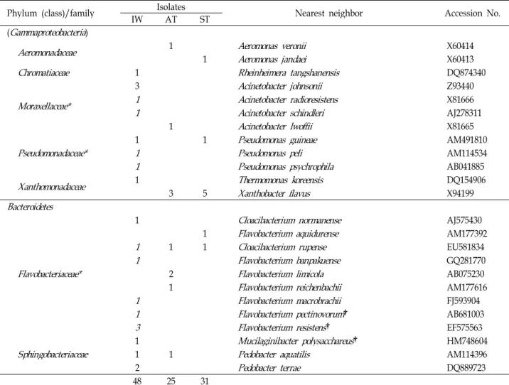
Table 3. Affiliation of the isolates from the natural dye wastewater treatment facility
Phylum (class)/family IW IsolatesAT ST Nearest neighbor Accession No.
Actinobacteria
Corynebacteriaceae
1Corynebacterium afermentans
subsp.afermentans
X82054Microbacteriaceae
1Microbacterium schleiferi
† Y172371
Frigoribacterium faeni
Y18807Micrococcaceae*
1Rothia dentocariosa
M590551
Kocuria gwangalliensis
EU2869641
Micrococcus luteus
AJ536198Nocardiaceae*
1Nocardia takedensis
AB158277Firmicutes
Bacillaceae*
2
Bacillus licheniformis
CP0000021
Bacillus simplex
AJ4390781
Bacillus thuringiensis
D162811 Bacillus stratosphericus
AJ831841Paenibacillaceae
1Paenibacillus urinalis
EF212892Staphylococcaceae*
1 1Staphylococcus caprae
AB0099351 Staphylococcus haemolyticus
X66100Streptococcaceae*
1Streptococcus salivarius
subsp.salivarius
AY188352Proteobacteria
(Alphaproteobacteria)
Bradyrhizobiaceae*
1 1Afipia birgiae
AF288304Caulobacteraceae
1
Brevundimonas mediterranea
AJ2278011 Brevundimonas nasdae
AB0719543 Brevundimonas vesicularis
AJ2277801
Brevundimonas bullata
D127851
Phenylobacterium haematophilum
AJ244650 1Phenylobacterium conjunctum
AJ227767Erythrobacteraceae
1Erythrobacter gaetbuli
AY562220Hyphomicrobiaceae
1 2 3Aquabacter spiritensis
FR733686Rhizobiaceae
1Rhizobium radiobacter
AB247615Rhodobacteraceae
1Rhodobacter capsulatus
† D16428Rhodospirillaceae*
1Elstera litoralis
EU678309Sphingomonadaceae*
1
Sphingomonas mali
Y096382
Sphingomonas wittichii
AB0214921
Sphingomonas histidinilytica
EF5302021
Sphingobium xenophagum
X940981
Sphingosinicella microcystinivorans
AB084247 1Novosphingobium resinovorum
EF0291101 Sphingopyxis panaciterrae
AB2453531
1Sphingopyxis soli
FJ599671Unclassified Rhodospirillales
1 2Reyranella massiliensis
EF394922 (Betaproteobacteria
)Burkholderiaceae
1Ralstonia solanacearum
† EF016361Comamonadaceae*
1
Acidovorax delafieldii
AF0787641 Acidovorax radicis
HM0275781
1Acidovorax soli
FJ5996722 Acidovorax temperans
AF0787663 Albidiferax ferrireducens
CP0002671
Hydrogenophaga defluvii
AJ5859931
Hydrogenophaga atypica
AJ585992Oxalobacteraceae
2Janthinobacterium lividum
Y08846Table 3. Continued
Phylum (class)/family IW IsolatesAT ST Nearest neighbor Accession No.
(
Gammaproteobacteria
)Aeromonadaceae
1Aeromonas veronii
X604141
Aeromonas jandaei
X60413Chromatiaceae
1Rheinheimera tangshanensis
DQ874340Moraxellaceae*
3
Acinetobacter johnsonii
Z934401 Acinetobacter radioresistens
X816661 Acinetobacter schindleri
AJ2783111
Acinetobacter lwoffii
X81665Pseudomonadaceae*
1 1
Pseudomonas guineae
AM4918101 Pseudomonas peli
AM1145341 Pseudomonas psychrophila
AB041885Xanthomonadaceae
1Thermomonas koreensis
DQ1549063 5
Xanthobacter flavus
X94199Bacteroidetes
Flavobacteriaceae*
1
Cloacibacterium normanense
AJ5754301
Flavobacterium aquidurense
AM1773921
1 1Cloacibacterium rupense
EU5818341 Flavobacterium banpakuense
GQ2817702
Flavobacterium limicola
AB0752301
Flavobacterium reichenbachii
AM1776161 Flavobacterium macrobrachii
FJ5939041 Flavobacterium pectinovorum
† AB6810033 Flavobacterium resistens
† EF575563Sphingobacteriaceae
1Mucilaginibacter polysacchareus
† HM7486041 1
Pedobacter aquatilis
AM1143962
Pedobacter terrae
DQ88972348 25 31
*
The taxa detected also by the DGGE profiles.†Candidate species (<97% 16S rRNA gene sequence similarity).
IW, influent water; AT, aeration tank; ST, settling tank.
염기서열이 혼합되어 존재하는 것을 알 수 있었다 . 3개의 조사 지점에서 모두 Proteobacteria 문이 가장 우점하였으며, Firmi- cutes 문, Actinobacteria 문과 Bacteroidetes 문에 속한 세균이 확 인되었다 (Table 4).
3개의 시료에서 진하게 나타난 band 4, 5, 6, 9그리고 10을 우점하는 군집으로 판단하여 염기서열을 확인 한 결과 band 4는 Sphingopyxis witflariensis, Acinetobacter guilouiae 와 Sphingopyxis flavimaris로 2개 종 이상이 혼합되었고, band 5는 Delftia acidovorans, 그리고band 6은 Sphingopyxis witflariensis 로 단일 균주임을 확인하였다 . Band 9는 Sphingopyxis wit- flariensis 와 Achromobacter marplatensis 이며, band 10은 Nocardia soli, Dongia mobilis 와 Limnohabitans parvus 가 혼합되 어 있었다 . 한편 band 15는 포기조와 침전조의 우점균으로 최근연종은 Rothia mucilaginosa 이었다.
쪽 추출물 분해
분리된 세균 25 균주를 독립적으로 배양할 경우 흡광도의 변화를 관찰할 수 없었다 . 접촉포기조 원액을 사용한 경우 색 도 제거율은 340 nm 에서는 10.5% 그리고 657 nm 에서는 18.8% 이었다.
고 찰
본 실험에 이용된 천연염색폐수처리시설에서 채취한 세 개
시료의 이화학적 특성은 색도를 제외한 모든 요인들은 큰 차
이가 나타나지 않았다 . 이것은 각각의 시료에 분포하는 세균
의 군집 구조도 큰 차이가 나타나지 않았던 것과 관련된다 .
그러나 배양이 가능한 세균을 이용한 실험에서는 폐수처리가
진행되는 과정에 따라 미생물들의 종 조성 변화를 확인 할
수 있었다 . 천염염색폐수 유입수에는 인간과 동식물에 기생하
는 세균 (Streptococcus, Staphylococcus and Acinetobacter) [5]이
Table 4. Sequence identification of clones obtained from DGGE bands Band
No. Source Phylum Family Nearest neighbor Accession No. Sequence
similarity (%)
1 IW, AT, ST Fir
Staphylococcaceae Staphylococcus caprae
AB009935 100.02 IW, AT, ST Pro
Bac Fir
Sphingomonadaceae Flavobacteriaceae Streptococcaceae
Sphingopyxis witflariensis Tamlana crocina Streptococcus gordonii
AJ416410 AM286230 AF003931
99.493.2 99.0 3 IW Fir
Rhodobacteraceae Oceanobacillus chironomi
DQ298074 100.04 IW, AT, ST Pro
Pro Pro
Sphingomonadaceae Moraxellaceae Sphingomonadaceae
Sphingopyxis witflariensis Acinetobacter guillouiae Sphingopyxis flavimaris
AJ416410 X81659 AY554010
99.499.5 98.2
5 IW, AT, ST Pro
Comamonadaceae Delftia acidovorans
AB021417 97.96 IW, AT, ST Pro
Sphingomonadaceae Sphingopyxis witflariensis
AJ416410 99.4 7 IW, AT, ST ProSphingomonadaceae Sphingopyxis witflariensis
AJ416410 99.48 IW Pro
Bac
Enterobacteriaceae
Chitinophagaceae Shigella sonnei
Terrimonas lutea
FR870445AB192292 99.5 96.39 IW, AT, ST Pro
ProPro
Sphingomonadaceae Alcaligenaceae Pseudomonadaceae
Sphingopyxis witflariensis Achromobacter marplatensis Pseudomonas hibiscicola
AJ416410 EU150134 AB021405
99.4 99.599.0
10 IW, AT, ST Pro
Pro
Rhodospirillaceae
Comamonadaceae Dongia mobilis
Limnohabitans parvus
FJ455532FM165536 98.8 95.4 11 AT ProSphingomonadaceae Sphingomonas xinjiangensis
FJ754464 100.0 12 AT ProBradyrhizobiaceae Salinarimonas rosea
EU878006 96.513 IW, AT, ST Act
Pro
Nocardioidaceae
Oxalobacteraceae Nocardioides dubius
Oxalicibacterium flavum
AY928902AY061962 93.0 96.914 IW, AT, ST Fir
ProPro
Planococcaceae Sphingomonadaceae Pseudomonadaceae
Bhargavaea cecembensis Sphingopyxis flavimaris Pseudomonas vranovensis
AM286423 AY554010 AY970951
99.5 100.099.4
15 AT, ST Act
Micrococcaceae Rothia mucilaginosa
X87758 99.416 ST Fir
Bacilliaceae Bacillus licheniformis
CP000002 100.0 17 ST BacFlavobacteriaceae Flavobacterium glycines
EU672803 98.4*Act,
Actinobacteria
; Bac,Bacteroidetes
; Fir,Firmicutes
; Pro,Proteobacteria
; IW, influent water; AT, aeration tank; ST, settling tank.다수 분포하였고 , 일부 세균은 폐수에서 자주 분리된 세균으 로 세포외 다당을 생산하며 색소를 띤 세균 (Flavobacterium, Pedobacter, Cloacibacterium)이었다[1]. 유입된 폐수가 포기조를 거쳐 침전조에 이르는 동안 오염균의 수가 감소하면서 유기물 과 방향족 화합물을 분해하는 세균 (Novosphingobium, Sphin- gobium, Sphingomona, Acidovora, Albidiferax) [27, 29, 34]이 증 가하는 경향을 확인하였다 .
DGGE band의 염기서열 분석을 통해 나타난 우점균 들은 폐수처리시설에 정상군총으로 존재하며 염료를 포함한 난분 해성 화합물의 분해에 관여한 세균들과 유사하였다 . DGGE를 통해 검출된 세균들의 분리원과 분해 기질 특이성은 아래와 같다 . Sphingopyxis witflariensis는 활성슬러지에서 분리된 종이 며 , 난분해성 화합물을 분해하며 세포외 다당을 합성하는 세 균이다 [16]. Delftia acidovorans는 acetamide로 오염된 토양에 서 [31], Dongia mobilis 는 염료인 malachite green 처리조에서
[22], 그리고 Achromobacter marplatensis 는 난분해성 방향족화 합물인 pentachlorophenol로 오염된 토양에서[12] 분리되었 다 . 이 외에도 세포외 다당을 형성하는 세균인 Rothia mucilagi- nosa [10]가 확인되었다.
합성염료 폐수처리에서 탈색에 관여한다고 알려진 세균으 로 Shewanella, Pseudomonas, Lactobacillus, Proteus, Enterococcus, Streptococcus, Bacillus와 Streptomyces 등이 알려져 있다[6, 7, 8, 11, 15, 26, 28, 33]. 본 연구 결과에서도 Pseudomonas, Streptococcus와 Bacillus 속의 세균들이 배양되었거나 DGGE를 통해 확인되었다 .
다른 천연염색폐수처리장의 미생물 군집에 관한 연구는 거
의 없으므로 본 연구의 미생물군집과 비교하기는 어렵다 . 다
만 생활하수 처리시설에서의 미생물 군집은 많은 경우 Proteo-
bacteria 가 우점하거나 몇몇 처리시설에서는 Bacteroidetes 가
우점하는 보고가 있다 . 이 두 문의 세균 외에 Acidobacteria,
Chloroflexi, Plantomycetes, Verrucomicrobia, Firmicutes와 Acti- nobacteria 순으로 점차 비율이 낮아진다고 하였다[14]. 본 연구 에서 확인된 세균 군집은 Proteobacteria, Bacteroidetes, Firmi- cutes와 Actinobacteria의 4가지 문이었다. 생활하수에 비해 유 기물의 함량이 낮고 색소 화합물이 많은 염색폐수이므로 세균 의 군집이 단순할 것으로 판단된다 . 그러나 nitroaromatic화합 물을 함유한 화학염색폐수의 활성슬러지에 분포하는 세균 군 집이 Proteobacteria와 Firmicutes의 2개의 문으로 단순했던 것 보다는 다양한 세균 군집이 확인되었다 [17]. 즉, 천연염색폐수 의 세균 조성은 화학염색폐수 보다는 다양하지만 생활하수 보다는 단순하였다 .
3개 지점의 세균 군집 간의 차이는 분리균의 군집 조성에 비해 DGGE 분석결과에서는 확실하게 나타나지 않았다. 그 이유 중 첫 번째는 세균을 분리할 때 각 시료별 분리균의 수가 동일하지 않았으며 , 가급적 형태적으로 다른 균을 선별하여 3개 시료에 공통으로 존재하는 세균들이 특정 시료에서는 분 리되지 않았을 수 있는 오류에 기인한다고 볼 수 있다 . 즉, 형태적으로 유사한 세균을 유입조에서 선택할 경우 나머지 두 시료에서는 배제 되었을 가능성이 매우 높다고 볼 수 있다 . 두번째는 DGGE band들 중 농도가 낮아 염기서열 분석에 이 용되지 않았던 세균 군집의 차이가 분리에 의한 종 조성과는 다른 결과를 나타냈다고 볼 수 있다 . 따라서 이와 같은 약점을 보완하기 위해서는 세균의 분리시 여러가지 배지와 다양한 배양 조건을 적용하고 , 가급적 많은 수의 세균을 분리해야 할 필요가 있다 . 또한 본 연구에 사용된 두 가지 방법 외에 시료에 존재하는 대부분의 세균의 16S rRNA 유전자들의 염기서열을 단시간에 대량으로 파악할 수 있는 pyrosequencing [14]과 같 은 기법을 적용해야 할 필요가 있다고 본다 .
한편 , 본 연구에 사용된 처리시설의 염색공정에서 가장 많 은 비율로 사용되는 염료는 쪽 추출물로서 indigo라고 한다.
Indigo (C
12H
8O
2N
2)는 물에 불용성이기 때문에 그대로는 염색 이 어려우므로 일반적으로 환원제와 알칼리를 병용하여 leu- co화하여 염색을 하게 된다[3]. Indigo 분해에 관여하는 세균 종을 탐색한 결과 분리된 단일 세균 종은 indigo의 분해에 큰 영향을 미치지 못하고 여러 종의 세균 집합체가 관여함을 알 수 있었다 . 이와 같은 결과는 타 연구와 유사하였다[4]. 폐수 처리시 색도의 제거가 세균들에 의한 분해 외에도 응집과 같 은 물리화학적인 방법이 미치는 영향에 후속 연구가 필요하 다 .
이와 같은 제한요인에도 불구하고 염색폐수 처리시설의 세 균 군집은 염료를 포함한 난분해성 화합물을 분해하는 세균 , 세포외 다당을 생산하는 세균 그리고 인간을 비롯한 동식물의 기생균과 유사한 종의 세균들이 우점하고 있음을 알 수 있었 으며 , 8개의 신종 후보 세균 또한 확보할 수 있었다. 이와 같이 천연염색 폐수처리시설에 서식하는 세균들 중에는 화학염색 폐수에서 염료를 비롯한 난분해성 화합물의 분해에 관여하는
세균들이 분포하고 있었다 . 또한 세포외 다당을 형성하는 세 균들이 분리되는 것을 확인하였으며 이 세균들은 유기물의 분해와 응집작용에 관여한다고 볼 수 있다 .
References
1. Allen, T. D., Lawson, P. A., Collins, M. D., Falsen, E. and Tanner, R. S. 2006.
Cloacibacterium normanense
gen. nov., sp.nov., a novel bacterium in the family
Flavobacteriaceae
iso- lated from municipal wastewater.Int J Syst Evol Microbiol
56, 1311-1316.2. American Public Health Association (APHA). 1998. Standard methods for the examination of water and wastewater, 20th ed., Washington D. C., USA.
3. Balan, D. S. L. and Monteiro, R. T. R. 2001. Decolorization of textile indigo dye by ligninolytic fungi.
J Biotechnol
89, 131-139.4. Banat, I. M., Nigam, P., Singh, D. and Marchant, R. 1996.
Microbial decolorization of textile-dye-containing effluents:
a review.
Bioresource Technol
58, 217-227.5. Bouvet, P. J. M. and Grimont, P. A. D. 1986. Taxonomy of the genus
Acinetobacter
with the recognition ofAcinetobacter baumannii
sp. nov.,Acinetobacter haemolyticus
sp. nov.,Acinetobacter johnsonii
sp. nov., andAcinetobacter junii
sp.nov. and emended descriptions of
Acinetobacter calcoaceticus
andAcinetobacter lwoffii
.Int J Syst Evol Microbiol
36, 228-240.6. Chang, J. S. and Lin, Y. C. 2000. Fed-batch bioreactor strat- egies for microbial decolorization of azo dye using a
Pseudomonas luteola
strain.Biotechnol Prog
16, 979-685.7. Chang, J. S., Chou, C., Lin, Y. C., Lin, P. J., Ho, J. Y. and Hu, T. L. 2001. Kinetic characteristics of bacterial azo-dye decolorization by
Pseudomonas luteola
.Water Res
35, 2841- 2850.8. Chen, K. C., Huang, W. T., Wu, J. Y. and Houng, J. Y. 1999.
Microbial decolorization of azo dyes by
Proteus mirabilis
.J Ind Microbiol Biotechnol
23, 686-690.9. Chun, J., Huq, A. and Colwell, R. R. 1999. Analysis of 16S-23S rRNA intergenic spacer regions of
Vibrio cholerae
andVibrio mimicus
.Appl Environ Microbiol
65, 2202-2208.10. Collins, M. D., Hutson, R. A., Båverud, V. and Falsen, E.
2000. Characterization of a
Rothia
-like organism from a mouse: description ofRothia nasimurium
sp. nov. and re- classification ofStomatococcus mucilaginosus
asRothia mucila- ginosa
comb. nov.Int J Syst Evol Microbiol
50, 1247-1251.11. Conneely, A., Smyth, W. F. and McMullan, G. 1999.
Metabolism of the phthalocyanine textile dye remazol tur- quoise blue by
Phanerochaete chrysosporium
.FEMS Microbiol Lett
179, 333-337.12. Gomila, M., Tvrzová, L., Teshim, A., Sedláček, I., González- escalona, N., Zdráhal, Z., Šedo, O., González, J. F., Bennasar, A., Moore, E. R. B., Lalucat, J. and Murialdo, S. E. 2011.
Achromobacter marplatensis
sp. nov., isolated from a penta- chlorophenol-contaminated soil.Int J Syst Evol Microbiol
61, 2231-2237.13. Gupta, V. K., Mittal, A. and Gajbe, V. 2005. Adsorption and
desorption studies of a water soluble dye, Quinoline Yellow, using waste materials.
J Colloid Interface Sci
284, 89-98.14. Hu, M., Wang, X., Wen, X. and Xia, Y. 2012. Microbial com- munity structures in different wastewater treatment plants as revealed by 454-pyrosequencing analysis.
Bioresour Technol
117, 72-79.15. Jian, H., Tso, W., Tso, M., Zhang, X., Xu, M., Deng, S. and Sun, G. 2000. Broad spectrum decolorizing bacterial strains and their functional plasmids, pp. 97-104. In: Healy. M., Wise, D. L. and Moo-Young, M. (eds.),
Environmental Monitoring and Biodiagnostics of Hazardous Contaminants.
Academic Publishers: Dordrecht, Netherlands.
16. Kämpfer, P., Witzenberger, R., Denner, E. B. M., Busse, H.
J. and Neef, A. 2002.
Sphingopyxis witflariensis
sp. nov., iso- lated from activated sludge.Int J Syst Evol Microbiol
52, 2029-2034.17. Kapley, A., Prasad, S. and Purohit, H. J. 2007. Changes in microbial diversity in fed-batch reactor operation with wastewater containing nitroaromatic residues.
Bioresour Technol
98, 2479-2484.18. Khelifi, E., Bouallagui, H., Touhami, Y., Godon, J. J. and Hamdi, M. 2009. Bacterial monitoring by molecular tools of a continuous stirred tank reactor treating textile wastewater.
Bioresour Technol
100, 629-633.19. Kim, O. S., Cho, Y. J., Lee, K., Yoon, S. H., Kim, M., Na, H., Park, S. C., Jeon, Y. S., Lee, J. H., Yi, H., Won, S. and Chun, J. 2012. Introducing EzTaxon-e: a prokaryotic 16S rRNA Gene sequence database with phylotypes that repre- sent uncultured species.
Int J Syst Evol Microbiol
62, 716-721.20. Lane, D. J. 1991. 16S/23S rRNA sequencing, pp. 115-175.
In: Stackebrandt, E. and Goodfellow, M. (eds.),
Nucleic Acid Techniques in Bacterial Systematics.
Wiley, Chichester, U.K.21. Lee, B. J., Wentzel, M. C. and Ekama, G. A. 2006.
Measurement and modelling of ordinary heterotrophic or- ganism active biomass concentrations in anoxic/aerobic ac- tivated sludge mixed liquor.
Water Sci Technol
54, 1-10.22. Liu, Y., Jin, J. H., Liu, Y. H., Zhou, Y. G. and Liu, Z. P.
2010.
Dongia mobilis
gen. nov., sp. nov., a new member of the familyRhodospirillaceae
isolated from a sequencing batch reactor for treatment of malachite green effluent.Int J Syst Evol Microbiol
60, 2780-2785.23. Liu, Y., Zhang, Y., Quan, X., Zhang, J., Zhao, H. and Chen, S. 2011. Effects of an electric field and zero valent iron on anaerobic treatment of azo dye wastewater and microbial community structures.
Bioresour Technol
102, 2578-2584.24. Massana, R., Murray, A. E., Preston, C. M. and Delong, E.
F. 1997. Vertical distribution and phylogenetic character-
ization of marine planktonic Archaea in the Santa Barbara Channel.
Appl Environ Microbiol
63, 50-56.25. Nigam, P., Singh, D. and Marchant, R. 1996. An inves- tigation of the biodegradation of textile dyes by aerobic and anaerobic microorganisms, pp. 278-287. In: Moo-Young, M., Amderson, W. A. and Chakrabarty, A. M. (eds.),
Environmental Biotechnology, Principles and Applications.
Academic Publishers: Dordrecht, Netherlands.
26. Phugare, S. S., Kagalkar, A. N., Govindwar, S. P. and Jadhav, J. P. 2011. A study on significant microbial inter- action leading to decolorization and degradation of textile dye Rubine 3GP.
J Basic Microbiol
51, 499-514.27. Ramana, C. V. and Sasikala, C. 2009.
Albidoferax
, a new ge- nus ofComamonadaceae
and reclassification ofRhodoferax fer- rireducens
(Finneranet al
., 2003) asAlbidoferax ferrireducens
comb. nov.J Gen Appl Microbiol
55, 301-304.28. Scheline, R. R., Nygaard, R. T. and Longberg, B. 1970.
Enzymatic reduction of azo dye, acid yellow, by extracts of
Streptococcus faecalis
isolated from rat intestine.Food Cosmet Toxicol
8, 55-58.29. Schulze, R., Spring, S., Amann, R., Huber, I., Ludwig, W., Schleifer, K. H. and Kämper, P. 1999. Genotypic diversity of
Acidovorax
strains isolated from activated sludge and de- scription ofAcidovorax defluvii
sp. nov.Syst Appl Microbiol
22, 204-214.30. Somerville, C. C., Knight, I. T., Straube, W. L. and Colwell, R. R. 1989. Simple, rapid method for direct isolation of nu- cleic acids from aquatic environments.
Appl Environ Microbiol
55, 548-554.31. Wen, A., Fegan, M., Hayward, C., Chakraborty, S. and Sly, L. I. 1999. Phylogenetic relationships among members of the
Comamonadaceae
, and description ofDelftia acidovorans
(den Dooren de Jong 1926 and Tamaokaet al
. 1987) gen. nov., comb. nov.Int J Syst Bacteriol
49, 567-576.32. Wright, E. S., Yilmaz, L. S. and Noguera, D. R. 2012.
DECIPHER, a search-based approach to chimera identi- fication for 16S rRNA sequences.
Appl Environ Microbiol
78, 717-725.33. Xu, M., Guo, J., Cen, Y., Zhong, X., Cao, W. and Sun, G.
2005.
Shewanella decolorationis
sp. nov., a dye-decolorizing bacterium isolated from activated sludge of a waste-water treatment plant.Int J Syst Evol Microbiol
55, 363-368.34. Yabuuchi, E., Yamamoto, H., Terakubo, S., Okamura, N., Naka, T., Fujiwara, N., Kobayashi, K., Kosako, Y. and Hiraishi, A. 2001. Proposal of
Sphingomonas wittichii
sp. nov.for strain RW1T, known as a dibenzo-