INTRODUCTION
tert-Butyl hydroperoxide (t-BHP), a short-chain lipid hy- droperoxide analogue, has been previously employed as a model to investigate the mechanism of cell injury initiated by acute oxidative stress (Jenner and Olanow 1998). This t- BHP can also be metabolized to free radical intermediates by cytochrome P450 in the hepatocytes (Minotti et al. 1986), which in turn may initiate lipid peroxidation, which affects cell integrity, and result in cell injury in hepatocyte cultures
and livers (Martin et al. 2001). Additionally, a number of pro-oxidant drugs, including t-BHP, have been implicated in the production of oxidative stress and cell injury resulting from the intracellular production of reactive oxygen species (ROS) in a variety of cell types (Goya et al. 2007; Kanupriya et al. 2007). Normally produced ROS are scavenged by endogenous antioxidants, which exist in abundance in the liver tissue (Yu 1994). However, liver damage can occur when large acute doses of or chronic exposure to toxic substances overpower the hepatic antioxidant defense sys- tem (Fruehauf and Meyskens 2007). These excess ROS inter- act readily with cellular macromolecules and structures, resulting in changes in membrane permeability, the activa-
─
─ 230 ──
Protective Effect of Allomyrina dichotoma Larva Extract on tert-butyl Hydroperoxide-induced Oxidative Hepatotoxicity
Kyung Jin Lee and Jong Bin Lee1,*
Cell Dynamics Research Center, Department of life Science, Gwangju Institute of Science and Technology, Gwangju 500-712, Korea
1Department of Biological Science, College of Natural Sciences, Chonnam National University, Gwangju 500-757, Korea
Abstract -- An extract of Allomyrina dichotoma larva (ADL), one of the insects used most frequen- tly in traditional Chinese medicine for the treatment of liver diseases such as hepatocirrhosis and hepatofibrosis, was assessed for antioxidant bioactivity in this study. In the current work, we have investigated the protective effects of ADL extracts on tert-butyl hydroperoxide (t-BHP)-induced hepatotoxicity in cultured hepa1c1c7 cells and in the mouse liver. The treatment of the hepa1c1c7 cells with ADL extracts induced a significant reduction of t-BHP-induced oxidative injuries, as determined by cell cytotoxicity, lipid peroxidation (LPO) and reactive oxygen species contents, in a dose-dependent manner. Moreover, ADL extracts evidenced a protective effect against t-BHP- induced oxidative DNA damage, as revealed by the results of the Comet assay in hepa1c1c7 cells.
ADL extracts also protected against hydroxyl radical-induced 2-deoxy-d-ribose degradation by ferric ion-nitrilotriacetic acid and H2O2. In addition, ADL extracts were shown to be able to quench 1,1-diphenyl-2-picrylhydrazyl (DPPH) free radicals. Our in vivo study revealed that ADL extracts pretreatment applied prior to t-BHP administration significantly prevented an increase in the ser- um levels of hepatic enzyme markers and reduced LPO in the mouse liver in a dose-dependent manner. Taken together, these results suggest that the protective effects of ADL extracts against t- BHP-induced hepatotoxicity may be attributable, at least in part, to its ability to scavenge free oxy- gen radicals, and to protect against DNA damage due to oxidative stress.
Key words : Allomyrina dichotoma larva, tert-butyl hydroperoxide, DNA damage, oxidative stress
* Corresponding author: Jong Bin Lee, Tel. 062-530-3395, Fax. 062-530-0306, E-mail. [email protected]
tion of proteases and nucleases, and altered gene expression in many tissues (Wu et al. 1999; Srinivasan et al. 2006;
Fruehauf and Meyskens 2007). This is one of the principal reasons that the liver is a target for toxic substances.
Allomyrina dichotoma larva (ADL) have been extensively employed in traditional Chinese medicine for the treatment of liver diseases, including hepatocirrhosis, hepatofibrosis, liver cancer, etc (Miyanoshita et al. 1996; Sagisaka et al.
2001). ADL, the Asian beetle, has been shown to harbor an antineoplastic agent similar to lectin, and the substance res- ponsible for this biological activity was detected principally in the larvae of the beetle (Yoshikawa et al. 1999). However, it remains to be determined whether ADL can prevent or alleviate liver injury induced by oxidative stress; no unequi- vocal evidence has been found to resolve this question.
Therefore, we attempted to assess the protective effects of ADL on t-BHP-induced oxidative hepatotoxicity and DNA dama-ge, and also elucidated the mechanism(s) underlying its protective effects in hepa1c1c7 cells and the rat liver.
MATERIALS AND METHODS
1. Chemicals
Chemicals and cell culture materials were acquired from the following sources: tert-butyl hydroperoxide (t-BHP), thio- barbituric acid, 1,1-diphenyl-2-picrylhydrazyl (DPPH), 2- deoxy-D-ribose and aspartate aminotransferase (AST), and alanine aminotransferase (ALT) diagnostic kits from Sigma Co.; lactate dehydrogenase (LDH) and WST-1 assay kit from Roche Co.; the Biotrin OxyDNA assay kit for the detection of oxidative DNA damage from Biotrin International Ltd; 5- chloromethl-2′ 7′-dichorodihdydro-fluorescein diacetate ethyl ester (CM-DCFDA) was obtained from Molecular Probes;
Dulbecco’s modified Eagle’s medium (DMEM), penicillin, streptomycin, neomycin, glutamine, and fetal bovine serum (FBS) were acquired from Life Technologies, Inc.; all other chemicals used were of the highest available commercial grade.
2. Preparation of ADL extracts
The Allomyrina dichotoma larva (ADL) extracts were obtained from Kachi-maeul Co., Chonnam, South Korea.
The ADL were prepared using the method described else-
where, and their compositions were published previously (Yoshikawa et al. 1999).
3. Cell culture and treatment
The hepa1c1c7 cell was obtained from the American Type Culture Collection (Bethesda, MA). The cells were maintain- ed in DMEM, 10% fetal bovine serum, 2 mM glutamine, and 1% penicillin/streptomycin in 75 cm2culture bottles in a 5%
CO2atmosphere at 37�C. The cells were then pretreated with ADL extracts for 6 h prior to the addition of t-BHP (250μM).
The control cells were treated with DMSO, the final concen- tration of which never exceeded 0.1%, and this concentration exerted no noticeable effects on the assay system.
4. Cell toxicity assay
Cell toxicity was assessed by means of the WST-1 assay and by measuring the release of LDH. The level of LDH release in the supernatants was measured using a LDH kit.
After the supernatant was removed for LDH determination, the cells were used for the WST-1 assay. The WST-1 and LDH assay were conducted in accordance with the manu- facturer’s instructions.
5. Lipid peroxidation assay
Malondialdehyde (MDA), the lipid peroxidation product in the cells, was assayed by a thiobarbituric acid fluoromet- ric method using 1,1,3,3-tetramethoxy-propane as the stan- dard. Control tests indicated that ADL extracts did not inter- fere with lipid peroxidation assays. The protein concentration was determined by the Bradford method, using bovine serum albumin as the standard.
6. Measurement of intracellular ROS production
The fluorescent probe, 5-chloromethly-2′ 7′-dichlorodihy- drofluorescein diacetate ethyl ester (CM-DCFDA), was used to monitor the intracellular generation of reactive oxygen species by t-BHP, as previously described (Lee et al. 2004).
7. Comet assay
Oxidative DNA damage was evaluated via a Comet assay.
Culture medium was a pirated from the hepa1c1c7 cell mo- nolayer and the cells were exposed to different concentrat-
ions of ADL extracts and t-BHP in PBS for 20 min on ice.
Following exposure to the oxidant, the cells were washed twice with ice-cold PBS. The cells were then detached from the culture dishes and processed for the Comet assay as pre- viously described (Lee and Jeong 2007).
8. Measurement of 2-deoxy-d-ribose degradation
To prepare the Fe3++-nitrilotriacetic acid (NTA), FeCl3was dissolved in 20 mM phosphate buffer to Fe3++ to an NTA molar ratio of 1 : 1, and the pH was adjusted to 7.4. A reac- tion mixture containing 2.8 mM 2-deoxy-d-ribose, 50 mM KH2PO4-KOH buffer at pH 7.4, 1 mM H2O2, and 30 mM Fe3++-NTA was prepared. The mixture was then incubated for 1 hour with or without ADL extracts at 37�C. The degra- dation of 2-deoxy-d-ribose was measured by determining the formation of the thiobarbituric acid-reactive material as described previously (You et al. 2002).
9. Assay of free radical-quenching activity
The free radical-quenching capacity of ADL was tested by a method involving the bleaching of stable DPPH (Lee et al. 2004).
10. Animal treatment and hepatotoxicity assessment
SD rats (200±5 g) were used for the experiments. The mice were allowed free access to Purina Rodent Chow and tap water, maintained in a controlled environment at 21±
2�C and 50±5% relative humidity with a 12 h : 12 h dark/
light photocycle, and acclimatized for at least l week before use. To study its protective effects against t-BHP-induced hepatotoxicity, ADL extracts in saline was intragastrically administered at 100~400 mg kg-1once daily for three con- secutive days. Three hours after the final treatment, the rats were treated with t-BHP (2 mmol, intraperitoneally, 100: l dissolved in saline). 24 hours after the administration of t- BHP, the rats were anesthetized with CO2, blood was re- moved via cardiac puncture to determine serum ALT and AST activity, after which the animals were sacrificed by cervical dislocation. After bleeding, the livers were weighed and a thin slice preserved in a buffered formalin solution to obtain the histological sections. The remaining livers were quickly frozen in dry ice/methanol and stored at -70�C for
lipid peroxidation analysis. Hepatotoxicity was assessed by quantifying the serum activities of ALT and AST and hepa- tic lipid peroxidation. The serum ALT and AST activities were measured using a spectrophotometric diagnostic kit.
11. Statistical analysis
All experiments were repeated at least three times. Results are expressed as the means±SD. ANOVA was utilized to evaluate the difference between multiple groups. If signifi- cance was observed between groups, the Student’s ‘t’ test was used to compare the means of two specific groups, with p⁄0.05 considered statistically significant.
RESULTS
1. Effect of ADL extracts on the t-BHP-induced cytotoxicity
The protective effects of ADL extracts against t-BHP-in- duced hepatotoxicity in hep1c1c7 cells were quantified via LDH and WST-1 assays. ADL, which is nontoxic even at high concentrations (400μg mL-1), afforded full protection from cell injury, as shown by the WST-1 assay (Fig. 1A).
To quantify more precisely the protective effects of ADL extracts toward t-BHP, we also conducted LDH assays, and the results of those assays were consistent with those obtain- ed from the WST-1 assay. The addition of ADL extracts to the cells effectively protected the cells against the cytotoxi- city induced by t-BHP, as shown by the leakage of LDH (Fig. 1B).
2. Effects of ADL extracts on t-BHP-induced lipid peroxidation and intracellular ROS production
ADL extracts alone induced no change in the degree of MDA formation as compared to the untreated controls. How- ever, 6 hours of exposure to t-BHP alone increased the amount of cell-associated MDA in hepa1c1c7 cells, and the presence of ADL extracts significantly prevented t-BHP- induced MDA production in a dose-dependent manner (Fig.
1C). In order to confirm that ADL extracts reduces t-BHP- induced oxidative stress in hepa1c1c7 cells, intracellular ROS production was evaluated by monitoring 5-chlorome- thyl-2′ 7′-dichlorodihydrofluorescein diacetate ethyl ester (CM-DCFDA) fluorescence. Rapid increases in intracellular
oxidant levels were noted in the cells after t-BHP treatment, but the oxidant burden observed after t-BHP exposure was reduced in the presence of ADL extracts in a dose-dependent
manner (Fig. 2). These results show that ADL extracts were able to inhibit membrane lipid peroxidation via the suppres- sion of intracellular ROS triggered by the injurious peroxy- radicals generated from t-BHP.
3. Effects of ADL extracts on t-BHP-induced oxidative DNA damage
The Comet assay is used to detect single-and double- stranded DNA breaks in naked supercoiled DNA. As t-BHP increased DNA damage via a mechanism that involved intracellular ROS production, t-BHP treatment applied for 20 minutes to the hepa1c1c7 cells caused a four-fold increase in the tail moments as compared to the control cells. How- ever, the tail moment increase caused by t-BHP-induced oxidative DNA damage was reduced in the presence of ADL extracts in a dose-dependent manner (Fig. 3). These results indicated that the increase in intracellular ROS pro- duction might mediate t-BHP-induced DNA damage to the cells.
4. Free radical-quenching activity of ADL extracts on the DPPH
In order to determine whether the protection afforded to the hepatocytes by ADL extracts against t-BHP-induced oxidative cytotoxicity and DNA damage might be the conse- quence of the free radical-quenching capacity of ADL ex- tracts, the bleaching of DPPH radical scavenging activity by Fig. 1. Cytoprotective effects of ADL extracts on t-BHP-induced
oxidative hepatotoxicity in hepa1c1c7 cells. The cells were plated on 24-well plates and various concentrations of ADL extracts were applied (250μM) for 24 h. Cell viability was estimated by (A) WST-1 assay and (B) LDH release assay.
(C) Lipid peroxidation was evaluated by malondialdehyde (MDA) formation as described in the Materials and Methods section. Each bar represents the mean±SD calculated from three independent experiments. *Significantly different from control at p⁄0.05.
0 5 10 15 20 25 30
0 2 4 6 8 10 12
* * *
*
*
*
*
*
*
Time (min)
Fold of intercellular ROS production
Control t-BHP
t-BHP+ ADL 100 μg mL-1 t-BHP+ ADL 200 μg mL-1 t-BHP+ ADL 400 μg mL-1
Fig. 2. Effects of ADL extracts on t-BHP-induced cellular ROS production. The Hepa1c1c7 cells were treated with ADL extracts and t-BHP (250μM) and the intracellular ROS was measured by monitoring fluorescence increases for 30 min.
*p⁄0.05, significantly different from t-BHP alone.
WST-1 assay(% of control)LDH relase assay(% of control)Lipid peroxidation(MDA: nmol mg-1protein) 120
100
80
60
40
20
0
5
4
3
2
1
0
10
8
6
4
2
0
Control 0 100 200 400 ADL (μg mL-1) t-BHP
Control 0 100 200 400 ADL (μg mL-1) t-BHP
Control 0 100 200 400 ADL (μg mL-1) t-BHP
*
*
*
*
*
*
(A)
(B)
(C)
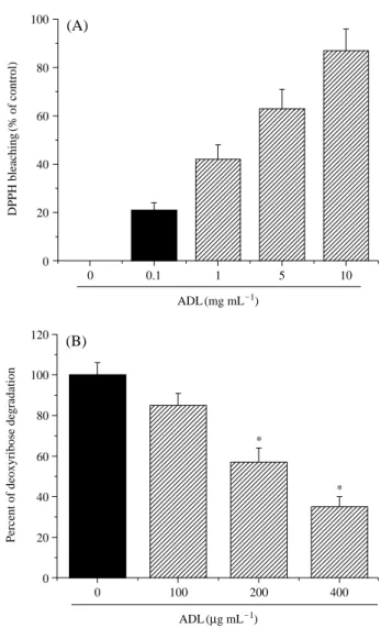
ADL extracts were measured. The results, which are sum- marized in Fig. 4A, showed that ADL extracts were able to quench the DPPH free radicals in a dose-dependent manner.
5. Effects of ADL extracts on hydroxyl radical- induced oxidative 2-deoxy-d-ribose degradation
To further assess the inhibitory effects of ADL extracts on oxidative DNA damage, Fe3++-NTA/H2O2-induced 2- deoxy-d-ribose degradation was assayed. The presence of ADL extracts were also determined to markedly inhibit the 2-deoxy-d-ribose degradation induced by Fe3++-NTA/H2O2
in a dose-dependent manner, thereby indicating that ADL extracts are potent scavengers of the hydroxyl radical, and confer protection against Fe3++-NTA/H2O2-mediated DNA damage (Fig. 4B). Therefore, these results show that ADL extracts have free radical-quenching activity.
6. Effect of ADL extracts on t-BHP-induced hepatotoxicity in rats
The hepatic enzymes, AST and ALT, were used as bio- chemical markers for early acute hepatic damage (Liu et al.
2002). A single dose of t-BHP (2 mmol kg-1) induced hepa- totoxicity in the rats, as indicated by the increase in their ALT and AST serum levels following t-BHP administration and ADL extracts pretreatment prevented the t-BHP-induced elevation of ALT and AST serum levels in a dose-dependent manner (Table 1). In a manner which was consistent with
the effect on the serum levels of ALT and AST, pretreatment with ADL extracts caused a significant reduction in t-BHP- induced lipid peroxidation in the liver (Table 1).
DISCUSSION
Liver injury induced by t-BHP is the best-characterized system of acute hepatic damage, and is a commonly utilized model for the screening of the hepatoprotective activities of
Control 0 100 200 400
0 1 2 3 4
* *
ADL (μg mL-1)
Tail movement (% of control)
t-BHP
Fig. 3. Effects of ADL extracts on t-BHP-induced oxidative DNA damage. The Hepa1c1c7 cells were treated with ADL ext- racts and t-BHP (250μM) and oxidative DNA damage was evaluated using the Comet assay by measurements of fluo- rescence intensity. *p⁄0.05 significantly different from t- BHP alone.
0 100 200 400
0 20 40 60 80 100 120
*
*
Percent of deoxyribose degradation
0 0.1 1 5 10
0 20 40 60 80 100
ADL (mg mL-1)
ADL (μg mL-1)
DPPH bleaching (% of control)
(A)
(B)
Fig. 4. Effects of ADL extracts on free radical quenching activity and 2deoxy-D-ribose degradation. (A) Radical quenching activity was measured by DPPH. ADL extracts were mixed with DPPH (10 mM, 30μL) in methanol (3 mL) as described in the Materials and Methods section. (B) 2-deoxy-D-ribose degradation was measured as described in the Materials and Methods section and the percentage of inhibition was calcu- lated from the difference in the presence and absence of ADL extracts. *p⁄0.05, significantly different from Fe3++ NTA/t-BHP alone.
drugs (Yen et al. 2004). The principal objective of this study was to evaluate the ability of ADL extracts to affect cellular and DNA damage in hepa1c1c7 cells, as well as hepatotoxi- city in mice associated with t-BHP-induced oxidative stress.
In the present study, ADL extracts effectively protected against t-BHP-induced hepatoxicity in hepa1c1c7 cells, as shown by measurements of cell viability and malondialde- hyde (MDA) formation. MDA is the major oxidative degra- dation product of membrane unsaturated fatty acids, and is considered to be an indirect measure of ROS generation (Blair 2001). Thus, we assessed CM-DCFDA fluorescent changes after t-BHP treatment as an additional sensitive means of intracellular ROS detection. It was observed that the levels of ROS in ADL extracts-treated cells were lower than in control cells following exposure to t-BHP. Moreover, it has been shown that increasing cellular ROS can break DNA strands within the cell (Termini 2000). DNA damage was assessed by the comet assay, a procedure which detects DNA strand breaks in individual cells, resulting from events including the direct scission of the DNA backbone by free radical attack (Johnson and Loo 2000). Thus, we determined that t-BHP exposure induced a greater level of DNA damage in the control cells than in the ADL extracts-treated cells.
Taken together, these data not only confirm the cytoprotec- tive and anti-lipid oxidation activities of ADL extracts, but also support the proposed functions of ADL extracts as a scavenger of radicals, which ultimately attenuates the asso- ciated cytotoxicity and genotoxicity. Consistent with our data, we suggest that ADL extracts are an effective ROS scavenger, which can protect cellular DNA against oxidative damage. However, the exact mechanisms underlying the antioxidant effects of ADL extracts remain unclear. There- fore, we attempted to determine whether the protection afforded to cells by ADL extracts against t-BHP-induced
oxidative hepatotoxicity and DNA damage might be a con- sequence of the free radical-quenching capacity of ADL.
DNA is a very sensitive target of hydroxyl radicals, and it has been demonstrated that in the presence of Fe3++NTA/
H2O2hydroxyl radicals are produced (Lee and Jeong 2007).
It has also been previously shown that Fe3++NTA/H2O2are capable of inducing 2-deoxy-D-ribose degradation (Lee et al. 2004). In the present data, ADL extracts inhibited the 2- deoxy-d-ribose degradation induced by Fe3++-NTA/H2O2, and this result indicates that ADL extracts are a potent scavenger of the hydroxyl radical. Additionally, ADL extracts also evi- dence a DPPH queuing effect. Overall, these data indicate that ADL extracts are an effective scavenger of ROS, and can protect cellular DNA against oxidative damage.
The results of our in vivo study also indicated that oral ADL extracts pretreatment significantly lowered t-BHP- induced serum levels of the hepatic enzyme markers (ALT and AST) and reduced oxidative stress in the liver, as deter- mined by our evaluation of lipid peroxidation.
In conclusion, the present study showed that ADL extracts protected against t-BHP-induced oxidative hepatotoxicity in Hep1c1c7 cells and in the mouse liver. Therefore, this study provides biological evidence that supports the notion that ADL extracts may prove useful for the treatment of liver disorders in living systems.
REFERENCES
Blair IA. 2001. Lipid hydroperoxide-mediated DNA damage.
Exp. Gerontol. 36:1473-1481.
Fruehauf JP and Meyskens FL Jr. 2007. Reactive oxygen spec- ies: a breath of life or death? Clin. Cancer Res. 13:789-794.
Goya L, C Delgado-Andrade, JA Rufian-Henares, L Bravo and FJ Morales. 2007. Effect of coffee melanoidin on human Table 1. Dose-dependent effects of ADL extracts on t-BHP-induced hepatotoxicity in rats
Treatment Serum ALT Serum AST Hepatic lipid peroxidation
(Karmen Units mL-1) (Karmen Units mL-1) (MDA, nmole g-1liver wt.)
Control 12±1* 56±14* 2.2±0.32*
t-BHP 172±44 270±52 5.6±0.72
ADL 100++t-BHP 153±12 241±21 4.9±0.42
ADL 200++t-BHP 97±10* 152±27* 3.5±0.42*
ADL 400++t-BHP 55±5* 97±8* 2.9±0.33*
Rats were pretreated with ADL extracts (100, 200 or 400 mg kg-1, i.g.) once daily for 3 consecutive days. Control mice were given saline. Three hours after the final treatment, mice were treated with t-BHP (2 mmol, i.p.). Hepatotoxicity was determined 18 h later by quantifying the serum activities of alanine aminotrans- ferase (ALT) and aspartate aminotransferase (AST) and hepatic lipid peroxidation. Each value represents the mean±SD of six mice. *Significantly different from t-BHP at p⁄0.05.
hepatoma HepG2 cells. Protection against oxidative stress induced by tert-butylhydroperoxide. Mol. Nutr. Food Res.
51:536-545.
Jenner P and CW Olanow. 1998. Understanding cell death in Parkinson’s disease. Annals of neurology 44:S72-84.
Johnson MK and G Loo. 2000. Effects of epigallocatechin gal- late and quercetin on oxidative damage to cellular DNA.
Mutat. Res. 459:211-218.
Kanupriya Prasad D, Sai Ram M, Sawhney RC, Ilavazhagan G and Banerjee PK. 2007. Mechanism of tert-butylhydropero- xide induced cytotoxicity in U-937 macrophages by altera- tion of mitochondrial function and generation of ROS.
Toxicol. Vitro 21:846-854.
Lee KJ, Choi CY, Chung YC, Kim YS, Ryu SY, Roh SH and Jeong HG. 2004. Protective effect of saponins derived from roots of Platycodon grandiflorum on tert-butyl hydropero- xide-induced oxidative hepatotoxicity. Toxicol. Lett. 147:
271-282.
Lee KJ and Jeong HG. 2007. Protective effects of kahweol and cafestol against hydrogen peroxide-induced oxidative stress and DNA damage. Toxicol. Lett. 173:80-87.
Liu CL, Wang JM, Chu CY, Cheng MT and Tseng TH. 2002.
In vivo protective effect of protocatechuic acid on tert-butyl hydroperoxide-induced rat hepatotoxicity. Food Chem.
Toxicol. 40:635-641.
Masaki N, Kyle ME, Serroni A and Farber JL. 1989. Mitochon- drial damage as a mechanism of cell injury in the killing of cultured hepatocytes by tert-butyl hydroperoxide. Arch.
Biochem. Biophys. 270:672-680.
Miyanoshita A, Hara S, Sugiyama M, Asaoka A, Taniai K, Yukuhiro F and Yamakawa M. 1996. Isolation and charac- terization of a new member of the insect defensin family from a beetle, Allomyrina dichotoma. Biochem. Biophys.
Res. Commun. 220:526-531.
Sagisaka A, Miyanoshita A, Ishibashi J and Yamakawa M.
2001. Purification, characterization and gene expression of a glycine and proline-rich antibacterial protein family from larvae of a beetle, Allomyrina dichotoma. Insect Mol. Biol.
10:293-302.
Termini J. 2000. Hydroperoxide-induced DNA damage and mutations. Mutat. Res. 450:107-124.
Yen GC, Yeh C and Chen YJ. 2004. Protective effect of Mesona procumbens against tert-butyl hydroperoxide-induced acute hepatic damage in rats. J. Agric. Food Chem. 52:4121-4127.
Yoshikawa K, Umetsu K, Shinzawa H, Yuasa I, Maruyama K, Ohkura T, Yamashita K and Suzuki T. 1999. Determination of carbohydrate-deficient transferrin separated by lectin affinity chromatography for detecting chronic alcohol abuse.
FEBS Lett. 458:112-116.
You HJ, Lee KJ and Jeong HG. 2002. Overexpression of human metallothionein-III prevents hydrogen peroxide-induced oxidative stress in human fibroblasts. FEBS Lett. 521:175- 179.
Yu BP. 1994. Cellular defenses against damage from reactive oxygen species. Physiol. Rev. 74:139-162.
Manuscript Received: March 11, 2008 Revision Accepted: May 21, 2009 Responsible Editor: Hak Young Lee