Introduction
Autoimmune hepatitis (AIH) is an autoimmune liver disease characterized by hypergammaglobulinemia, the presence of se- rum autoimmune antibodies, and interface hepatitis. AIH is treat- ed with immunosuppressants, including glucocorticoids, with or without azathioprine [1-3]. The diagnosis of AIH is usually made according to a scoring system, such as the Revised International Autoimmune Hepatitis Group (IAIHG) scoring system, due to heterogeneous clinical manifestations [4]. Recently, a new disease entity called Immunoglobulin G4 (IgG4)-associated AIH, which is different from classic AIH, has been described [5]. IgG4-associ- ated AIH, as a subtype of AIH, is a rare disease characterized by the hepatic accumulation of IgG4-expressing plasma cells with markedly elevated serum IgG4 levels [6].
Differential diagnosis involves observation of markedly elevated serum IgG levels, presence of rouleaux formation in the peripheral
smear, and low albumin/globulin ratios expressed in mono or polyclonal gammopathy in order to differentiate the condition from lymphoproliferative disorders such as multiple myeloma [7].
We report on a 47-year-old female with autoantibody-negative IgG4-related AIH that mimicked a lymphoproliferative disorder.
Case
All authors declare that written informed consent was obtained from the patient for publication of this case report and accompa- nying images. This study was approved by the Institutional Re- view Board of the Yeungnam University Hospital (IRB No: 2020- 03-026).
A 47-year-old female visited our clinic for evaluation of abnor- mal liver function tests on a health-check examination. She had a previous history of a contrast allergy. She did not have any signifi- cant symptoms. There were no abnormal findings on physical ex-
Anti-nuclear antibody-negative immunoglobulin G4- associated autoimmune hepatitis mimicking
lymphoproliferative disorders
Min Kyu Kang 1 , Jung Gil Park 1 , Joon Hyuk Choi 2
1
Department of Internal Medicine, Yeungnam University College of Medicine, Daegu, Korea
2
Department of Pathology, Yeungnam University College of Medicine, Daegu, Korea
Immunoglobulin G4 (IgG4)-associated autoimmune hepatitis (AIH) is a very rare subtype of auto- immune hepatitis and characterized by marked elevated serum IgG and hepatic infiltration of IgG4-expressing plasma cells. Pathologic confirmation of hepatic IgG4-expressing plasma cells is usually required for the final diagnosis of IgG4-associated AIH. Herein, we report the case of a 47-year-old female diagnosed with autoantibody-negative IgG4-associated AIH mimicking lymphoproliferative disorders.
Keywords: Autoimmune hepatitis; Immunoglobulin G; Immunoglobulin G4-related disease; Plas- ma cells; Prednisolone
Received: February 20, 2020 Revised: March 6, 2020 Accepted: March 9, 2020 Corresponding author:
Jung Gil Park
Department of Internal Medicine, Yeungnam University College of Medicine, 170 Hyeonchung-ro, Nam-gu, Daegu 42415, Korea Tel: +82-53-620-3316 Fax: +82-53-654-8386 E-mail: [email protected]
Yeungnam Univ J Med 2020;37(2):136-140 https://doi.org/10.12701/yujm.2020.00066
Copyright © 2020 Yeungnam University College of Medicine
This is an Open Access article distributed under the terms of the Creative Commons Attribution Non-Commercial License (http://creativecommons.org/licenses/by-nc/4.0/)
which permits unrestricted non-commercial use, distribution, and reproduction in any medium, provided the original work is properly cited.
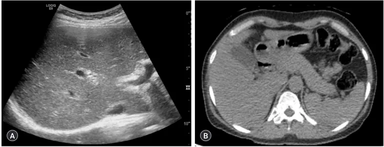
amination. Abdominal ultrasound and non-contrast computed tomography revealed coarsened hepatic echotexture, without any biliary duct abnormalities, and no evidence of pancreatitis such as peripancreatic swelling or a sausage-like lesion (Fig. 1). Blood chemistry revealed the following: white blood cells, 3,650/μL;
hemoglobin, 12 g/dL; platelets, 294× 10
3/μL; serum total pro- tein, 11.03 g/dL; serum albumin, 3.63 g/dL; total bilirubin, 1.02 mg/dL; serum aspartate aminotransferase (AST), 610 IU/L; ala- nine aminotransferase (ALT), 221 IU/L; alkaline phosphatase (ALP), 102 IU/L; gamma-glutamyl transferase (GGT), 176 IU/
L; amylase, 65 IU/L; lipase, 54 IU/L; and prothrombin time-in- ternational normalized ratio, 1.12.
Additional serologic tests, including serum hepatitis B surface antigen, hepatitis C antibody, anti-nuclear antibody (ANA), an- ti-smooth muscle antibody (SMA), anti-mitochondria antibody, anti-neutrophil cytoplasmic antibody, anti-liver kidney microso- mal type 1 antibody, and other viral tests—including Epstein-Barr virus and cytomegalovirus—were all negative. However, she ex- hibited a markedly elevated serum IgG level at 6,614 mg/dL (range, 700–1,600 mg/dL). Peripheral blood smear revealed the rouleaux formation of the red blood cells (RBC).
The patient was referred to the Department of Hematology to evaluate the possibility of a lymphoproliferative disorder, such as multiple myeloma (MM). Serum and urine protein electrophore- sis, free light chain ratio, skeletal survey, and bone marrow aspira- tion were performed. All were normal with the exception of an el- evated serum IgG4 subclass level, which was 221.3 mg/dL (range, 6–120 mg/dL).
A percutaneous ultrasound-guided liver biopsy was performed.
The pathologic results revealed moderate spotty necrosis and in- terface hepatitis with highly elevated IgG4-positive plasma cells (10–15/high power field [HPF]), consistent with AIH (Fig. 2).
The pretreatment revised IAIHG scoring was 17, consistent with a definite diagnosis of AIH. Detailed IAIHG scoring was as follows:
histological feature, 4; no drug and alcohol history, 3; negative find- ings of viral markers, 3; hypergammaglobulinemia, 3; ALP/AST ra- tio <1.5, 2; and female, 2.
Initially, she was treated with 60 mg prednisolone daily, which was tapered by 10 mg prednisolone weekly for 3 weeks. After 3 weeks, her laboratory test results were much improved as follows:
AST, 25 IU/L; ALT, 23 IU/L; GGT, 67 IU/L; and IgG, 2,067 mg/dL. Subsequently, 50 mg azathioprine was added for mainte- nance treatment, and 30 mg prednisolone was tapered by 5 mg weekly for 4 weeks. After 4 weeks, her laboratory test results were slightly worsened as follows: AST, 57 IU/L; ALT, 65 IU/L; GGT, 103 IU/L; and IgG, 1,367 mg/dL. Considering the risk of possi- ble worsening after early prednisone withdrawal, we increased the dose of prednisolone up to 20 mg and then slowly tapered every 2 weeks. After 1 month, all laboratory test results normalized (Fig. 3).
Discussion
In our case, the initial presumptive diagnosis was monoclonal or polyclonal gammopathy, such as MM or other lymphoprolifera- tive diseases, based on the initial serological results including those for inverted albumin/globulin ratio (A/G) ratio (0.33), ele- vated serum IgG, rouleaux formation of RBC, and negative sero- logic autoimmune markers. However, based on the results of the
A B
Fig. 1. Abdominal sonography and computed tomography showing (A) increased echogenicity of the liver parenchyma with heterogeneous echotexture and (B) normal pancreas without significant evidence of autoimmune pancreatitis.
Yeungnam Univ J Med 2020;37(2):136-140
liver biopsy and elevated serum IgG4 levels, the final diagnosis was IgG4-associated AIH which was treated with prednisolone and azathioprine.
Previous studies of IgG4-associated AIH are summarized in Table 1 [6,8-11]. Recently, a new disease entity called IgG4-asso- ciated AIH, which is different from classic AIH, has emerged [5].
Umemura et al. [5,6] had proposed that IgG4-AIH, as a subtype of AIH, is a rare disease characterized by the hepatic accumula- tion of IgG4-expressing plasma cells (≥10 IgG4 plasma cells per HPF) with markedly elevated serum IgG levels (≥135 mg/dL).
Of the 60 patients with AIH in Japan, only two (3%) were diag-
nosed with IgG4-associated AIH [6]. The median values of IgG and IgG4 levels were 4,015 mg/dL and 560 mg/dL, respectively.
These levels were higher than those of classic AIH (IgG, 2,940 mg/dL and IgG4, 22 mg/dL, respectively) [6]. Also, two pa- tients were diagnosed with IgG4-associated AIH, with positive ANA and SMA (1 of 2 patients), which was defined as a subtype of AIH [6]. In a Western study, based on Umemura’s histologic criterion, the prevalence of IgG4-associated AIH was 25% (7 pa- tients). The presence of the autoimmune antibodies was not sig- nificantly different from IgG4-associated AIH and classic AIH [8].
In our case, serum IgG and IgG4 levels were 6,614 mg/dL and 221.3 mg/dL, respectively. These markedly elevated IgG levels were four times greater than the normal upper level, which satis- fied Umemura’s serologic criterion. In the liver, IgG4-bearing plasma cells of 10–15 HPF also met Umemura’s histologic crite- rion for diagnosis of IgG4-associated AIH. Owing to the initially all-negative autoimmune antibody serology, marked elevated hy- pergammaglobulinemia, and inverted A/G ratio in our case, we preferentially considered a lymphoproliferative disorder such as MM. Though most IgG4-associated AIH had positive ANA or SMA [6,8], the frequency of autoantibody-negative AIH in pa- tients with acute and acute-severe clinical features reached 7% in one study [12]. Therefore, though patients show negative ANA, hypergammaglobulinemia, and elevated liver enzymes, addition- al serum IgG4 tests would be needed for differential diagnosis of IgG4-associated AIH. Then, when IgG4 is elevated, a subsequent liver biopsy is required for the definite diagnosis of IgG4-associ-
A B
Fig. 2. Histologic findings of liver. (A) Moderate interface hepatitis with lymphocytes and plasma cells infiltration in the portal tract is present (hematoxylin-eosin stain, x200). (B) Immunoglobulin G4-positive plasma cells are seen (immunohistochemical stain, x400).
800 700 600 500 400 300 200 100 0
8,000 7,000 6,000 5,000 4,000 3,000 2,000 1,000 0
Serum AST , AL T, GGT (IU/L) Serum lgG, (mg/dL)
AST ALT GGT
3 6 10 14 wk
lgGSteroid 30 mg + Azathioprine
50 mg Steroid 15 mg
→ 20 mg
Steroid 60 mg Biopsy