Introduction
Squatting is considered the best way to help re- store the strength of the quadriceps muscle in overall activities of daily living. The squatting movement increases knee extensor muscle strength, which then indirectly improves the quality of life in the field of strength and conditioning (Schoenfeld, 2010). Muscle forces also vary depending on the angle of the knee joint assumed during a squat (Barton et al, 2014).
The lower limb muscle activities also differ accord-
ing to the movement of the arms and length-tension relationship in squat exercises (Dionisio et al, 2008).
Many muscle groups can be activated simulta- neously when squatting is performed, as it is a mul- ti-joint task. Several studies have shown that vari- ous squat exercises resulted in altered quadriceps muscle activity, depending on the foot position, sur- face stability, intensity of load, and range of motion (Aspe and Swinton, 2014; Paoli et al, 2009). As a multi-joint exercise, the knee extensors are consid- ered to be the prime movers during a squat. In par- Corresponding author: Seung-chul Chon [email protected]
Comparison of the Electromyographic Changes in the Vastus Medialis Oblique and Vastus Lateralis Muscles According to the Knee Joint
Angle During Squat Exercise Using a Gym Ball
Hee-won Jeong, MSc, PT, Seung-chul Chon, PhD, PT Dept. of Physical Therapy, College of Medical Science, Konyang University
Abstract1)
Background: Squatting is one of the best functional exercises to strengthen the quadriceps muscle in independent gait and activities of daily living. Although the use of a gym ball during squat exercise is the most common way of strengthening the vastus medialis oblique (VMO) muscle, published data on this subject are limited.
Objects: The purpose of this study was to compare the sequential muscle activation of the VMO and vastus lateralis (VL) muscles during squat exercise using a gym ball at different knee flexion angles.
Methods: Forty healthy adults were randomly divided into experimental (squat exercise using a gym ball) and control (squat exercise alone) groups, in which squats were performed at 45° and 90° knee flexion. Electromyographic (EMG) activity data were collected over 10 seconds under the 2 angles of knee flexion (45° and 90°).
Results: There was significant group and time interaction effect for VMO and VL muscle activation at 45° knee flexion. This was similarly demonstrated at 90° knee flexion. No significant group main effect and time main effect for VMO and VL muscle activation were noted at 45° knee flexion, respectively. In contrast, there was significant group main effect and time main effect for VMO and VL muscle activation at 90° knee flexion. These significant differences were demonstrated through two-way analysis of variance over repeated measurements, suggesting that the EMG activity of the VMO muscle during squatting with a gym ball showed remarkable improvement compared to that of the VL muscle.
Conclusion: This research suggests that squat exercise using a gym ball may be more beneficial in improving the activity of VMO than of the VL muscle at both 45° and 90° of knee flexion, respectively.
We highly recommend squat exercises with a gym ball for selective strengthening of the VMO muscle in knee rehabilitation.
Key Words : Electromyography; Gym ball; Squat exercise; Vastus lateralis; Vastus medialis oblique.
ticular, the use of a gym ball is already widely ac- cepted as a common way to selectively strengthen the vastus medialis oblique (VMO) muscle in several clinical conditions (Oh, 2013). As the VMO and vastus lateralis (VL) muscles are antagonistic in terms of the knee joint position, the VMO and VL must be am- plified in a coordinated manner to ensure efficient neuromuscular function of the quadriceps muscle.
Knee joint problems such patellofemoral pain syn- drome alters the biomechanical imbalance between the VMO and VL muscles.
Numerous studies have been reported about the electromyographic (EMG) activity of the VMO and VL muscles (Cowan et al, 2001; Sheehy et al, 1998).
For instance, Boling et al (2006) reported that the VMO muscle activity increased during squat exercise with a gym ball because the activity of the hip ad- ductor muscle is promoted. And also, it was reported that squatting performance required hip adduction as well as knee flexion (Boling et al, 2006). Irish et al (2010) referred that VMO/VL muscle activity ratio for selective strengthening VMO is recommended to 1.2:1 as the closed-kinetic chain exercise.
In contrast, Dionisio et al (2008) reported that the VMO muscle activity is higher than that of the VL, regardless of the angle of the knee joint during squat exercises, which indicates that the movement strength of the quadriceps muscle increases by lumbar stabili- zation irrespective of the angle of the knee joints.
Park et al (2013) reported that below 100 degree of the knee joint only revealed increasing muscle activ- ity of the quadriceps muscle through repeated squat exercises. Koh et al (2011) reported that the force of hip adduction could increase the VL muscle activation during squatting. Kim (2012) suggested that foot po- sitioning and tibial torsion could affect the muscle activity of both the VMO and VL. Lastly, Barton et al (2014) reported that the activity of the gluteus maximus and gluteus medius muscles increased when squat exercise was performed using a gym ball.
Although evidence suggests that knee joint posi- tion drives the quadriceps muscle’s performance dur-
ing squatting (Oh, 2013; Park et al, 2013), little is known about the neuromuscular changes of the VMO and VL muscles that occur from a muscle ac- tivation standpoint. Elucidating how muscle activation patterns change in the knee extensors during squat- ting at different knee angles would enhance our un- derstanding of how one could capitalize on max- imizing muscle activation to specific evaluations and EMG normalization. The purpose of this study was to evaluate the muscle activity of the VMO and VL muscles during dynamic squatting using a gym ball compared with the existing static measures at two different knee joint angle positions.
Methods
Subjects
Forty healthy adults were recruited from a local community, and all of the subjects gave their in- formed consent. Randomization was done with sealed envelopes. The sealed letters for the experimental group (squat exercise using a gym ball) and the con- trol group (squat exercise alone) were arranged by the experimenter. The experimenter prepared group allocation on a sheet of paper and blindly gave it to the subjects. Subjects were allocated before the initial measurement. All of them participated in the measurements. The experimenter undertaking the measurement was also blinded to the group allocation.
The study subjects were free from any known medi- cal problems. Subjects with any neuromuscular path- ologies or history of spinal surgery were excluded from the study. The demographic characteristics of the study subjects are shown in Table 1.
Procedures
Surface EMG was used to record the amplitudes of the contractions of the VMO and VL muscles. These measurements were collected for the experimental and control groups to determine the sequential activation of these muscles during squatting. In order to reduce im-
pedance, each subject’s skin was shaved and an elec- trode gel was applied. A pair of active electrodes (inter-electrode distance of 2 ㎝) was placed in parallel over the muscle bellies to be tested. Tape and elastic straps were used to eliminate cable movement artifacts and to fix the electrode patches onto the skin.
The Myo-Research software (TeleMyo 2400T DTS, Noraxon Inc., Scottsdale, USA) was used to acquire the EMG signals at a sampling frequency of 1024 ㎐, and processed with a bandpass (20∼450 ㎐) and a 60 ㎐ notch filter. The root mean square (RMS) EMG amplitude for these muscles was calcu- lated using the average time during the squat ex- ercise, while the sequential activations of the VMO and VL muscle activities were displayed on a com- puter monitor. Electrode patches with a diameter of 1.1 ㎝ were placed on the VMO and VL muscles at standardized sites, as described by Gilleard et al (1998). To normalize the EMG signals recorded for each muscle, the RMS of 5 seconds maximal volun- tary isometric contraction (MVIC) was calculated for each muscle at the manual muscle testing positions, as recommended by Kendall et al (2005); all of the
EMG data were averaged over three repetitions of each measurement. The EMG signals collected were then expressed as a percentage of the calculated RMS of the MVIC (%MVIC).
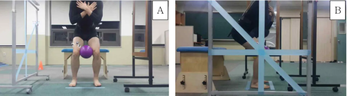
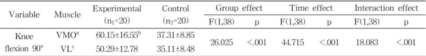
During the squat exercise with a gym ball of di- ameter 180 ㎜, subjects were asked to adopt the standing position, and a 10 seconds set that ranges from standing to sitting was performed with the legs spread shoulder-length apart, and the heels planted firmly on the ground, while bending to the instructed knee flexion angles (Figure 1). Subjects were then asked to squat with a gym ball between their knees while adducting the hips in order to suspend the ball. An automatic auditory cue via a verbal in- struction and visual feedback via a mirror was pro- vided by the experimenter. The order of testing was randomly determined for each subject in order to avoid the learning effect. The squat exercise was performed for 10 seconds at 2 different angles of knee flexion (45° and 90°), and the RMS EMG am- plitudes were collected as mean values.
The activation of the VMO and VL muscle was displayed on a computer monitor. Subjects were in- Parameters Experimental group (n1=20) Control group (n2=20) t p
Sex (female/male) 10/10 10/10
Age (year) 20±1.2a 20±1.1 -.271 .586
Height (㎝) 166.4±8.3 165.5±7.8 -.360 .574
Weight (㎏) 63.9±11.0 62.6±11.1 -.379 .953
amean±standard deviation.
Table 1. Demographic characteristics of the subjects (N=40)
A B
Figure 1. Squat positions (A: anterior view, B: lateral view).
structed to squat through contraction of the quad- riceps muscles, including the VMO and VL muscles, followed by low abdominal contraction using a gym ball, and then rest before the next measurement. An automatic auditory cue was used to trigger each squatting event, which lasted for 10 seconds over a trial period. Based on this protocol, an immediate collection of the muscle activity data was displayed on the monitor and stored for further analysis. Data that were unacceptable due to movement artifact were discarded, and the collection was then repeated.
The speed of the squat exercise was controlled to a comfortable 10 seconds to avoid fatigue of the lower limb muscles; a rest interval was given between each consecutive trial (Gefen et al, 2002).
Statistical analysis
The results are expressed as mean±standard deviation.
The normal distribution of the sample was tested using the Kolmogorov-Smirnov test, which showed a normal distribution for all variables. Two-way analysis of var- iance with repeated measures was used to assess the main effects (group and time effects) and the inter- action effects on each activation of the VMO and the VL muscles during squatting at 45° and 90° knee flexion, respectively. The collected data were ana- lyzed using a SPSS ver. 18.0 (SPSS Inc., Chicago, IL, USA). The level of statistical significance was set at a p value of <.05.
Results
There was no significant group effect (F=4.005, p=.530) and time effect (F=.307, p=0.058) for VMO
and VL muscle activation at 45° knee flexion.
However, at this angle, there was significant group and time interaction effect (F=9.316, p=.004) for VMO and VL muscle activation (Table 2), indicating that the EMG activity of the VMO muscle showed re- markable improvement compared to that of the VL muscle, attributable to squatting using a gym ball.
Similarly, there was significant group effect (F=26.025, p<.001) and time effect (F=44.715, p<.001) for VMO and VL muscle activation at 90° knee flexion. Additionally, at this angle, there was also significant group and time interaction effect (F=18.083, p<.001) for VMO and VL muscle (Table 3), suggest- ing that squat exercises using a gym ball showed notable improvement, compared with the controls.
Discussion
Several studies have investigated the effects of squat exercises on the performance of a variety of activities on the muscles around the knee joint (Aspe and Swinton, 2014; Paoli et al, 2009; Schoenfeld, 2010). Different knee joint positions influence the quadriceps muscle forces during squat exercises;
however, in spite of several results, little is known regarding the neuromuscular changes that occur from a quadriceps muscle activation standpoint. In this study, we evaluated the muscle activity of the VMO and VL muscles at two angles of knee joint flexion:
at 45° and 90°, respectively. The major finding of this investigation was that during squat exercises with a gym ball, both knee joint flexion positions demonstrated high VMO muscle activation; in con- trast, squat exercises performed on its own were as-
Variable Muscle Experimental (n1=20)
Control (n2=20)
Group effect Time effect Interaction effect
F(1,38) p F(1,38) p F(1,38) p
Knee flexion 45°
VMOa 22.26±6.54b 16.94±6.64
4.005 .530 .307 .058 9.316 .004
VLc 20.86±6.46 18.96±3.68
avastus medialis oblique,bmean±standard deviation, cvastus lateralis.
Table 2. Muscle activity of the vastus medialis oblique and the vastus lateralis at 45° knee flexion
sociated with lower VMO muscle activation values.
This result indicates that squat exercises using a gym ball was effective in strengthening the VMO muscle for quadriceps imbalance, considered one of the main contributing factors based on the neuro- muscular changes observed in patients with knee joint problems (Park et al, 2013).
During the squat exercise, the collected EMG data for VMO and VL muscles were methodologically normalized as percentage values of %MVIC. The re- sulting data from direct comparisons obtained for different subjects showed less reliability as absolute amplitudes of EMG activity vary broadly among subjects (Cram et al, 1998). Therefore, the EMG ac- tivity for the subjects in this study was expressed as the percentage of the activity performed during maximum voluntary contraction; the %MVIC value obtained thus indicated the efficacy of the muscle contraction. In order to analyze the EMG change of both VMO and VL during squatting, we collected EMG data for 10 seconds. The activity of the VMO and VL muscles begins from the start to the end of a squat. The EMG data collected during this 10 sec- onds squatting exercise therefore mainly represent the muscular efforts associated with the whole proc- ess, which activates the VMO and VL to properly adapt the knee joints to gravitational force.
To quantify the mechanical movements of knee pathologies, most studies utilized the VMO:VL ratio to reflect the relative contributions of the VMO and VL muscles (Sheehy et al, 1998). However, apart from these two muscles, several other muscles and joints interact around the knee joints. In this study, we focused on the activation of the VMO and VL muscles according to the changing angle of the knee
flexion in the sagittal plane. Additionally, variables such as knee joint height level, foot positions and body positions were well controlled during measure- ment process in this study.
The present EMG data are consistent with pre- vious findings investigating the effects of the appli- cation of a gym ball on VMO muscle activation dur- ing squatting. At 45° and 90° knee joint flexion, the amplitude of the root mean square VMO muscle EMG data during the squat exercise in combination with a gym ball increased by approximately 38% and 62%, compared with that of the squat exercise alone, from 16.94 to 22.26 %MVIC and from 37.31 to 60.15
%MVIC, respectively. Our results confirm that squat exercises performed with a gym ball at two different knee joint flexion positions can increase hip adductor muscle activity associated with lumbar stabilization, resulting in increased activity of the VMO muscle.
Additionally, although there were no statistically sig- nificant results between the 45° and 90° knee joint flexion, the VMO muscle activation tended to in- crease at the 90° flexion during squat exercises with a gym ball. These findings further indicate that low levels of knee flexion conditions may have produced recruitment of the hip adductor muscles, which effi- ciently stimulated the lumbar stabilization against the gym ball, leading to augmented squat exercise.
Certainly, these results have important clinical im- plications, as they demonstrated that squat exercise in combination with a gym ball is beneficial for se- lective recruitment of the VMO muscle and its closed-kinetic mechanism of action around the knee joint, and that the mechanism of stronger muscu- lar-fascial chaining can be further augmented by hip adductor muscles. Previous evidence on the clinical Variable Muscle Experimental
(n1=20)
Control (n2=20)
Group effect Time effect Interaction effect
F(1,38) p F(1,38) p F(1,38) p
Knee flexion 90°
VMOa 60.15±16.55b 37.31±8.85
26.025 <.001 44.715 <.001 18.083 <.001 VLc 50.29±12.78 35.11±8.48
avastus medialis oblique,bmean±standard deviation, cvastus lateralis.
Table 3. Muscle activity of the vastus medialis oblique and the vastus lateralis at 90° knee flexion
management of knee joint problems suggests that support and protection of the VMO muscle is essen- tial to stabilize the knee joints during selective strength training of the VMO muscle, thereby mini- mizing clinical complaints about patellofemoral pain syndrome, osteoarthritis, and knee joint instability.
Several shortcomings were identified in this re- search, which may be considered to enhance more robust and larger clinical studies in the future. First, this research represents a short-term period intended to investigate the immediate effects of squat exercises in combination with a gym ball among healthy subjects. Future studies examining the long-term ef- fects of the intervention in pathological populations, such as those suffering from patellofemoral pain syn- drome, osteoarthritis, and knee joint instability will be beneficial. Second, our sample size was admittedly small; a larger study will be required to corroborate our results. Third, muscle activity was evaluated ex- clusively with surface EMG, thereby opening the possibility of cross-talk or movement artifacts from the adjacent muscles during the squatting. Fourth, we did not consider more detailed kinematic or kinetic factors when measuring the effects of squatting.
Kinematic data detected by measurements such as motion analysis could provide more detailed move- ment pattern data. However, the emphasis of this re- search was to identify the changes in VMO and VL muscle activation patterns that may be considered a primary cause of the majority of knee pathologies.
Conclusion
This study showed that squat exercises combined with a gym ball is useful in enhancing muscle activa- tion in the VMO muscle. It offers clinical insights into the additive effects of a gym ball in selectively-stim- ulating the VMO muscle, and suggests that it may be used as an alternative squat exercise for the manage- ment of patients with patellofemoral pain syndrome, osteoarthritis, and knee joint instability.
References
Aspe RR, Swinton PA. Electromyographic and kinetic comparison of the back squat and overhead squat.
J Strength Cond Res. 2014;28(10):2827-2836.
https://doi.org/10.1519/JSC.0000000000000462 Barton CJ, Kennedy A, Twycross-Lewis R, et al.
Gluteal muscle activation during the isometric phase of squatting exercises with and without a Swiss ball. Phys Ther Sport. 2014;15(1):39-46.
https://doi.org/10.1016/j.ptsp.2013.02.006
Boling MC, Bolgla LA, Mattacola CG, et al.
Outcomes of a weight-bearing rehabilitation program for patients diagnosed with patellofe- moral pain syndrome. Arch Phys Med Rehabil.
2006;87(11):1428-1435.
Cowan SM, Bennell KL, Hodges PW, et al. Delayed onset of electromyographic activity of vastus medialis obliquus relative to vastus lateralis in subjects with patellofemoral pain syndrome.
Arch Phys Med Rehabil. 2001;82(2):183-189.
Cram JR, Kasman GS, Holtz J. Introduction to Surface Electromyography. 1st ed. Gaitheresburg, Aspen Publishers, 1998:408.
Dionisio VC, Almeida GL, Duarte M, et al.
Kinematic, kinetic and EMG patterns during downward squatting. J Electromyogr Kinesiol.
2008;18(1):134-143.
Gefen A, Megido-Ravid M, Itzchak Y, et al. Analysis of muscular fatigue and foot stability during high-heeled gait. Gait Posture. 2002;15(1):56-63.
Gilleard W, McConnell J, Parsons D. The effect of patellar taping on the onset of vastus medialis obliquus and vastus lateralis muscle activity in persons with patellofemoral pain. Phys Ther.
1998;78(1):25-32.
Irish SE, Millward AJ, Wride J, et al. The effect of closed-kinetic chain exercises and open-kinetic chain exercise on the muscle activity of vastus medialis oblique and vastus lateralis. J Strength Cond Res. 2010;24(5):1256-1262. https://doi.org/
10.1519/JSC.0b013e3181cf749f.
Kendall FP, McCreary EK, Provance PG, et al.
Muscle: Testing and function, with posture and pain. 5th ed. Baltimore, Lippincott Williams &
Wilkins, 2005:317-330.
Kim BJ. Comparison of quadriceps femoris muscle activations during wall slide squats. J Korean Soc Phys Med. 2012;7(4):541-550.
Koh EK, Lee KH, Jung DY. The effect of isometric hip adduction and abduction on the muscle ac- tivities of vastus medialis oblique and vastus lateralis during leg squat exercises. Korean Journal of Sports Biomechanics. 2011;21(3):361-368.
Oh TY. The effects of squatting exercise with gym ball and wall on lower extremity muscles activation. J Korean Soc Phys Med. 2013;8(4):
647-653.
Paoli A, Marcolin G, Petrone N. The effect of stance width on the electromyographical activity of eight superficial thigh muscles during back squat with different bar loads. J Strength Cond Res. 2009;23(1):246-250.
Park SJ, Choi GR, Kim CK. Comparison and analysis of muscle activities on angles of knee joint dur- ing squat exercise. Journal of Sport and Leisure Studies. 2013;53(2):879-887.
Schoenfeld BJ. Squatting kinematics and kinetics and their application to exercise performance.
J Strength Cond Res. 2010;24(12):3497-3506.
https://doi.org/10.1519/JSC.0b013e3181bac2d7 Sheehy P, Burdett RG, Irrgang JJ, et al. An electro-
myographic study of vastus medialis oblique and vastus lateralis activity while ascending and de- scending steps. J Orthop Sports Phys Ther.
1998;27(6):423-429.
This article was received September 19, 2016, was reviewed September 19, 2016, and was accepted November 7, 2016.