Identification of C4orf32 as a Novel Type I Endoplasmic Reticulum Resident Membrane Protein
Seung-Hwan Lee1, Sang-Won Park1, Jin-A Lee2* and Deok-Jin Jang1*
1Department of Ecological Science, College of Ecology and Environment, Kyungpook National University, 2559, Gyeongsang-daero, Sangju-si, Gyeongsangbuk-do, 37224, Korea
2Department of Biological Science and Biotechnology, College of Life Science and Nano Technology, Hannam University, 1646, Yuseong-daero, Yuseong-gu, Daejeon 34054, Korea
Received July 17, 2019 /Revised September 5, 2019 /Accepted September 9, 2019
Membrane topology is a key characteristic of membrane proteins. We previously reported the cloning of the chromosome 4 open-reading frame 32 (C4orf32) gene as a potential membrane protein; how- ever, the cellular localization and membrane topology of C4orf32 was as yet unknown. In this study, we found that green fluorescent protein (GFP) fused to the C-terminus of C4orf32 (C4orf32-GFP) was localized to the endoplasmic reticulum (ER). We applied three tools to identify determinants of C4orf32 topology: protease protection, fluorescence protease protection (FPP), and an inducible system using the ternary complex between FK506 binding protein 12 (FKBP), rapamycin, and the rapamy- cin-binding domain of mTOR (FRB) (the FRB-rapamycin-FKBP system). Using protease protection and FPP assays, we found that the GFP tag in C4orf32-GFP was localized to the cytoplasmic surface of the ER membrane of HeLa cells. Protease protection and FPP assays are useful and complimentary tools for identifying the topology of GFP fusion membrane proteins. The FRB-rapamycin-FKBP system was also used to study the topology of C4orf32. In the absence of rapamycin, a monomeric red fluo- rescent protein–FKBP fusion (mRFP-FKBP) and C4orf32-GFP-FRB were localized to the cytoplasm and the ER membrane, respectively. However, in the presence of rapamycin, the mRFP-FKBP was shifted from the cytoplasm to the ER and colocalized with the C4orf32-GFP-FRB. These results in- dicate that the FRB moiety is facing the cytoplasmic surface of ER membrane. Overall, our results clearly suggest that C4orf32 belongs to the family of type I ER resident membrane proteins.
Key words : C4orf32, FPP assay, FRB-rapamycin-FKBP complex, protease protection assay, Type I ER membrane protein
*Corresponding authors
*Tel : +82-54-530-1213, Fax : +82-54-530-1218
*E-mail : [email protected] (Deok-Jin Jang)
*Tel : +82-42-629-8785, Fax : +82-42-629-8789
*E-mail : [email protected] (Jin-A Lee)
This is an Open-Access article distributed under the terms of the Creative Commons Attribution Non-Commercial License (http://creativecommons.org/licenses/by-nc/3.0) which permits unrestricted non-commercial use, distribution, and reproduction in any medium, provided the original work is properly cited.
Journal of Life Science 2019 Vol. 29. No. 9. 949~954 DOI : https://doi.org/10.5352/JLS.2019.29.9.949
Introduction
Membrane proteins play crucial roles in various cellular functions including cell signaling and intracellular traffick- ing between different membranous organelles within cells.
Transmembrane proteins can be classified into type I, II, and multi-membrane-spanning proteins [8]. Type I and II trans- membrane proteins have a single transmembrane domain.
In type I transmembrane proteins, the N-terminus of the pro- tein faces the extracellular side of the plasma membrane or
the luminal side of intracellular organelles. In type II trans- membrane proteins, the C-terminus of the protein faces the extracellular side in the plasma membrane or the luminal side of intracellular organelles.
To elucidate the topology of membrane proteins in mem- branous organelles, various methods are available including fluorescence-based and Western blotting assays [4]. Protease protection and fluorescence protease protection (FPP) assays are based on the principle that externally added protease can cleave only the accessible portions of membrane pro- teins; therefore, luminal and membrane portions are pro- tected [7]. In a protease protection assay, the plasma mem- brane is mechanically disrupted, while in an FPP assay, the cholesterol-rich plasma membrane is permeabilized by brief digitonin treatment before adding proteinase K or trypsin to cleave the cytoplasm-facing portions of membrane pro- teins. Peptides are then detected by Western blotting in pro- tease protection assays or by confocal microscope in FPP assays. Therefore, for the study of GFP-fused membrane pro-
teins, these two tools are useful for identifying membrane topology.
Another tool is an inducible system using the ternary complex between FK506 binding protein 12 (FKBP), rapamy- cin, and the rapamycin-binding domain of mTOR (FRB) [10].
FRB binds to FKBP in the presence of rapamycin. Normally, FRB is fused to the membrane protein, which is localized to membranous organelles, and monomeric red fluorescent protein (mRFP) is fused to FKBP (mRFP-FKBP), which is lo- calized in the cytoplasm. However, in the presence of rapa- mycin, if the FRB moiety is localized to the cytoplasmic sur- face of a membranous organelle by virtue of the protein it is fused with, then the mRFP-FKBP becomes co-localized with that membranous organelle. We have used this system in previous studies to verify the topology of other membrane proteins [2, 3].
We previously reported the cloning of C4orf32, but its cellular localization and topology was as yet unclear [2]. In this study, we found that C4orf32 was localized to the ER membrane. Using three different tools, including protease protection, FPP, and FRB-rapamycin-FKBP assays, we showed that the human chromosome 4 open reading frame 32 gene (C4orf32) was localized to the ER. We show that these three methods are useful tools for identifying the topology of GFP-fused membrane proteins in intracellular organelles in- cluding ER and mitochondria.
Material and Methods
DNA constructs
Human C4orf32 was cloned by RT-PCR from HeLa cell cDNA using a nested polymerase chain reaction (PCR) with the following specific primer sets: hC4orf32-S (GAGTTGG CTGCGGGATGT)/hC4orf32-A1 (GGCATGGGGATAGAGG TGTA) (first-round PCR) and hC4orf32-S/hC4orf32-A2 (AC AGCCAGCACATGATTTGA) (second-round PCR). The am- plified fragment was inserted into an N3-pEGFP vector to generate N3-C4orf32-pEGFP using the restriction enzymes HindⅢ and Kpn1. N3-C4orf32-3xFLAG and N3-C4orf32- pEGFP-FRB were amplified by PCR using FLAG-S (GACGG TACCGACTACAAAGACCATGAC), FLAG-A (ATAAGAA TGCGGCCGCTTACTTGTCATCGTCATCCTT), pEGFP-S (CGCGGATCCGTGAGCAAGGGCGAGGAG), FRB-A (AT AAGAATGCGGCCGCCTACTTTGAGATTCGTCGGAA) and inserted in the N3-C4orf32-pEGFP vectors using the re- striction enzymes Kpn1 and Not1. N3-mPRMT8 (N20)-GFP
and SP-GFP were amplified by PCR using mPRMT8-S (CGCCCAAGCTTGCCACCATGGCGGAGAATGCAGTC), mPRMT8-A (GACGGTACCTGCATTCTCCGCCATTTT), SP- S2 (CTAGGGCTAGCGCCACCATGT TATTGCAAGCTTTT TTATTTCTGC), SP-S1 (AAGCTTTTTTATTTCTGCTGGCA GGT TTTGCAGCAAAGAT), SP-A (GACGGTACCAGAGG CAGAAATCTTTGCTGCAAAACCT) and inserted in the N3-pEGFP vectors using the restriction enzymes HindIII, Kpn1 and Nhe1. Lyn11 (#79572), Sec61 (#49155) were ob- tained from Addgene (Cambridge, MA, USA).
Cell culture
Mouse embryonic fibroblast (MEF) cells, HEK293T cells, and HeLa cells were grown in Dulbecco’s modified Eagle’s medium (DMEM) supplemented with 10%(v/v) fetal bovine serum (FBS) and penicillin/streptomycin in a humidified at- mosphere of 5%(v/v) CO2 at 37℃. Cells were seeded in a sticky-slide 8-well system (#80828; Ibidi, Martinsried, Germany) to obtain 40-60% conflence on the day of imaging and in 10 cm cell culture dishes (#20100, Spl Life Science, Pocheon, South Korea) for other assays. Cells were trans- fected with plasmid DNA constructs using calcium phos- phate (Takara Bio, Kusatsu, Shiga, Japan) or Lipofectamine 2000 (Life Technologies, Carlsbad, CA, USA) 24-26 hr before imaging. The relative amount of each construct was empiri- cally determined based on the relative expression of each construct combination. Cells were observed under an in- verted Zeiss LSM-700 scanning laser confocal microscope operated by ZEN software (Carl Zeiss, Oberkochen, Ger- many). The laser lines for excitation and the emission wave- lengths for the fluorochromes were 488 with 508-543 nm for GFP and 561 with 578-649 nm for mRFP, respectively.
Appropriate GFP (500-550 nm) and mRFP (575-625 nm) emission fllters were used during the sequential imaging of each fluorescent protein. Most images were taken with live cells.
Fluorescence Protease Protection (FPP) assay HeLa cells were transfected with lipofectamine and grown for 36 hr, then the cell culture media was removed, and cells were washed three times with KHM buffer (110 mM Potassium acetate, 20 mM pH 7.5, and 2 mM MgCl2). The 8-well slide containing cells in KHM buffer was recorded by fluorescence microscope for the first image, which repre- sents the control, or the “pre-permeabilization” stage. To permeabilize the plasma cell membrane, 0.0025% digitonin
A
B
C
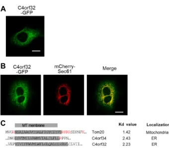
Fig. 1. ER localization of hC4orf32 in HeLa cells. (A) Cellular localization of C4orf32-GFP in HeLa cells. C4orf32- GFP is expressed in the network structure within the cytoplasm. Scale bar, 20 μm. (B) Co-localization of C4 orf32-GFP with mCherry-Sec61, an ER marker in HeLa cells. Scale bar, 20 μm. (C) Sequence alignment of the transmembrane domains and their flanking regions for Tom20, C4orf34, and C4orf32, and their Kd values.
MT, mitochondria; ER, endoplasmic reticulum.
(D141, SIGMA, Saint Louis, USA) in KHM buffer was added to the cells for 90 sec. Efficient permeabilization of most cells occurred within 1~2 min. Images of the cells in the “post- permeabilization” stage were then taken. Cells were washed with KHM buffer again and added KHM buffer with 7.5 μg/ml of PK for 90 sec. Images were taken to determine whether fluorescent signals persisted or disappeared.
FRB/rapamycin/FKBP assay
For determination of membrane protein topology in vivo, FRB-FKBP DNA constructs were co-transfected by lipofect- amine into MEF cells and grown for 24 hr. 2 μM rapamycin (R8781, SIGMA, Saint Louis, USA) was treated to form the FRB-FKBP complex. After rapamycin treatment, the cell morphology changed rapidly, so images were recorded within 3 min after rapamycin treatment.
Protease protection assay
For protease protection assays, DNA constructs were transfected into HEK293T cells with calcium phosphate and grown for 36 hr. Cells were harvested, washed twice with PBS, sedimented (500x g for 5 min at 4℃), and resuspended in proteinase K (PK) buffer (20 mM Hepes pH 7.6, 220 mM Mannitol, 70 mM Sucrose). Cells were homogenized by 10 passages through a 27-gauge needle. To obtain supernatant, homogenized cells were sedimented by centrifugation at 500x g for 10 min at 4℃. The supernatant was collected and divided equally into three tubes. Samples were treated with 25 μg/ml PK on ice for 30 min in the presence or absence of 1% Triton-100 (TX-100). The reaction was stopped by the addition of 1 mM PMSF (Proteinase K inhibitor) (#P7626, SIGMA, Saint Louis, USA) for 30 min on ice. Samples were precipitated with trichloroacetic acid (TCA), and then wash- ed by 100% acetone. Precipitated samples were dried for 18 hr. Pellets were resuspended in SDS sample buffer and ana- lyzed by SDS-PAGE.
Western Blot
Sample of protease protection assay were separated by SDS-PAGE, and immunoblotted with the following primary anti-ERP57 antibodies (#SC-23886, Santa Cruz, Heidelberg, Germany), anti-green fluorescent protein (GFP) antibodies (#D1717, Santa Cruz, Heidelberg, Germany), and anti-B-ac- tin antibodies (#SC47778, Santa Cruz, Heidelberg, Ger- many), overnight at 4℃. After several washes in TBST buffer (20 mM Tris pH 7.5, 150 mM NaCl, and 0.05% Tween),
Secondary goat anti-mouse IgG-HRP (#SC2005, Santa Cruz, Heidelberg, Germany) antibodies were used for 30 min at room temperature. Advansta WesternBright ECL (K-12045- D50) were used in visualizing PVDF membrane.
Results and Discussion
ER localization of C4orf32
As reported previously, human C4orf32 has 132 putative amino acids and one potential transmembrane domain (TMD) [6]. C4orf32 has no putative N-terminal signal se- quences that are known to target proteins to intracellular organelles. Many transmembrane proteins have no a signal sequence for organelle targeting; rather, a TMD itself acts as the targeting sequence [6].
To examine its cellular localization, C4orf32-GFP was ex- pressed in HeLa cells. The expression of C4orf32-GFP showed a network-like pattern including the nuclear enve- lope in HeLa cells (Fig. 1A). ER usually exhibits an intra- cellular network pattern within cells; therefore, to determine whether C4orf32 was localized to the ER, we expressed C4orf32-GFP together with mCherry-Sec61, where the Sec61 protein is a known ER marker in HeLa cells. Human C4orf32-GFP was co-localized with mCherry-Sec61 (Fig. 1B),
A B
Fig. 2. Identification of C4orf32 topology using pro- tease protection assay and FPP assay. (A) Western blot analysis of C4orf32-GFP using protease protection assay. Endogenous ERP57 and actin were used as control markers. (B) FPP assay indicates the top- ology of C4orf32-GFP. Individual images were taken before and after treatment with 0.0025% digitonin and 7.5 μg/ml of Protei- nase K as indicated. Scale bar, 20 μm.
SP-GFP, signal peptide-GFP; PK, protei- nase K; Digi, digitonin.
confirming the ER localization of C4orf32-GFP. Overall, our data suggest that C4orf32 is targeted to ER in HeLa cells.
It is well known that membrane proteins with strong hy- drophobicity are mainly recruited to the ER, whereas mem- brane proteins with moderate hydrophobicity are recruited to the mitochondria via their binding to unidentified protein complexes [6, 11]. Therefore, we calculated hydrophobicity values for the predicted TMD of C4orf32 by using the Kyte–
Doolittle (Kd) hydrophobicity scale. As a control, we exam- ined the Kd value of Tom20 as a mitochondrial targeting protein, and of C4orf34 as an ER targeting protein. The Kd values of Tom20 and C4orf34 are 1.42 and 2.43, respectively.
As shown in Figure 1C, the Kd value of C4orf32 is 2.23, which is similar to that of C4orf34. Taken together, C4orf32 has strong hydrophobicity in its TMD domain and is lo- calized to ER membrane.
Determination of C4orf32 topology using protease protection and FPP assays
Next, to confirm the topology of C4orf32, we used a pro- tease protection assay in HEK293T cells expressing C4orf32- GFP. As controls, endogenous ERp57 was the ER luminal protein reference and endogenous actin was the cytosol lo- calization reference. As shown in Fig. 2A, C4orf32-GFP was degraded by protease protection in the absence or presence of non-ionic detergent TX-100 similar to endogenous actin.
In contrast, endogenous ERp57, an ER lumen-resident pro- tein was degraded by PK only in the presence of TX-100.
These results suggest that GFP domain in C4orf32-GFP is located to the cytoplasmic surface of ER membrane, indicat- ing that C4orf32-GFP belongs to type I ER transmembrane
protein.
To further confirm the topology of C4orf32-GFP, we used an FPP assay. As a control, we used signal peptide-GFP (SP-GFP) which is expressed on the luminal side of the ER and PRMT8 (N20)-GFP, which is expressed on the cytoplas- mic surface of the plasma membrane and the luminal side of mitochondria [9]. In C4orf32-GFP- or PRMT8 (N20)-GFP- expressing HeLa cells, digitonin was first applied for 50 sec and then replaced with PK solution for additional 80 sec.
As shown Figure 2B, significant GFP signal remained in the plasma membrane and mitochondria in PRMT8 (N20)-GFP-, SP-GFP-, or C4orf32-GFP-expressing HeLa cells. After PK solution was added, GFP signal in the plasma membrane was greatly reduced, but GFP signal in mitochondria or in ER still was remained in PRMT8 (N20)-GFP-expressing HeLa cells. On the other hand, GFP signal in ER membrane was greatly reduced in C4orf32-GFP-expressing HeLa cells.
These results indicate that the GFP domain in C4orf32-GFP is facing the cytoplasmic surface of ER membrane.
Collectively, our results from both the proteinase protection FPP assays suggest that C4orf32 is a type I ER membrane protein.
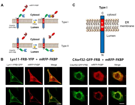
Determination of topology of C4orf32 using FRB- rapamycin-FKBP complex
To further confirm the topology of C4orf32, we used an inducible system of the ternary complex between FKBP, ra- pamycin, and FRB [2, 10]. In FRB-rapamycin-FKBP system, FRB cannot bind to FKBP in the absence of rapamycin. FRB can form a ternary structure with rapamycin and FKBP only in the presence of rapamycin. In our experiments, to perform
A
B
C
Fig. 3. Identification of C4orf32 topology using FRB-rapamycin-FKBP. (A) A schematic diagram of an experimental model of the GFP-FRB/rapamycin/mRFP-FKBP system. If the C-terminus of C4orf32 is exposed to the cytoplasmic surface of the ER membrane, then FRB fusion to the exposed C-termini of C4orf32-GFP should be able to recruit cytosolic mRFP-FKBP to ER membrane in the presence of rapamycin (upper). On the other hand, if the N-terminus of C4orf32 is exposed to the cytoplasmic surface of the ER membrane (Type II), then FRB fusion to the exposed C-termini of C4orf32-GFP should not be able to recruit mRFP-FKBP to ER membrane in the presence of rapamycin. (B) The cellular localization of C4orf32-GFP-FRB or Lyn11-FRB-YFP with mRFP-FKBP in the absence or presence of rapamycin in HEK293T cells. The localization of mRFP-FKBP was shifted from cytoplasm to ER or the plasma membrane by adding rapamycin in HEK293T cells co-expressing with C4orf32-GFP-FRB or Lyn11-FRB-YFP Ly, respectively. Rapa, 1 μM rapamycin. Scale bar, 20 μm.
the FRB-rapamycin-FKBP assay, the FRB domain was in- serted into the C-terminus of C4orf32-GFP (C4orf32-GFP- FRB). We used mRFP fused to FKBP (mRFP-FKBP). If the C-terminus of C4orf32 is exposed to the cytoplasmic surface of the ER membrane (Type I ER resident membrane protein), then FRB fusion to the exposed C-termini of C4orf32-GFP should be able to recruit mRFP-FKBP to ER membrane in the presence of rapamycin (Fig. 3A). On the other hand, if the N-terminus of C4orf32 is exposed to the cytoplasmic sur- face of the ER membrane (Type II ER membrane protein), then FRB fusion to the exposed C-termini of C4orf32-GFP should not be able to recruit mRFP-FKBP to ER membrane in the presence of rapamycin (Fig. 3A). We then co-trans- fected C4orf32-GFP-FRB with mRFP-FKBP into MEF cells.
As a control, Lyn11-FRB-YFP was used. As shown in Figure 3B, mRFP-FKBP and C4orf32-GFP-FRB were expressed in cy- toplasm or ER in the absence of rapamycin in HeLa cells,
respectively. However, in the presence of 1 μM rapamycin, mRFP-FKBP was shifted from cytoplasm to ER in C4orf32- GFP-FRB expressing cells (Fig. 3B). With the control con- structs, mRFP-FKBP and Lyn11-FRB-YFP were expressed in cytoplasm and plasma membrane, respectively, in the ab- sence of rapamycin. However, in the presence of 1 μM rapa- mycin, mRFP-FKBP was shifted from cytoplasm to plasma membrane in Lyn11-FRB-YFP-expressing cells (Fig. 3B). This result indicates that the FRB moiety is facing the cytoplasmic surface of ER, in which cytoplasmically expressed mRFP- FKBP is associated with C4orf32-GFP-FRB in the presence of rapamycin (Fig. 3B). Taken together, our results suggest that the N-terminus of C4orf32 is facing the luminal side of the ER and that the C-terminus of C4orf32 is facing the cytoplasm (Fig. 3C).
As shown above, C4orf32 is localized to ER membrane.
For membrane proteins of the secretory pathway, including
초록:Type I 소포체 목표화 막단백질에 속하는 새로운 C4orf32 막단백질의 동정 이승환1․박상원1․이진아2*․장덕진1*
(1경북대학교 생태환경대학 생태과학과, 2한남대학교 생명나노과학대학 생명시스템과학과)
세포막 단백질의 topology는 막단백질의 중요한 특징이다. 우리는 이전에 C4orf32단백질을 클로닝 하였으나, 이 단백질의 세포내 위치나 topology는 알지 못했다. 이번 연구를 통해 C4orf32는 세포내에서 소포체에 위치되는 막단백질임을 알게 되었다. C4orf32의 topology를 알기 위해 protease protection assay, fluorescence protease protection (FPP) assay, FRB/rapamycin/FKBP system을 활용하였다. Protease protection assay와 FPP assay를 적용한 결과 C-말단에 GFP를 붙인 C4orf32-GFP의 경우 GFP가 소포체의 세포질 표면에 위치함을 확인할 수 있었 다. 또한, FRB/rapamycin/FKBP시스템을 이용한 실험에서 rapamycin이 처리되지 않은 경우는 mRFP-FKBP가 세 포질에 위치하다가 rapamycin이 처리되면 C4orf32-GFP-FR가 위치하는 소포체로 이동함을 확인할 수 있었다. 이 러한 사실은 C4orf32의 C-말단이 소포체의 세포질쪽 면에 위치한다는 사실을 말해준다. 이러한 연구를 통해 C4orf32는 Type I 소포체 막단백질에 속한다는 사실을 확인할 수 있었다.
the ER and Golgi complex, N-linked glycosylation-based as- says are used including glycosylatable GFP (gGFP) or in- troduction of N-linked glycosylation sites by site-directed mutagenesis [2, 5]. Normally, oligosaccharyl transferase rec- ognizes an N-X-(T/S) (X is not proline) and adds oligo- saccharides onto the asparagine residue within the endoplas- mic reticulum (ER) lumen [1]. However, glycosylation effi- ciency is dependent on various factors and therefore, it is difficult to determine whether potential N-glycosylation sites are in fact glycosylated in cells. Thus, the combination of an FPP assay, a protease protection assay and an FRB-ra- pamycin-FKBP complex assay is a useful strategy for identi- fying the topology of GFP-fused membrane proteins in vari- ous membranous organelles in cells.
Acknowledgement
This work was supported by the Kyungpook National University Research Fund, 2017.
References
1. Dempski, R. E. Jr. and Imperiali, B. 2002. Oligosaccharyl transferase: gatekeeper to the secretory pathway. Curr. Opin.
Chem. Biol. 6, 844-850
2. Jun, M. H., Jun, Y. W., Kim, K. H., Lee, J. A. and Jang, D. J. 2014. Characterization of the cellular localization of C4orf34 as a novel endoplasmic reticulum resident protein.
BMB Reports 47, 563-568.
3. Jun, Y. W., Park, H., Lee, Y. K., Kaang, B. K., Lee, J. A.
and Jang, D. J. 2016. D-AKAP1a is a signal-anchored protein in the mitochondrial outer membrane. FEBS Lett. 590, 954- 961
4. Lee, H. and Kim, H. 2014. Membrane topology of trans- membrane proteins: determinants and experimental tools.
Biochem. Biophys. Res. Commun. 453, 268-276
5. Lee, H., Lara, P., Ostuni, A., Presto, J., Johansson, J., Nilsson, I. and Kim, H. 2014. Live-cell topology assessment of URG7, MRP6102 and SP-C using glycosylatable green fluorescent protein in mammalian cells. Biochem. Biophys. Res. Commun.
450, 1587-1592
6. Lee, J., Kim, D. H. and Hwang, I. 2014. Specific targeting of proteins to outer envelope membranes of endosymbiotic organelles, chloroplasts, and mitochondria. Front Plant Sci.
5, 173
7. Lorenz, H., Hailey, D. W., Wunder, C. and Lippincott- Schwartz, J. 2006. The fluorescence protease protection (FPP) assay to determine protein localization and membrane topology. Nat. Protoc. 1, 276-279
8. Ott, C. M. and Lingappa, V. R. 2002. Integral membrane protein biosynthesis: why topology is hard to predict. J. Cell Sci. 115, 2003-2009
9. Park, S. W., Jun, Y. W., Choi, H. E., Lee, J. A. and Jang, D. J. 2019. Deciphering the molecular mechanisms under- lying the plasma membrane targeting of PRMT8. BMB re- ports, in press
10. Suh, B. C., Inoue, T., Meyer, T. and Hille, B. 2006. Rapid chemically induced changes of PtdIns(4,5)P2 gate KCNQ ion channels. Science 314, 1454-1457
11. Walther, D. M. and Rapaport, D. 2009. Biogenesis of mi- tochondrial outer membrane proteins. Biochem. Biophys. Acta 1793, 42-51.